Spine Surgery and Related Research
Online ISSN : 2432-261X
ISSN-L : 2432-261X
Volume 1, Issue 2
Displaying 1-9 of 9 articles from this issue
REVIEW ARTICLE
  • Yasuaki Tokuhashi, Hiroshi Uei, Masashi Oshima
    2017 Volume 1 Issue 2 Pages 44-55
    Published: April 20, 2017
    Released on J-STAGE: December 20, 2017
    JOURNAL OPEN ACCESS

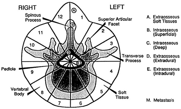
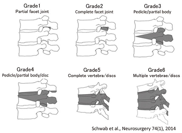
    Introduction: Accurate evaluation of metastasis and life prognosis is essential for selecting a suitable therapeutic strategy for metastatic spine tumors owing to limitations in treatment options. For this purpose, various classification, evaluation, and scoring systems have been developed.

    Methods: Classification, evaluation, and scoring systems for metastatic spine tumors reported to date were identified by performing a literature search on PubMed. We reviewed the most cited classifications and scorings before 2009, and all classifications and scorings reported after 2010 from the search results.

    Results: Six classifications and 23 scorings were reviewed. The classification/evaluation methods are divided into 1) anatomical classification/evaluation methods, 2) evaluation methods for neurological symptoms/instability, and 3) scoring systems for predicting life expectancy. The first 2 were useful for the planning and evaluation of surgical indications. Scoring systems for life prognosis also permitted rough prediction of the outcomes and were useful for the selection of a suitable treatment. However, variation of the patient background, diversity of adopted prognostic factors, and the absence of scoring systems that could predict the outcome with an accuracy of 90% or higher introduced some limitations.

    Conclusion: The identified classification, evaluation, and scoring systems have been generally useful for treatment strategies. However, we emphasize the necessity of multidisciplinary development and revision of classification and evaluation methods to adapt to the prolongation of survival associated with increased diversity and improvement of treatment options.

    Download PDF (575K)
  • Yukihiro Matsuyama
    2017 Volume 1 Issue 2 Pages 56-60
    Published: April 20, 2017
    Released on J-STAGE: December 20, 2017
    JOURNAL OPEN ACCESS

    Planning of surgical treatment requires determination of whether osteoarthritis of the spine with lumbar pain should be treated as spinal canal stenosis with or without deformity or as lumbar degenerative scoliosis and clarification of whether treatment should be provided for both. When sagittal spinal alignment is not appropriate, symptoms of adult spinal deformity may be observed. To correct this deformity, appropriate pelvic tilt should be obtained similar to that in healthy adults, and, especially, the retroversion of the pelvis should be corrected.

    Download PDF (476K)
  • Yawara Eguchi, Hirohito Kanamoto, Yasuhiro Oikawa, Munetaka Suzuki, Ha ...
    2017 Volume 1 Issue 2 Pages 61-71
    Published: April 20, 2017
    Released on J-STAGE: December 20, 2017
    JOURNAL OPEN ACCESS

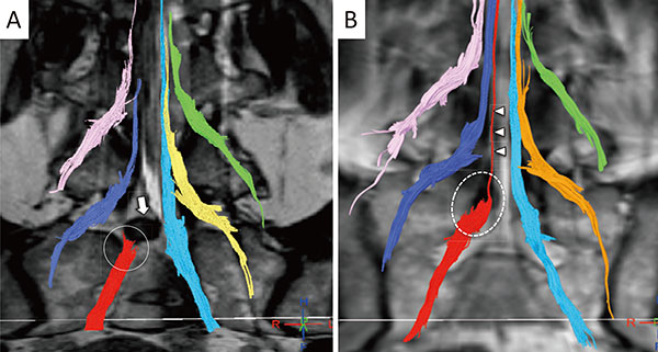
    Much progress has been made in neuroimaging with Magnetic Resonance neurography and Diffusion Tensor Imaging (DTI) owing to higher magnetic fields and improvements in pulse sequence technology. Reports on lumbar nerve DTI have also increased considerably.

    Many studies have shown that the use of DTI in lumbar nerve lesions, such as lumbar foraminal stenosis and lumbar disc herniation, makes it possible to capture images of interruptions of tractography at stenotic sties, enabling the diagnosis of stenosis. DTI can also reveal significant decreases in fractional anisotropy (FA) with significant increases in apparent diffusion coefficient (ADC) values in compression lesions.

    FA values have higher accuracy than ADC values. Furthermore, strong correlations exist between FA values and indications of neurological severity, including the Japanese Orthopedic Association (JOA) score, the Oswestry Disability Index (ODI), and the Roland-Morris Disability Questionnaire (RDQ) in patients with lumbar disc herniation-induced radiculopathy.

    Most lumbar DTI has become 3T; 3T MRI has made it possible to take high-resolution DTI measurements in a short period of time. However, increased motion artifacts in the magnetic susceptibility effect lead to signal irregularities and image distortion. In the future, high-resolution DTI with reduced field-of-view may become useful in clinical applications, since visualization of nerve lesions and quantification of DTI parameters could allow more accurate diagnoses of lumbar nerve dysfunctions. Future translational studies will be necessary to successfully bring MR neuroimaging of lumbar nerve into clinical use.

    Download PDF (1533K)
ORIGINAL ARTICLE
  • Tsutomu Akazawa, Toshiaki Kotani, Tsuyoshi Sakuma, Shohei Minami, Sumi ...
    2017 Volume 1 Issue 2 Pages 72-77
    Published: April 20, 2017
    Released on J-STAGE: December 20, 2017
    JOURNAL OPEN ACCESS

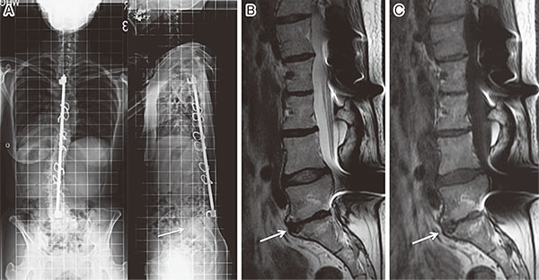
    Introduction: The purpose of this study was to investigate the long-term incidence of lumbar disc degeneration and Modic changes in the non-fused segments of patients with adolescent idiopathic scoliosis (AIS) who previously underwent spinal fusion.

    Methods: Study subjects consisted of 252 patients with AIS who underwent spinal fusion between 1968 and 1988. Of 252 patients, 35 subjects underwent lumbar spine MRI and whole spine X-ray examination. The mean patient age at the time of follow-up was 49.8 years, with an average follow-up period of 35.1 years. We classified the subjects into two groups based on the lowest fused vertebra: H group whose lowest fused vertebra was L3 or higher levels and L group whose lowest fused vertebra was L4 or lower levels.

    Results: The L group had significantly advanced disc degeneration on MRI. There was no significant difference between two groups in Modic changes. The L group showed less lumbar lordosis than the H group (H group: 48.1 degrees; and L group: 32.1 degrees) and greater SVA (H group: 1.2 cm; and L group: 5.5 cm).

    Conclusions: In AIS patients, 35 years after spinal fusion surgery on average, we evaluated lumbar disc degeneration and Modic changes of the non-fused segments. In patients with the lowest fusion level at L4 or lower, there were reduced lumbar lordosis, considerable SVA imbalance, and severe disc degeneration compared with those with the lowest fusion level at L3 or higher. The lowest fusion level at L3 or higher is recommended to reduce disc degeneration in midlife.

    Download PDF (202K)
  • Ryo Sugawara, Katsushi Takeshita, Yasuhisa Arai, Masashi Takaso, Jun T ...
    2017 Volume 1 Issue 2 Pages 78-81
    Published: April 20, 2017
    Released on J-STAGE: December 20, 2017
    JOURNAL OPEN ACCESS

    Introduction: The Japanese Scoliosis Society (JSS) planned to make a longitudinal survey of the mortality and morbidity (M&M) of spinal deformity surgery and established the M&M Committee in 2012. We reported the analysis of the surgical complication (M&M) survey in 2012.

    Methods: A request to participate in this survey was mailed to all JSS members. Questionnaires were sent by email to members who agreed to cooperate, and their answers were obtained. Diagnosis was grouped into idiopathic scoliosis, congenital scoliosis, neuromuscular scoliosis, spondylolisthesis, pediatric kyphosis, and adult spinal deformity. Complications were grouped into death, blindness, neurological deficit, infection, massive bleeding, hematoma, pneumonia, cardiac failure, DVT/PE, gastrointestinal perforation, and instrumentation failure.

    Results: A total of 2,906 patients were reported from sixty-eight hospitals: idiopathic 488, congenital 91, neuromuscular 82, others 214, spondylolisthesis 1,241, pediatric kyphosis 41, and adult spinal deformity 749. Complications were death in 3, neurological deficit in 49, early infection in 37, late infection in 14, massive bleeding in 91, hematoma in 18, pneumonia in 6, cardiac failure in 1, DVT/PE in 9, gastrointestinal perforation in 2, and instrumentation failure in 73. The complication rate of having a neurological deficit, massive bleeding, and instrumentation failure was 4.88%, 7.32%, and 4.88% respectively in patients with pediatric kyphosis, and 3.07%, 8.01%, and 5.21% respectively in patients with an adult spinal deformity. The complication rate of early infection was 4.88% in the patients with pediatric kyphosis.

    Conclusions: The complication rates of pediatric kyphosis and adult spinal deformity were high.

    Download PDF (59K)
  • Yoshihito Sakai, Sadayuki Ito, Tetsuro Hida, Kenyu Ito, Hiroyuki Koshi ...
    2017 Volume 1 Issue 2 Pages 82-89
    Published: April 20, 2017
    Released on J-STAGE: December 20, 2017
    JOURNAL OPEN ACCESS

    Introduction: Several studies have demonstrated improvement in low back pain (LBP) after decompression surgery for lower extremity symptoms in lumbar spinal stenosis (LSS); however, the influence of neuropathic disorders on LBP is uncertain. Aim of this study is to identify the features of motion-induced and walking-induced LBP in patients with LSS and to assess whether neuropathic LBP develops.

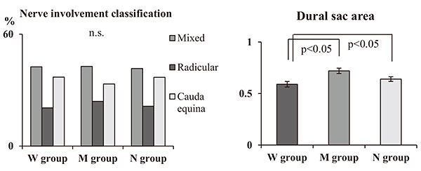
    Methods: In total, 234 patients with LSS including L4/5 lesion were asked to identify their LBP. Subjects were classified into three groups: walking-induced LBP that aggravated during walking (W group), motion-induced LBP that aggravated during sitting up (M group), and no LBP (N group). Cross-sectional areas of the dural sac, lumbar multifidus, and the erector spinae were measured. Intramuscular oxygenation was evaluated with near-infrared spectrophotometer. Surface electromyography (EMG) and mechanomyography (MMG) were performed on the lumbar multifidus. Morphological, hemodynamic, and electrophysiological differences in the onset of LBP were evaluated.

    Results: The prevalence of W, M, and control groups was 31.2%, 32.1%, 36.8%, respectively. Concordance between the laterality of LBP and leg symptoms including pain and numbness was 86.3% in the W group and 47.0% in the M group. Dural sac area was lower in the W group than in the M and control groups. In the hemodynamic evaluation, the oxygenated hemoglobin level was significantly lower in the W group than in the M and N groups. In electrophysiological evaluation of lumbar multifidus, the mean power frequency in EMG was significantly higher in the W group than in the N group. Amplitude in MMG was significantly lower in the W group than in the N group.

    Conclusions: Neurologic disturbance in patients with LSS may be attributed to "neuropathic LBP." Neuropathic multifidus disorder plays a role in walking-induced LBP.

    Download PDF (112K)
  • Eiki Unoki, Naohisa Miyakoshi, Eiji Abe, Takashi Kobayashi, Toshiki Ab ...
    2017 Volume 1 Issue 2 Pages 90-95
    Published: April 20, 2017
    Released on J-STAGE: December 20, 2017
    JOURNAL OPEN ACCESS

    Introduction: Sacroiliac joint pain (SIJP) after lumbar fusion surgery has recently gained attention as a source of low back pain after lumbar fusion. There are two risk factors for postoperative SIJP, i.e., fusion involving the sacrum and multiple-segment fusion. In this study, we examined whether SIJP could occur more frequently in patients with two risk factors (multiple-segment fusion to sacrum). Further, we examined SIJP after multiple-segment (≥3) lumbar fusion, focusing on the difference between floating fusion (non-fused sacrum) and fixed fusion (fused sacrum).

    Methods: Ninety-one patients who underwent multiple-segment lumbar fusion were included. Patients without preoperative clinical SIJP were considered. Of these, 17 developed new-onset SIJP. We investigated postoperative SIJP development, duration from surgery to SIJP onset, and postoperative treatment outcomes of SIJP patients using Japanese Orthopaedic Association (JOA) scores. We compared the findings between floating fusion group and fixed fusion group.

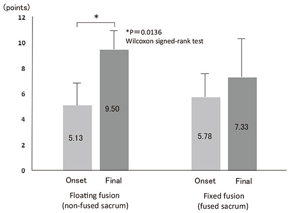
    Results: The incidence of SIJP was significantly higher with fixed fusion (32.1%) than with floating fusion (12.7%). The mean time of onset of sacroiliac joint pain was at 8.63 (2-13) months after surgery in the floating fusion group and 3.78 (1-10) months after surgery in the fixed fusion group, indicating that incidence occurred significantly earlier in the fixed fusion group. Our treatment outcome indicated that the mean JOA score significantly improved in the floating fusion group from 5.13 at the time of onset to 9.50 at the time of final follow-up; however, in the fixed fusion group, it improved from 5.78 at the time of onset to 7.33 at the time of final follow-up, indicating no significant improvement.

    Conclusions: In multiple-segment lumbar fusion, fixed fusion (fused sacrum) has a very high risk of SIJP. In addition, the onset of SIJP in such cases may occur earlier. This aspect deserves consideration, given the difficulty of pain treatment.

    Download PDF (133K)
  • Shurei Sugita, Hideki Murakami, Noritaka Yonezawa, Satoru Demura, Saka ...
    2017 Volume 1 Issue 2 Pages 96-99
    Published: April 20, 2017
    Released on J-STAGE: December 20, 2017
    JOURNAL OPEN ACCESS

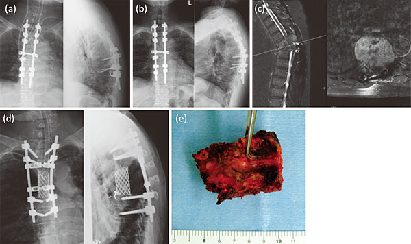
    Introduction: We often experience recurrence of spinal metastases after palliative surgery, even with radiotherapy. We examined the clinical outcome of radical surgery containing en bloc corpectomy for patients with recurrent spinal metastasis.

    Methods: Seven patients underwent en bloc corpectomy for recurrent spinal metastases. We assessed the prognosis scores (Tomita, Tokuhashi), pre- and postoperative Frankel scale scores, operation time, intraoperative blood loss, and perioperative complications.

    Results: The preoperative estimated prognosis was less than six months (two patients), six months to one year (two patients), and over one year (three patients), according to Tokuhashi score. Major perioperative complications were dura mater injury and pleural injury. Neurological improvement was seen in four patients. All patients were ambulatory at discharge and lived longer than the preoperatively estimated life expectancy (range: seven months to four years).

    Conclusions: Radical surgery consisting of en bloc corpectomy may be a therapeutic choice for patients with recurrent spinal metastases.

    Download PDF (260K)
CASE REPORT
  • Shotaro Fujino, Masayuki Miyagi, Shuichiro Tajima, Takayuki Imura, Ryo ...
    2017 Volume 1 Issue 2 Pages 100-106
    Published: April 20, 2017
    Released on J-STAGE: December 20, 2017
    JOURNAL OPEN ACCESS

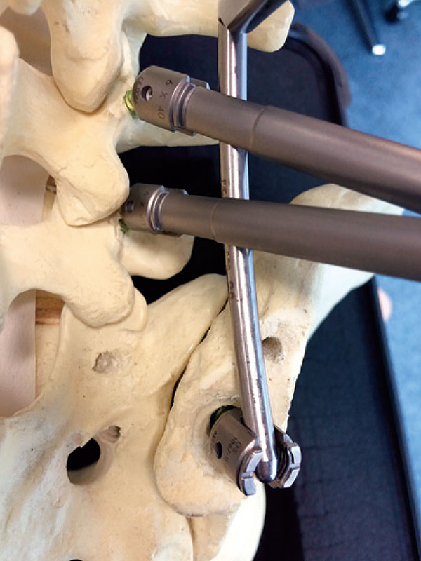
    Introduction: Suicidal jumper's fracture (unstable sacral fracture) is characterized not only by multiple fractures including thoracolumbar fractures, but also major chest and abdominal injuries. Early stabilization of these fractures and early ambulation are required for the treatment and management of chest and abdominal injuries. We present 3 cases of suicidal jumper's fracture with thoracolumbar burst fracture, treated with minimally invasive posterior fixation surgery, which is a combination of percutaneous pedicle screws (PPS) and the mini-open Galveston technique.

    Case reports: Case 1. A 50-year-old woman was injured by a fall from the 5th floor of a building as the result of a suicide attempt. Computed tomography revealed an H-shaped unstable sacral fracture and thoracolumbar fractures with major chest and abdominal injuries. For early stabilization of spinopelvic instability and early ambulation, we treated the patient with PPS and the mini-open Galveston technique. Her early postoperative emergence from bedrest contributed to the improvement of her general condition. One year after surgery at the final follow-up, she was able to walk with a T-cane without any motor, bladder, or bowel dysfunction (BBD) and achieved almost complete healing of the fractures. Cases 2 and 3. A 25-year-old woman (Case 2) and a 43-year-old woman were injured in falls. They had multiple injuries including unstable sacral fractures, and thoracolumbar fractures with major chest and abdominal injuries. We treated these patients with PPS and the mini-open Galveston technique. One year after surgery, they were able to walk with a T-cane and achieved almost complete healing of thoracolumbar fractures, but delayed healing of an unstable sacral fracture in Case 2, and remaining BBD in Case 3.

    Conclusion: PPS and the mini-open Galveston technique is a good approach to fixation because they are minimally invasive and provide moderately rigid fixation, especially in patients with multiple trauma whose general condition is poor.

    Download PDF (956K)
feedback
Top