Spine Surgery and Related Research
Online ISSN : 2432-261X
ISSN-L : 2432-261X
Volume 1, Issue 4
Displaying 1-11 of 11 articles from this issue
REVIEW ARTICLE
  • Shigeru Hirabayashi
    2017 Volume 1 Issue 4 Pages 158-163
    Published: October 20, 2017
    Released on J-STAGE: November 27, 2017
    JOURNAL OPEN ACCESS

    Ossification of the ligamentum flavum (OLF or OYL: ossification of the yellow ligament) usually occurs at the lower thoracic level and causes various types of neurological symptoms in accordance with the compression level of the spinal cord, the nerve roots, the conus medullaris, and the cauda equina. Although the greatest compression level to the nerve tissues is thought to be the most pathognomonic one, it is difficult to diagnose it in some cases. The effectiveness of conservative treatment such as applying a corset is small. Early surgical treatment is strongly recommended for the patients with spastic gait, severe decrease of muscle power in the lower extremities, bladder-bowel disturbance, combined ossification of the posterior longitudinal ligament at the same level, and severe compression to the spinal cord due to thickly hypertrophied ossification. However, numbness of the lower extremities and spastic gait tend to remain even after surgery.

    Download PDF (73K)
  • Keitaro Matsukawa, Yoshiyuki Yato
    2017 Volume 1 Issue 4 Pages 164-173
    Published: October 20, 2017
    Released on J-STAGE: November 27, 2017
    JOURNAL OPEN ACCESS

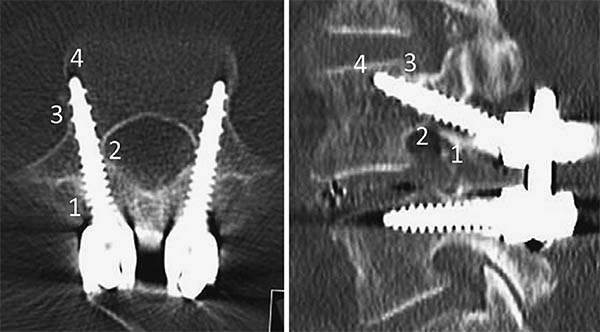
    Over the past few decades, many attempts to enhance the integrity of the bone-screw interface have been made to prevent pedicle screw failure and to achieve a better clinical outcome when treating a variety of spinal disorders. Cortical bone trajectory (CBT) has been developed as an alternative to the traditional lumbar pedicle screw trajectory. Contrary to the traditional trajectory, which follows the anatomical axis of the pedicle from a lateral starting point, CBT starts at the lateral part of the pars interarticularis and follows a mediolateral and caudocranial screw path through the pedicle. By markedly altering the screw path, CBT has the advantage of achieving a higher level of thread contact with the cortical bone from the dorsal entry point to the vertebral body. Biomechanical studies demonstrated the superior anchoring ability of CBT over the traditional trajectory, even with a shorter and smaller CBT screw. Furthermore, screw insertion from a more medial and caudal starting point requires less exposure and minimizes the procedure-related morbidity, such as reducing damage to the paraspinal muscles, avoiding iatrogenic injury to the cranial facet joint, and maintaining neurovascular supply to the fused segment. Thus, the features of CBT, which enhance screw fixation with limited surgical exposure, have attracted the interest of surgeons as a new minimally invasive method for spinal fusion.

    The purpose of this study was: 1) to identify the features of the CBT technique by reviewing previous anatomical and biomechanical literature, and 2) to describe its clinical application with a focus on the indications, limitations, surgical technique, and clinical evidence.

    Download PDF (1680K)
ORIGINAL ARTICLE
  • Yoshiharu Kawaguchi, Masato Nakano, Taketoshi Yasuda, Shoji Seki, Kayo ...
    2017 Volume 1 Issue 4 Pages 174-178
    Published: October 20, 2017
    Released on J-STAGE: November 27, 2017
    JOURNAL OPEN ACCESS

    Introduction: In more than 20 years' follow-up after cervical laminoplasty, some patients died at an early stage, within 5 years postoperatively. The details remain unclear. This study was conducted to elucidate the clinical features in patients who died at an early stage after cervical laminoplasty to determine possible preventive measures against early death after surgery.

    Methods: A total of 74 patients who died with the follow-up period were included. The patients were divided into two groups: patients with a short survival period (S group, died ≤5 years after surgery) and patients with a long survival period (L group, died >5 years after surgery). Diseases, gender, age, causes of the death, general complications before surgery, and the pre- and postoperative JOA scores were compared between the two groups.

    Results: Eleven patients (15%) died within 5 years after laminoplasty. The average age at death in the S group was 68.7 years which was considerably younger than that in the L group (80.2 years). The ratio of pneumonia was higher in the S group, compared to that in the L group. Postoperative JOA score in the S group was lower than that in the L group.

    Conclusions: 11 patients out of 74 patients (15%) died within 5 years after laminoplasty. The average age at the death in the S group was much younger than that in the L group. The postoperative JOA score in the S group was lower than that in the L group. As pneumonia was more prevalent in the S group, it might be reasonable to give the information for the protection of pneumonia after cervical laminoplasty.

    Download PDF (268K)
  • Koji Tamai, Hidetomi Terai, Akinobu Suzuki, Hiroaki Nakamura, Masaomi ...
    2017 Volume 1 Issue 4 Pages 179-184
    Published: October 20, 2017
    Released on J-STAGE: November 27, 2017
    JOURNAL OPEN ACCESS

    Introduction: With an aging population, the proportion of patients aged ≥80 years requiring cervical surgery is increasing. Surgeons are concerned with the high incidence of complications in this population, because "age" itself has been reported as a strong risk factor for complications. However, it is still unknown which factors represent higher risk among these elderly patients. Therefore, this study was conducted to identify the risk factors related to surgical complications specific to elderly patients by analyzing the registry data of patients aged ≥80 years who underwent cervical surgery.

    Methods: We retrospectively studied multicenter collected registry data using multivariate analysis. Sixty-six patients aged ≥80 years who underwent cervical surgery and were followed up for more than one year were included in this study. Preoperative patient demographic data, including comorbidities and postoperative complications, were collected from multicenter registry data. Complications were considered as major if they required invasive intervention, caused prolonged morbidity, or resulted in prolongation of hospital stay. Logistic regression analysis was performed to analyze the risk factors for complications. A p-value of <0.05 was considered as statistically significant.

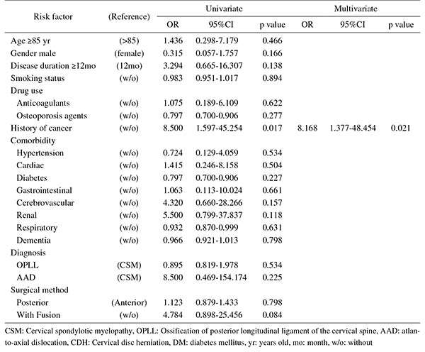
    Results: The total number of patients with complications was 21 (31.8%), with seven major (10.6%) and 14 minor (21.2%) complications. Multivariate logistic regression analysis, after adjusting for age, revealed two significant risk factors: preoperative cerebrovascular disorders (OR, 6.337; p=0.043) for overall complications and cancer history (OR, 8.168; p=0.021) for major complications. Age, presence of diabetes mellitus, and diagnosis were not significant predictive factors for complications in this study.

    Conclusions: Preoperative cerebrovascular disorders and cancer history were risk factors for complications after cervical surgery in patients over 80 years old. Surgeons should pay attention to these specific risk factors before performing cervical surgery in elderly patients.

    Download PDF (111K)
  • Naoyuki Nakamura, Yutaka Inaba, Shinya Kato, Takako Momose, Shunsuke Y ...
    2017 Volume 1 Issue 4 Pages 185-190
    Published: October 20, 2017
    Released on J-STAGE: November 27, 2017
    JOURNAL OPEN ACCESS

    Introduction: This study aimed to assess treatment outcomes and caregivers' satisfaction regarding scoliosis surgery for handicapped children.

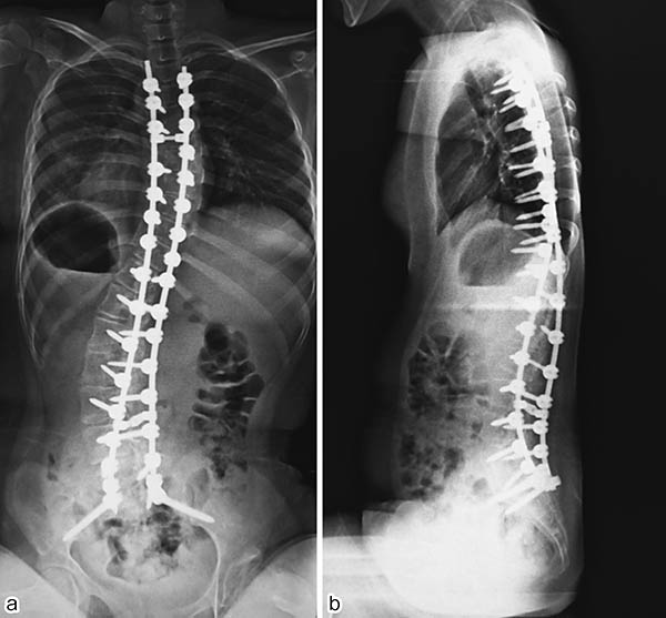
    Methods: Handicapped children are, by definition, noncommunicatory and/or nonambulatory. We recruited 26 handicapped children who were followed-up for >1 year after a scoliosis surgery. We recruited 40 patients with adolescent idiopathic scoliosis (AIS) who underwent a surgery during the same period as controls. We used a posterior approach in all the children. We determined preoperative body mass index (BMI), main Cobb angle, Cincinnati correction index (CCI), and fusion level; intraoperative time and blood loss per level; and postoperative complications. We also assessed caregivers' satisfaction with surgical treatments for these patients using the modified Bridwell's questionnaire.

    Results: We have described the results as handicapped children/AIS. Median preoperative BMI was 16.1/18.6 kg/m2. Preoperative and final Cobb angles were 94.2°/59.7° and 39.7°/17.0°, respectively and CCI was 2.0/1.7. The number of fusion levels was 14.6/9.0. The operative time and blood loss per level were 40.1/44.1 minutes and 264/138 ml, respectively. Postoperative complications in handicapped children were adynamic ileus in 8 cases, dysphagia in 5, pneumonia in 3, urinary tract infection in 2, and superior mesenteric artery syndrome (SMA), surgical site deep infection, infectious enteritis, agitation, and liver dysfunction in 1 each. However, in the AIS group, there was only 1 case of SMA. Median caregivers' satisfaction score on the 0-10 visual analog scale was 9. Caregivers for 19 of the 26 handicapped cases (73%) recommended surgical treatment to caregivers of other children with the same disease.

    Conclusions: Surgical treatment for neuromuscular and syndromic scoliosis was associated with a high rate of postoperative complications. However, the caregivers' satisfaction score after surgery was high.

    Download PDF (268K)
  • Toru Asari, Shuichi Aburakawa, Gentaro Kumagai, Sunao Tanaka, Yasuyuki ...
    2017 Volume 1 Issue 4 Pages 191-196
    Published: October 20, 2017
    Released on J-STAGE: November 27, 2017
    JOURNAL OPEN ACCESS

    Introduction: Lumbar spinal canal stenosis (LSS) is a very common disease. When the responsible level is considered to be L4/5 despite the appearance of double-level (L3/4 and L4/5) stenosis on magnetic resonance imaging (MRI), it is difficult for spinal surgeons to decide whether prophylactic decompression should be performed at the L3/4 level. The purpose of this study was to investigate the relationship between the dural sac cross-sectional area (DCSA) at the L3/4 level and clinical symptoms in patients with double-level stenosis.

    Methods: Thirty-five patients with double-level stenosis were registered in this study. All patients underwent decompression surgery at the L4/5 responsible level. The severity of patients' symptoms was evaluated by the Japanese Orthopaedic Association (JOA) score and its rate of recovery. A measurement program on MRI was used to determine the DCSA.

    Results: The clinical course of LSS according to the JOA score recovery rate at the final follow-up revealed that the good group (≥50%) included 27 patients, and the poor group (<50%) included 8 patients. In the good group, the mean DCSA at the L3/4 level was 72.3 ± 32.1 mm2 preoperatively and 71.3 ± 29.0 mm2 at the final follow-up. In contrast, in the poor group, the mean DCSA at the L3/4 level was 49.1 ± 23.8 mm2 preoperatively and 40.6 ± 14.1 mm2 at the final follow-up. Significant differences were observed in the preoperative and final follow-up DCSAs at the L3/4 level between two groups.

    Conclusions: Considering the present results, prophylactic decompression surgery at the L3/4 level should be performed for patients with double-level stenosis and DCSA <50 mm2 at the L3/4 level.

    Download PDF (204K)
  • Yasuhiro Shiga, Sumihisa Orita, Kazuhide Inage, Jun Sato, Kazuki Fujim ...
    2017 Volume 1 Issue 4 Pages 197-202
    Published: October 20, 2017
    Released on J-STAGE: November 27, 2017
    JOURNAL OPEN ACCESS

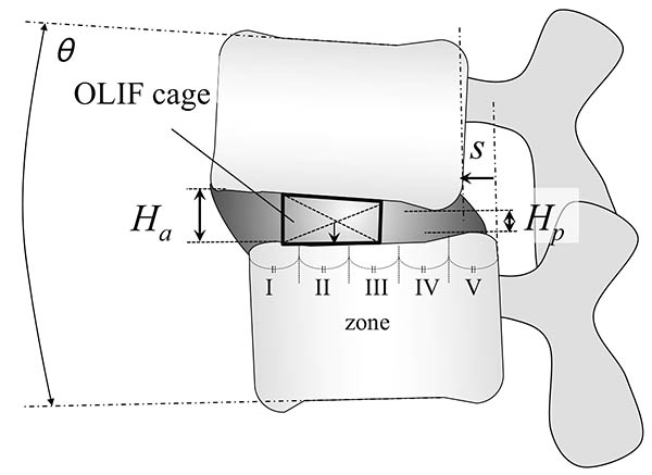
    Introduction: Oblique lateral interbody fusion (OLIF) can achieve recovery of lumbar lordosis (LL) in minimally invasive manner. The current study aimed to evaluate the location of lateral intervertebral cages during OLIF in terms of LL correction.

    Methods: The subjects were patients who underwent OLIF for lumbar degenerative diseases, including lumbar spinal stenosis, spondylolisthesis, and discogenic low back pain. Their clinical outcome was evaluated using visual analogue scale on lower back pain (LBP), leg pain and numbness. The following parameters were retrospectively evaluated on plain radiographic images and computed tomography scans before and at 1 year after OLIF: the intervertebral height, vertebral translation, and sagittal angle. The cage position was defined by equally dividing the caudal endplate into five zones (I to V), and its association with segmental lordosis restoration was analyzed. Subjects were also evaluated for a postoperative endplate injury.

    Results: Eighty patients (121 fused levels) with lumbar degeneration who underwent OLIF were included. There were no significant specific distribution in preoperative disc pathology such as disc angle, height, and translation. After OLIF, sagittal alignment was improved with an average correction angle of 3.8º at the instrumented segments in a level-independent fashion. All cases showed significant improvement in clinical outcomes, and had improvement in the radiological parameters (P<0.05). A detailed analysis of the cage position showed that the most significant sagittal correction and the most postoperative endplate injuries occurred in the farthest anterior zone (I). Cages with a 12-mm height were associated with more endplate injuries compared with shorter cages (8 or 10 mm).

    Conclusions: OLIF improves sagittal alignment with an average correction angle of 3.8º at the instrumented segments. We suggest that the optimal cage position for better lordosis correction and the fewest endplate injuries is zone II with a cage height of up to 10 mm.

    Download PDF (232K)
  • Kotaro Satake, Tokumi Kanemura, Hiroaki Nakashima, Hidetoshi Yamaguchi ...
    2017 Volume 1 Issue 4 Pages 203-210
    Published: October 20, 2017
    Released on J-STAGE: November 27, 2017
    JOURNAL OPEN ACCESS

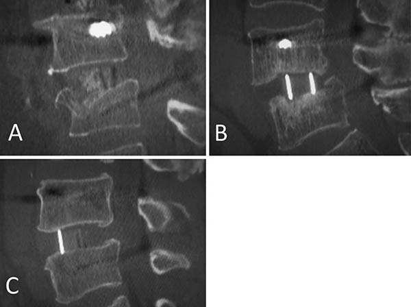
    Introduction: Few studies have investigated the influence of cage subsidence patterns (intraoperative endplate injury or late-onset cage settling) on bony fusion and clinical outcomes in lateral interbody fusion (LIF). This retrospective study was performed to compare the fusion rate and clinical outcomes of cage subsidence patterns in LIF at one year after surgery.

    Methods: Participants included 93 patients (aged 69.0±0.8 years; 184 segments) who underwent LIF with bilateral pedicle screw fixation. All segments were evaluated by computed tomography and classified into three groups: Segment E (intraoperative endplate injury, identified immediately postoperatively); Segment S (late-onset settling, identified at 3 months or later); or Segment N (no subsidence). We compared patient characteristics, surgical parameters and fusion status at 1 year for the three subsidence groups. Patients were classified into four groups: Group E (at least one Segment E), Group S (at least one Segment S), Group ES (both Segments E and S), or Group N (Segment N alone). Visual analog scales (VASs) and the Japanese Orthopedic Association Back Pain Evaluation Questionnaire (JOABPEQ) were compared for the four patient groups.

    Results: 184 segments were classified: 31 as Segment E (16.8%), 21 as Segment S (11.4%), and 132 as Segment N (71.7%). Segment E demonstrated significantly lower bone mineral density (-1.7 SD of T-score, p=0.003). Segment S demonstrated a significantly higher rate of polyetheretherketone (PEEK) cages (100%, p=0.03) and a significantly lower fusion rate (23.8%, p=0.01). There were no significant differences in VAS or in any of the JOABPEQ domains among the four patient groups.

    Conclusions: Intraoperative endplate injury was significantly related to bone quality, and late-onset settling was related to PEEK cages. Late-onset settling demonstrated a worse fusion rate. However, there were no significant differences in clinical outcomes among the subsidence patterns.

    Download PDF (155K)
  • Taiki Mori, Yoshihito Sakai, Mitsunori Kayano, Akio Matsuda, Keisuke O ...
    2017 Volume 1 Issue 4 Pages 211-217
    Published: October 20, 2017
    Released on J-STAGE: November 27, 2017
    JOURNAL OPEN ACCESS

    Introduction: Molecular pathways involved in ligamentum flavum (LF) hypertrophy are still unclarified. The purpose of this study was to characterize LF hypertrophy by microRNA (miRNA) profiling according to the classification of lumbar spinal stenosis (LSS).

    Methods: Classification of patients with LSS into ligamentous and non-ligamentous cases was conducted by clinical observation and the morphometric parameter adopting the LF/spinal canal area ratio (LSAR) from measurements of magnetic resonance imaging (MRI) T2 weighed images. LF from patients with ligamentous stenosis (n=10) were considered as the degenerative hypertrophied samples, and those from patients with non-ligamentous LSS (n=7) and lumbar disc herniation (LDH, n=3) were used as non-hypertrophied controls. Profiling of miRNA from all samples was conducted by Agilent microarray. Microarray data analysis was performed with GeneSpring GX, and pathway analysis was performed using Ingenuity Pathway Analysis.

    Results: The mean LSAR in the ligamentous group was significantly higher than that in the control group (0.662±0.154 vs 0.301±0.068, p=0.0000171). Ten significantly differentially expressed miRNA were identified and taken as a signature of LF hypertrophy: nine miRNA showed down-regulated expression, and one showed up-regulated expression in the ligamentous LF. Among those, miR-423-5p (rs=-0.473, p<0.05), miR-4306 (rs=-0.628, p<0.01), miR-516b-5p (rs=-0.629, p<0.01), and miR-497-5p (rs=0.461, p<0.05) were correlated to the LSAR. Pathway analysis predicted aryl hydrocarbon receptor signaling (p<0.01), Wnt/β-catenin signaling (p<0.01), and insulin receptor signaling (p<0.05) as canonical pathways associated with the miRNA signature.

    Conclusions: Classification based on quantification of the MRI axial image is useful for studying hypertrophy of the LF. Aryl hydrocarbon receptor and Wnt/β-catenin signaling may be involved in LF hypertrophy.

    Download PDF (334K)
TECHNICAL NOTE
  • Yoshihisa Sugimoto, Takahiro Hayashi, Takamitsu Tokioka
    2017 Volume 1 Issue 4 Pages 218-221
    Published: October 20, 2017
    Released on J-STAGE: November 27, 2017
    JOURNAL OPEN ACCESS

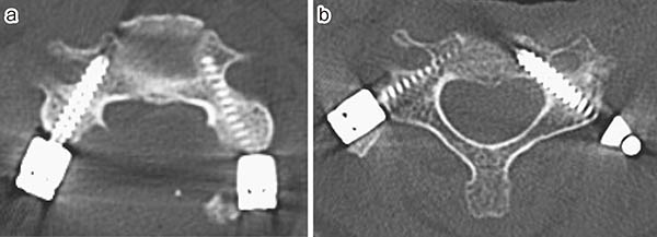
    Background: To avoid lateral misplacement of midcervical pedicle screws, we developed a method for Minimally Invasive Cervical Pedicle Screw (MICEPS) fixation via a posterolateral approach. This intramuscular approach allows for horizontal pedicle screw insertion and reduced intraoperative bleeding. We reviewed our initial experience with MICEPS fixation for patients with cervical metastases.

    Methods: This study included 18 consecutive patients who received cervical spinal surgery for metastatic tumor. We treated 12 patients with conventional cervical pedicle screw fixation, and 6 patients with the MICEPS fixation technique. Average follow-up was 14 months (range 3 to 34). We inserted 117 pedicle screws using the navigation system. Average fusion area was 4.9 vertebrae (range 3 to 8). Alpha-angles between a line perpendicular to the posterior cortex of the vertebral body and the screw trajectory in the transverse plane were also measured.

    Results: The average surgical time was 250 min (range 151 to 420 min) with the conventional pedicle screw fixation and 234 min (range 154 to 300 min) with the MICEPS fixation. The average total blood loss was 780 mL (range, 180-1430 mL) in the conventional pedicle screw fixation group and 180 mL (range, 70-400 mL) in the MICEPS fixation group. At the level of midcervical (C3-5), average alpha-angles was 52 degrees (range 43 to 62) in MICEPS fixation group, and 39 degrees (range 19 to 55) in conventional cervical pedicle screw group.

    Conclusions: The MICEPS fixation technique uses an intramuscular approach, which is minimally invasive and reduces intraoperative bleeding. Intramuscular approach allows for horizontal pedicle screw insertion, and reduced critical screw deviation.

    Download PDF (181K)
CASE REPORT
  • Kenyu Ito, Noriaki Kawakami, Taichi Tsuji, Tetsuya Ohara, Toshiki Sait ...
    2017 Volume 1 Issue 4 Pages 222-224
    Published: October 20, 2017
    Released on J-STAGE: November 27, 2017
    JOURNAL OPEN ACCESS

    Introduction: Gaucher's disease is a congenital metabolic disorder characterized by the accumulation of glucocerebroside in the reticuloendothelial system. Its clinical manifestations include splenomegaly, osteopenia, and pathological fractures. Cases of patients with kyphotic deformities caused by pathological vertebral compression fractures associated with Gaucher's disease are well reported. However, there has been no report regarding surgical treatment of kyphotic deformity caused by Gaucher's disease without compression fractures. In the present report, we describe surgical treatment for kyphotic deformity caused by Gaucher's disease without a past history of vertebral compression fractures.

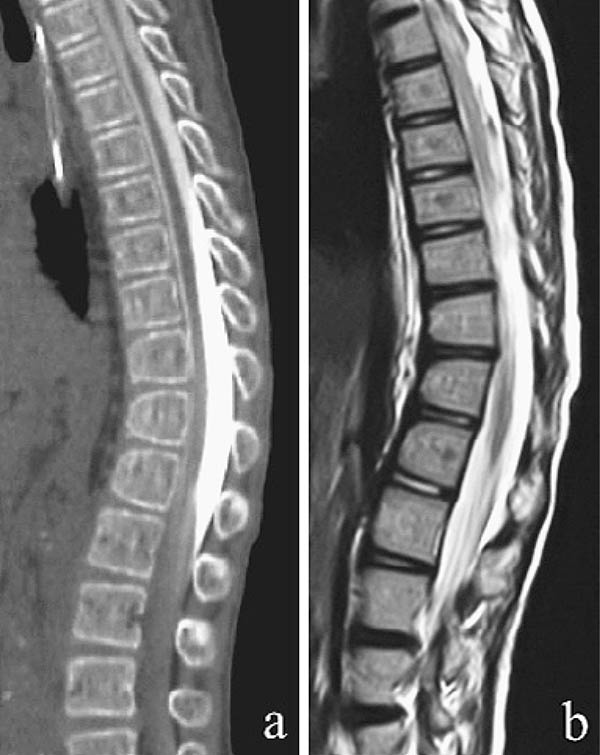
    Case Report: The patient was diagnosed with Gaucher's disease at the age of 15 months. The patient was a 10-year-old girl with progressive kyphosis (84° between T6 and L3, with T12 as the apical vertebra) without compression fractures. Although the patient had been treated using a brace since the age of 3 years, the kyphosis progressed to the point where corrective surgery was required. We initially performed T3-L3 posterior spinal fusion, followed by anterior fusion 3 months later, which corrected the kyphosis to 35°. Postoperatively, the patient suffered fractures of the upper and lower extremities but did not have spinal fractures.

    Conclusions: Two-stage anterior/posterior combined correction and fusion was performed in a patient with kyphotic deformity caused by Gaucher's disease without compression fractures. Because of bone fragility in Gaucher's disease, careful selection of the fusion range and postoperative therapy was necessary.

    Download PDF (292K)
feedback
Top