Spine Surgery and Related Research
Online ISSN : 2432-261X
ISSN-L : 2432-261X
Volume 7, Issue 2
Displaying 1-15 of 15 articles from this issue
REVIEW ARTICLE
  • Jean Dubousset, Bassel G. Diebo
    2023 Volume 7 Issue 2 Pages 120-128
    Published: March 27, 2023
    Released on J-STAGE: March 27, 2023
    Advance online publication: June 28, 2022
    JOURNAL OPEN ACCESS

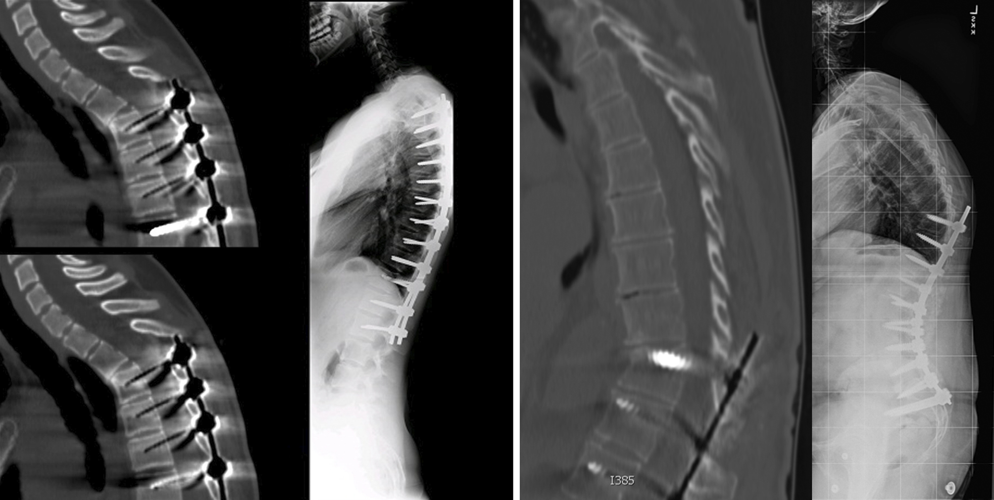
    In the past few decades, proximal junctional kyphosis (PJK) has emerged as a new complication after instrumented spinal fusion in adult and pediatric spinal deformities. This phenomenon has occurred concomitantly with the rise of robust instrumentation techniques and enhancement of our abilities to obtain greater spinal deformity correction. The goal of this paper is to review the mechanical and biological causes of PJK and recommend prevention strategies.

    Download PDF (594K)
  • Yang Chen, Zhichao Gao
    2023 Volume 7 Issue 2 Pages 129-135
    Published: March 27, 2023
    Released on J-STAGE: March 27, 2023
    Advance online publication: February 13, 2023
    JOURNAL OPEN ACCESS

    Discography is an important method for diagnosing discogenic low back pain (LBP) and replicating the effects of pain. However, its development is not smooth due to its safety and reliability, which have not been completely confirmed. Beginning with the clinicians using discography, there remains constant controversy. With the continuous progress of related research on discography, clinicians and scholars' understanding of discography is constantly improving. This article reviews the background, clinical application, and safety of discography.

    Download PDF (95K)
ORIGINAL ARTICLE
  • Takuya Yamanaka, Katsushi Takeshita, Takao Mochizuki, Hirokazu Inoue, ...
    2023 Volume 7 Issue 2 Pages 136-141
    Published: March 27, 2023
    Released on J-STAGE: March 27, 2023
    Advance online publication: October 28, 2022
    JOURNAL OPEN ACCESS

    Introduction: We often treat patients with peripheral neuropathic pain due to spine diseases with mirogabalin as an alternative to pregabalin because of adverse events or insufficient efficacy associated with pregabalin treatment. However, there have been few reports on the safety and efficacy of mirogabalin in such cases. This study aimed to evaluate the safety and efficacy of switching from pregabalin to mirogabalin in patients with peripheral neuropathic pain due to spine diseases.

    Methods: Between January 2019 and July 2021, we treated 106 patients (47 men and 59 women) with peripheral neuropathic pain due to spine diseases. All patients had switched from pregabalin to mirogabalin due to adverse events or lack of efficacy. We evaluated the retention rate, incidence of adverse events, and response rate of mirogabalin during the treatment course.

    Results: The mean age of the patients was 67.5 years (range, 33-93 years), and the average dose of mirogabalin was 13.8 mg (range, 2.5-30 mg) at the final follow-up. The average duration of mirogabalin treatment was 148.7 days (range, 3-463 days). The retention rate of mirogabalin was 78.3%, the incidence of adverse events after mirogabalin administration was 28.3%, and the response rate of mirogabalin was 66%. Somnolence with pregabalin or mirogabalin administration in the mirogabalin discontinuation group was increased compared with that in the mirogabalin continuation group (pregabalin: 52.2% vs. 19.3%, mirogabalin: 26.1% vs. 7.2%). The patients who responded to mirogabalin had a lower average age, higher retention rate, and longer drug administration period than those who did not respond to it.

    Conclusions: This study indicated that mirogabalin treatment might be continued in patients with peripheral neuropathic pain due to spinal diseases who could not continue pregabalin treatment.

    Download PDF (88K)
  • Yu Chung Wong, Tsun Kit Lau, Wai Wang Chau, Kin On Kwok, Sheung Wai La ...
    2023 Volume 7 Issue 2 Pages 142-148
    Published: March 27, 2023
    Released on J-STAGE: March 27, 2023
    Advance online publication: October 28, 2022
    JOURNAL OPEN ACCESS

    Introduction: Aspirin is commonly used for the primary and secondary prevention of cardiovascular disease and stroke. Controversy exists concerning whether and when is the optimal time to stop aspirin before spinal surgery. Previous studies on this topic mainly focused on patients who received thoracolumbar spine surgeries. There are only a few literatures concerning the safety of aspirin use in cervical spine surgery patients.

    Methods: This pilot study recruited patients who received cervical laminoplasty from January 2010 to December 2021. The operation time, intraoperative blood loss, and postoperative complications of the patients who had taken aspirin during the perioperative period were compared with age, sex, and comorbidity-matched control patients. Propensity score matching was utilized in the selection of control to minimize bias.

    Results: Twenty-one patients who have received cervical laminoplasty while taking aspirin during the perioperative period were included. The control group included 21 age, sex, and comorbidity-matched patients who have not taken aspirin. The operation time and intraoperative and postoperative blood loss were higher in patients taking aspirin but were not statistically significant. There was no statistically significant difference in the wound infection rate. No case of epidural hematoma was found.

    Conclusions: In patients undergoing cervical laminoplasty, a continuation of aspirin is safe and does not increase the difficulty of the surgery, wound complication, or hemorrhage.

    Download PDF (464K)
  • Norimitsu Wakao, Yoshihito Sakai, Naoaki Osada, Takaya Sugiura, Hiroki ...
    2023 Volume 7 Issue 2 Pages 149-154
    Published: March 27, 2023
    Released on J-STAGE: March 27, 2023
    Advance online publication: October 13, 2022
    JOURNAL OPEN ACCESS

    Introduction: Although thoracic spondylotic myelopathy (TSM) without ossification or disc disorder has been associated with some dynamic factors in the thoracolumbar area, a detailed investigation is yet to be published. Thus, in this study, we investigated the segmental motion and sagittal alignment of the thoracolumbar area in patients with and without TSM.

    Methods: Patients with TSM who were treated from 2013 to 2020 were enrolled in this study. The non-TSM group consisted of sex- and age-matched patients with spinal disorders other than TSM. Segmental mobility from T10-L2 during passive maximum flexion and extension following myelography and the sagittal cobb angles of T10-L1 and L1-L5 in the standing position were measured using multidetector computed tomography (CT). The mobility of each segment was set as the difference in the angles between the two positions.

    Results: In total, 10 patients (8 males and 2 females, mean age 65.8 years) with TSM and 20 without TSM were enrolled. The most stenotic level was observed at T10-T11 in four cases and T11-T12 in six. The average mobility at this segment in the TSM group (5.8°) was significantly greater than that in the non-TSM group (2.1°) (p<0.001). In the TSM group, the cobb angles of T10-L1 and L1-L5 were 2.3° and 17.4° of lordosis, respectively, which differed significantly from those in the non-TSM group, which were 8° of kyphosis and 32.2° of lordosis, respectively (p<0.001 and p=0.001, respectively).

    Conclusions: Compared with those without TSM, patients with TSM were found to have greater segmental mobility at the most stenotic level, thoracolumbar lordosis, and decreased lumbar lordosis.

    Download PDF (1345K)
  • Takeshi Sasagawa
    2023 Volume 7 Issue 2 Pages 155-160
    Published: March 27, 2023
    Released on J-STAGE: March 27, 2023
    Advance online publication: October 13, 2022
    JOURNAL OPEN ACCESS

    Introduction: Thoracic percutaneous pedicle screw (PPS) fixation is technically challenging because of the complexity of the spinal anatomy involved. Furthermore, owing to the proximity of critical neurovascular structures, serious complications have been reported because of misplaced thoracic pedicle screws. Therefore, it is important to know the factors associated with the misplacement of thoracic PPS, but there have been few reports to date.

    Methods: The present study included 663 PPSs inserted from T4 to T12 in 127 patients. The accuracy of pedicle screw placement was assessed using computed tomography (CT) scans conducted within two weeks postoperatively. We compared the screws in the misplaced group (Group M) and the optimal placed group (Group O) for sex, age, body mass index, the consecutive surgery numbers, type of disease, instrumented level, laterality, the pedicle diameter, the inclination angle of the transverse process, and Hounsfield units (HU) at the base of the transverse processes of the instrumented vertebrae.

    Results: Screw misplacement was observed in 28 (4%) of 663 screws on CTs conducted within two weeks postoperatively. In univariate analysis, there was a statistically significant difference between Group M (n=25) and Group O (n=638) for insertion level, the pedicle diameter, and the HU value of the transverse process. In multivariate logistic regression analysis, T4-6 level (T4-6; odds ratio [OR]=12.083, 95% confidence interval [CI]: 3.219-45.355) and greater HU value at the transverse process (OR=1.009, 95% CI: 1.004-1.014) were identified as independent factors associated with the misplacement of thoracic PPS.

    Conclusions: The misplacement of thoracic PPS was observed in 28 (4%) of 663 screws. The vertebral level (T4-6) and greater HU values at the base of the transverse process were identified as independent factors associated with the misplacement of thoracic PPS.

    Download PDF (700K)
  • Ikenna H. Ifearulundu, G. Michael Mallow, Josha Woodward, Emilia Ferre ...
    2023 Volume 7 Issue 2 Pages 161-169
    Published: March 27, 2023
    Released on J-STAGE: March 27, 2023
    Advance online publication: October 13, 2022
    JOURNAL OPEN ACCESS

    Introduction: This study aimed to identify demographic, clinical, and operative factors associated with increased postoperative compliance of patient-reported outcome (PRO) assessments following lumbar spine surgery.

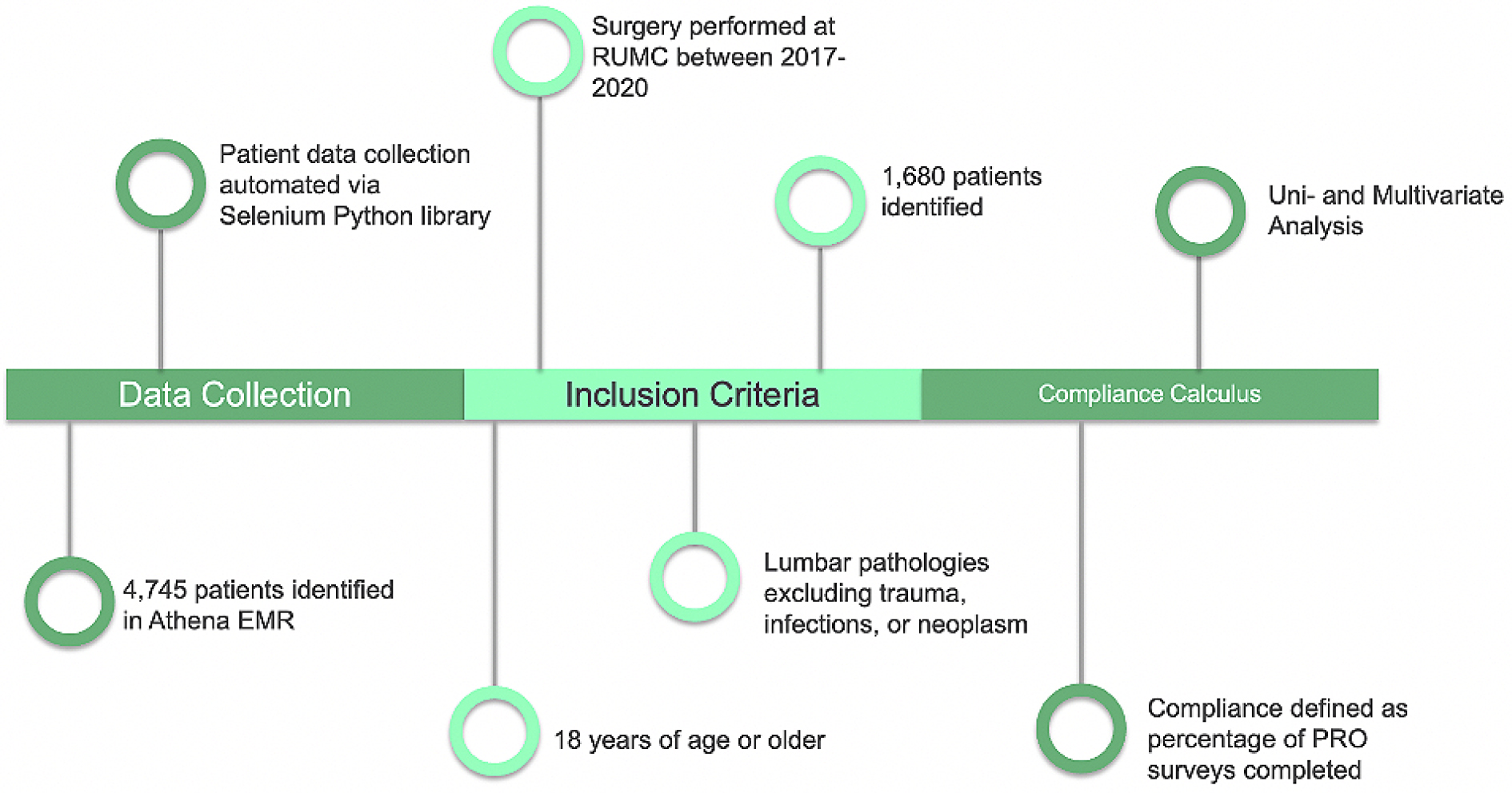
    Methods: A retrospective study of prospectively collected data of 1,680 consecutive adult patients who underwent elective lumbar surgery at a single institution from 2017-2020. Digital assessment questionnaires were used to assess PROs (i.e., VAS-back, VAS-leg, Oswestry Disability Index, Short Form (SF-12) mental & physical health, VR-12 mental and physical, and VR6D scores) and patient compliance, defined as the percentage of questionnaires completed preoperatively, at 3 months and 1 year after surgery. Multivariate logistic regression was used to assess the association between PRO compliance and patient characteristics.

    Results: A total of 1,680 patients (53.1% male, mean age: 57.7 years) had a mean PRO compliance of 64.7%. Compliance decreased continuously from initial preoperative rates (84.5%) to lower rates at 3 months (54.4%) and 12 months (45.6%), respectively, with 33.2% of patients completing zero assessment questionnaires at 12 months, postoperatively. Factors associated with significantly increased PRO compliance included being employed (preop: odds ratio [OR]=2.58, p=0.002; 3-month postop: OR=1.25, p=0.095; 12-month postop: OR=1.34, p=0.028). Factors associated with decreased compliance included preoperative smoking status (3-month postop: OR=0.63, p=0.029; 12-month postop: OR=0.60, p=0.016).

    Conclusions: Patients who completed greater than 50% of their PROs demonstrated significantly different rates of being employed compared with those who completed less than 50% throughout 1 year of follow-up. Preoperative smoking status was associated with decreased compliance, whereas a history of employment was associated with increased compliance throughout follow-up. To validate our findings and explore additional parameters that affect postoperative compliance of PROs, further investigation is required.

    Download PDF (396K)
  • Masatsune Sato, Masaaki Uesugi, Takuya Kawai, Yosuke Kobayashi, Yoichi ...
    2023 Volume 7 Issue 2 Pages 170-178
    Published: March 27, 2023
    Released on J-STAGE: March 27, 2023
    Advance online publication: October 28, 2022
    JOURNAL OPEN ACCESS

    Introduction: Patients with adult spinal deformity (ASD) develop compensatory mechanisms. The Cobb angle between T4 and T12 has been used as a standard to define thoracic kyphosis (TK) because radiological evaluation is difficult in upper thoracic region. The upper thoracic compensatory mechanism has not been sufficiently explored, and focal thoracic compensation remains poorly understood. This study aimed to determine upper sagittal thoracic compensation and features of thoracic compensation in ASD patients.

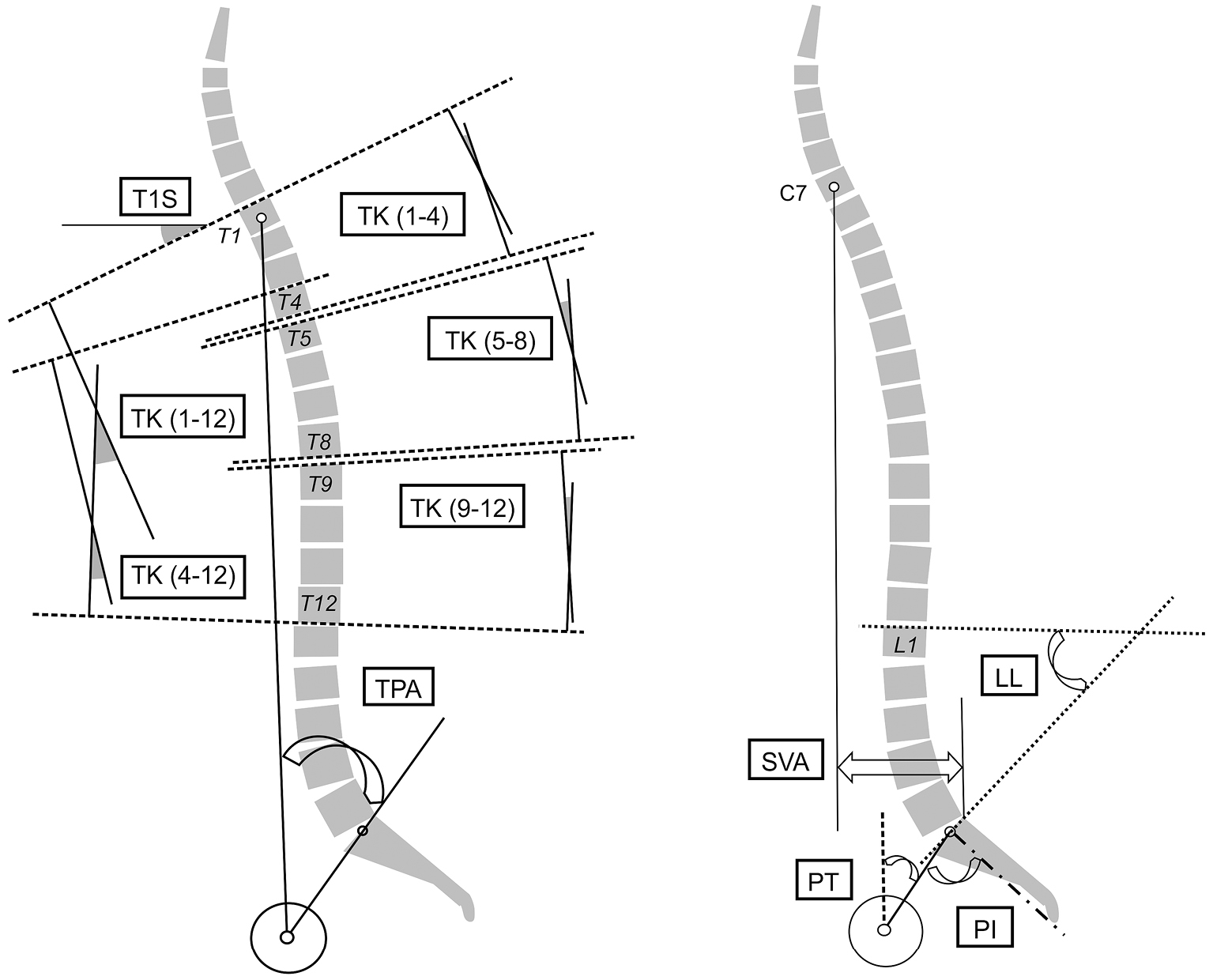
    Methods: The records of 218 consecutive patients who underwent full-standing EOS imaging were retrospectively examined. Spinopelvic parameters-including the T1-T12, T4-T12, T1-T4, T5-T8, and T9-T12 angles-were measured using a dedicated and validated software. The relationship between the thoracic and spinopelvic parameters was statistically evaluated. Thoracic compensation was compared among four typical types of spinal deformity.

    Results: A total of 127 ASD patients met the inclusion criteria. TK (1-12) was negatively correlated with the sagittal vertical axis (SVA) (r=−0.35), T1 pelvic angle (TPA) (r=−0.29), and pelvic incidence minus lumbar lordosis (PI-LL) (r=−0.60). TK (1-4) showed a stronger correlation with the SVA and TPA than TK (5-8) or TK (9-12) (r=−0.39, −0.38, respectively). TK (1-4), TK (5-8), and TK (9-12) had a similar negative correlation with PI-LL; however, TK (5-8) had the strongest correlation (r=−0.38). Both age and TK (9-12) were significantly associated with decreased TK (1-4), and vertebral fracture was significantly associated with increased TK (9-12). Both patients with high SVA and those in the hyperthoracic kyphosis group had lower TK (1-4) angles.

    Conclusions: In ASD patients, upper thoracic spine compensation plays an important role in countering global malalignment. The middle part of the thoracic spine has a strong correlation with LL. This study findings can help clinicians better manage ASD patients.

    Download PDF (866K)
TECHNICAL NOTE
  • Jung-Suk Kim, Yong-Min Kim, Seung-Rim Yi, In Hee Kim, Minwook Kang, Do ...
    2023 Volume 7 Issue 2 Pages 179-182
    Published: March 27, 2023
    Released on J-STAGE: March 27, 2023
    Advance online publication: January 12, 2023
    JOURNAL OPEN ACCESS

    Introduction: The strut iliac bone graft has been widely used to achieve fusion in various anterior cervical spinal surgeries but some complications often remain, such as pain and gross deformity. Considering these, we designed a new technique to restore the iliac ridge, using the outmost part of the iliac crest. We aim to assess the efficacy of our new restoration technique of the iliac ridge after harvesting strut bone graft for anterior cervical fusion. The clinical and radiological outcomes of our hinged roof method were evaluated.

    Technical Note: A retrospective review was conducted of 29 patients who underwent hinged roof reconstruction of the iliac ridge after harvesting a bicortical strut bone graft for anterior cervical fusion using a cervical plate system. The clinical outcome for pain and gross appearance and radiological results were evaluated. Three months after the surgery, pain at the donor site became minimal or absent in all cases. At 1 year follow-up, no patient had reported pain and palpable discomfort, such as step-off on the donor site. Final X-ray and follow-up computed tomography revealed a bony union of the reconstructed iliac ridge to both margins.

    Conclusions: By showing good clinical and radiological outcomes, the authors' hinged roof reconstruction of the iliac crest after harvesting strut bone graft seemed to be a simple and effective technique that can reduce complications, such as pain and deformity on the donor iliac crest.

    Download PDF (444K)
CLINICAL CORRESPONDENCE
LETTER TO THE EDITOR
ERRATUM
feedback
Top