All issues

Successor
Volume 34, Issue 9
Displaying 1-10 of 10 articles from this issue
- |<
- <
- 1
- >
- >|
Preface
-
Atsushi IKAI2013 Volume 34 Issue 9 Pages 475
Published: September 10, 2013
Released on J-STAGE: September 21, 2013
JOURNAL FREE ACCESSDownload PDF (209K)
Special Issue: SPM Analysis for Biomaterial and Cell Surfaces
-
Takeshi FUKUMAArticle type: Current Topic
2013 Volume 34 Issue 9 Pages 476-481
Published: September 10, 2013
Released on J-STAGE: September 21, 2013
 JOURNAL FREE ACCESSRecent progress in frequency modulation atomic force microscopy (FM-AFM) has enabled its operation in liquid with true atomic resolution. However, the information contained in a two-dimensional height image obtained by FM-AFM is often insufficient for understanding the structures and phenomena at a solid/liquid interface. In this study, we have developed a method referred to as 3D scanning force microscopy (3D-SFM). Combined with FM-AFM, the method enables to visualize 3D distribution of water and fluctuating surface structures at solid/liquid interfaces. Here we present basic principle and applications of 3D-SFM. The 3D-SFM image obtained at a mica/water interface shows 3D distribution of the hydration layers and adsorbed water molecules with subnanometer-scale resolution. In addition, the 3D-SFM image obtained at a lipid/water interface shows 3D distribution of the fluctuating lipid headgroups.View full abstractDownload PDF (1325K)
 -
Tatsuo USHIKI, Masato NAKAJIMA, Futoshi IWATAArticle type: Current Topic
2013 Volume 34 Issue 9 Pages 482-487
Published: September 10, 2013
Released on J-STAGE: September 21, 2013
 JOURNAL FREE ACCESSScanning ion conductance microscopy (SICM), first introduced by Hansma et al. (1989), can obtain topographic images of the sample surface and has been expected to be useful for biological studies. In this paper, we explained the principle of the SICM and showed some results of the application of SICM to biology. We showed SICM images of collagen fibrils, chromosomes and cultivated cells in liquid. We then applied SICM for imaging the surface of tissue blocks (e.g., trachea, etc.). Sequential time-lapsed SICM images of live cells were also shown for revealing the movement of cellular processes on the time scale of minutes. The advantages of SICM imaging in biology were discussed by comparison with atomic force microscopy (AFM) and/or scanning electron microscopy (SEM) images.View full abstractDownload PDF (1748K)
 -
Yasufumi TAKAHASHI, Hitoshi SHIKU, Tomokazu MATSUEArticle type: Current Topic
2013 Volume 34 Issue 9 Pages 488-493
Published: September 10, 2013
Released on J-STAGE: September 21, 2013
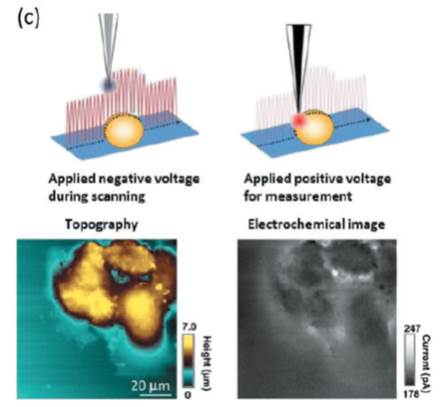 JOURNAL FREE ACCESSThe micro/nanoelectrode has unique characteristics, which are not expected for conventional electrodes, such as the capability to perform localized measurements, low double layer charging currents, low ohmic drops, and fast mass transport. Scanning electrochemical microscopy (SECM) with a micro/nanoelectrode for detecting electroactive chemical species and is an effective tool for the investigation of the localized chemical properties of sample surfaces and interfaces. Because SECM has high temporal resolution under physiological conditions, it is particularly well suited for quantitative measurements of chemicals like neurotransmitters, nitric oxide, reactive oxygen species, and oxygen, which are released/consumed by living cells. This article presents an overview of the recent progress of SECM.View full abstractDownload PDF (836K)
 -
Yuki KOIDE, Kosuke KUBO, Taito SEKINE, Yoshiki MIZUSHITA, Narangere ...Article type: Current Topic
2013 Volume 34 Issue 9 Pages 494-499
Published: September 10, 2013
Released on J-STAGE: September 21, 2013
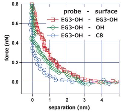 JOURNAL FREE ACCESSThe mechanism underlying the bioinertness of nonfouling self-assembled monolayers was investigated with protein adsorption experiments, platelet adhesion tests, and surface force measurements with an atomic force microscope. Our force measurements revealed strong repulsion operating between nonfouling SAMs in the range of 4 to 6 nm in PBS buffer solution. In addition, we found that the protein-adsorbing and cell-adhering SAMs did not exhibit such repulsion. We concluded that the repulsion originated from structured interfacial water molecules. Considering the correlation among the above results, we propose that the structured interfacial water with a thickness of 2 to 3 nm (half of the range of the repulsion observed in the surface force measurements) plays an important role in deterring proteins and cells from adsorption or adhesion.View full abstractDownload PDF (740K)
 -
Takeomi MIZUTANI, Kazushige KAWABATAArticle type: Current Topic
2013 Volume 34 Issue 9 Pages 500-504
Published: September 10, 2013
Released on J-STAGE: September 21, 2013
 JOURNAL FREE ACCESSAtomic force microscope (AFM) has advantages in the measurement of mechanical properties of soft materials. Here, we introduced applications of AFM to elucidate a single cell mechanics and multi-cellular dynamics. Comparison of elasticity map of fibroblasts with distribution of intracellular cytoskeletal fibers showed correspondence of high elasticity regions andactin fiber distributions. A representative morphology of cellular membrane ruffling was clearly observed and elasticity of the ruffling regions was higher than that of the outside of the ruffling regions. Elasticity map of epithelial cells showed cell-cell contact point is high elasticity. Cellular elasticity measurements by AFM will be applied to medical usages and some modifications of AFM will open a new door to study dynamics of soft materials.View full abstractDownload PDF (901K)
Planning Series
Surface Science for Energy Issues
-
Kenji TSUCHIYA2013 Volume 34 Issue 9 Pages 505-506
Published: September 10, 2013
Released on J-STAGE: September 21, 2013
 JOURNAL FREE ACCESSDownload PDF (593K)
Science Café
Research Abroad
-
Takayuki SUZUKI2013 Volume 34 Issue 9 Pages 507-508
Published: September 10, 2013
Released on J-STAGE: September 21, 2013
JOURNAL FREE ACCESSDownload PDF (443K)
News & Trends
-
2013 Volume 34 Issue 9 Pages 509
Published: September 10, 2013
Released on J-STAGE: September 21, 2013
JOURNAL FREE ACCESSDownload PDF (182K)
Qualifying Examination for Surface Science Engineers
-
2013 Volume 34 Issue 9 Pages 510-511
Published: September 10, 2013
Released on J-STAGE: September 21, 2013
JOURNAL FREE ACCESSDownload PDF (2375K)
- |<
- <
- 1
- >
- >|