All issues

Successor
Volume 36, Issue 4
Displaying 1-14 of 14 articles from this issue
- |<
- <
- 1
- >
- >|
Preface
-
Takashi SEKIGUCHI2015Volume 36Issue 4 Pages 157
Published: April 10, 2015
Released on J-STAGE: April 21, 2015
JOURNAL FREE ACCESSDownload PDF (170K)
Special Issue: Frontier Technologies of Scanning Electron Microscopy
-
Masaaki SUGIYAMA, Akira TANIYAMAArticle type: Review
2015Volume 36Issue 4 Pages 158-165
Published: April 10, 2015
Released on J-STAGE: April 21, 2015
 JOURNAL FREE ACCESSConventional scanning electron microscope has been remarkably progressed on the electron optics and instrumentations concerning with several kinds of detector systems. The recent appearance of low voltage SEM indicates to the necessity of a correct understanding and interpretation of incident electrons trajectories in a sample. Based on the literature, four categories of secondary electrons such as SE1, SE2, SE3 and SE4 are reviewed, and some of applications of the low voltage SEM and backscattering electron image techniques to the field of microstructure analysis in steels are introduced. The progress of BSE detectors enables us to see single dislocation images in a bulk sample of steel, whose observation condition is strongly depended on the incident electron direction to the sample. Using the ECCI technique, the analysis of dispersed hard phase microstructure such as an island martensite is examined with a combination of conventional EBSD analysis. As an advanced technique, furthermore, the high temperature EBSD measurement technique has been developed up to 1000°C on the special heating stage, which will proceed the in situ microstructure analysis during the phase transformation under the steel processing at high temperature in near future.View full abstractDownload PDF (1608K)
 -
Seiichi TAKAMI, Takanari TOGASHI, Tsutomu AIDA, Daisuke HOJO, Nobuaki ...Article type: Current Topic
2015Volume 36Issue 4 Pages 166-171
Published: April 10, 2015
Released on J-STAGE: April 21, 2015
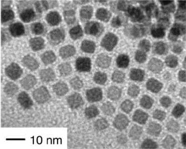 JOURNAL FREE ACCESSWe have studied hydrothermal synthesis of organic-modified metal oxide nanoparticles and found that the assemblies of nanoparticles were formed when we synthesized metal oxide nanoparticles in the presence of organic molecules with two functional groups. The structure of synthesized nanoassemblies was analyzed by scanning electron microscopes (SEM) and other analytical methods. The use of SEM revealed the three-dimensional nanostructure of the assemblies as well as their surface morphology. In this review, we will show how our study on the synthesis of metal oxide nanoparticles was accelerated by the use of SEM.View full abstractDownload PDF (1503K)
 -
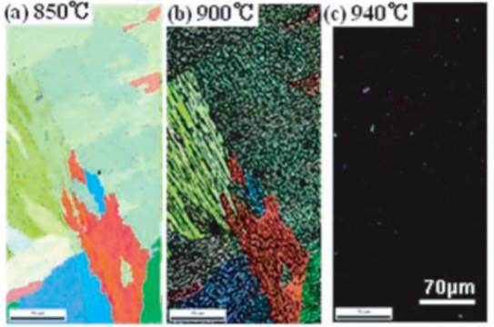
Hisashi SATO, Takanori SONODA, Tomokazu MORITANI, Stefan ZAEFFERER ...Article type: Current Topic
2015Volume 36Issue 4 Pages 172-177
Published: April 10, 2015
Released on J-STAGE: April 21, 2015
 JOURNAL FREE ACCESSElectron backscatter diffraction (EBSD) is effective method to investigate crystal orientation distribution in materials. Especially, EBSD has given a lot of novel knowledge for phase transformation of Fe and Fe alloys. In this study, microstructural analysis of martensite (α′) with butterfly shape in Fe-Ni alloys are performed by conventional EBSD observation and in-situ EBSD observation during tensile test. By using EBSD, it is found that crystal orientation in the butterfly-type α′ is locally changed. Moreover, the butterfly-type α′ has both features of lath and lenticular α′. This comes from the change of accommodation process for shape strain of the butterfly-type α′ during its growth. Furthermore, the tensile deformation influences the change of accommodation process for shape strain of the butterfly-type α′. From the obtained results, formation process of the butterfly-type α′ is discussed.View full abstractDownload PDF (1236K)
 -
Yoshikazu HOMMA, Hiroki KATOArticle type: Current Topic
2015Volume 36Issue 4 Pages 178-183
Published: April 10, 2015
Released on J-STAGE: April 21, 2015
 JOURNAL FREE ACCESSWe discuss the mechanisms of secondary electron image formation for single-walled carbon nanotubes (CNTs) and monolayer graphene. For CNTs suspended between micro structures, direct imaging is possible by using an electron beam with energy around 1 keV, while those on a conducting substrate are hard to be observed because of accumulation of carbon contamination caused by electron irradiation. On the other hand, CNTs on an insulator substrate, thick images are formed due to electron-beam induced current on the insulator surface around CNTs. For graphene on metal surface, the secondary electron yield difference between oxidized metal surface and non-oxidized graphene-covered surface makes it possible to obtain clear contrast of graphene. The same mechanism as that for CNT on insulator contributes bright secondary electron images of graphene on an insulator surface.View full abstractDownload PDF (740K)
 -
Masami TERAUCHI, Takashi IMAZONO, Masato KOIKEArticle type: Current Topic
2015Volume 36Issue 4 Pages 184-188
Published: April 10, 2015
Released on J-STAGE: April 21, 2015
 JOURNAL FREE ACCESSElectron beam induced soft-X-ray emission spectroscopy (SXES) that uses a grating spectrometer has been introduced to a conventional scanning electron microscope (SEM) for characterizing desired specimen areas of bulk materials. The spectrometer has a grazing-incidence flat-field optics by using aberration corrected (varied-line-spacing) gratings, which has already been applied to transmission electron microscopes. The best resolution was confirmed as 0.13 eV at Mg L-emission (50 eV), which is comparable with that of recent dedicated electron energy-loss spectroscopy instruments. Apparent band structure effects have been observed in Mg-L, Si-L, B-K, and Ti-L emission spectra obtained from bulk materials using SEM-SXES instrument.View full abstractDownload PDF (535K)
 -
Tatsuo USHIKIArticle type: Current Topic
2015Volume 36Issue 4 Pages 189-194
Published: April 10, 2015
Released on J-STAGE: April 21, 2015
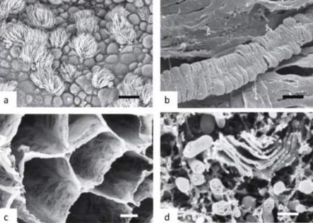 JOURNAL FREE ACCESSSince the introduction of scanning electron microscopy (SEM) to biology in the late of 1960’s, numerous researchers have studied various preparation techniques suitable for SEM observation of biological samples, including cells and tissues. In this paper, the standard preparation techniques for histology was briefly reviewed and, biological applications of SEM were introduced, especially in relation to the advances in technology of SEM. This paper also showed the recent advances in biological application of low-vacuum SEM, atmospheric SEM, real-time stereoscopic SEM, manipulation SEM, and 3D reconstruction for SEM.View full abstractDownload PDF (1190K)
 -
Susumu KUWABATA, Tetsuya TSUDA, Eiko MOCHIZUKI, Tsukasa TORIMOTOArticle type: Current Topic
2015Volume 36Issue 4 Pages 195-200
Published: April 10, 2015
Released on J-STAGE: April 21, 2015
 JOURNAL FREE ACCESSIonic liquid, which is salt melted even at room temperature, can be observed by scanning electron microscopes without charging. This fact implies that ionic liquid can be utilized as a liquid material to put conductivity to insulating samples for their scanning electron microscope (SEM) observation. This method is, in particular, effective for SEM observation of the soft materials like biological materials and polymer materials. As ionic liquid can reach to any place even at lumpy or fluffy surfaces, charge up of the samples is completely suppressed. Furthermore, keeping wet condition prevents change in the sample shape under vacuum condition. Such the advantage raises the precision of SEM images of soft materials.View full abstractDownload PDF (1552K)
 -
Takahiko HARIYAMA, Yasuharu TAKAKU, Hiroshi SUZUKI, Daisuke ISHII, Mas ...Article type: Current Topic
2015Volume 36Issue 4 Pages 201-206
Published: April 10, 2015
Released on J-STAGE: April 21, 2015
 JOURNAL FREE ACCESSScanning electron microscopy (SEM) has become an essential tool for observing the biological materials, however, various complex procedures preclude observation of living organisms to date. Here we present a new method to observe living organisms by a SEM. We observed the active movements of animals in vacuo (10−5—10−7 Pa) by coating them with a thin polymer membrane, NanoSuit®, made by polyoxyethylene (20) sorbitan monolaurate and found that the surface fine structure of the living organism is very different from that of traditionally fixed samples. After observation of the larvae of mosquitos, despite the high vacuum, we could rear them in normal culture conditions. We also found that the NanoSuit® and self-standing biocompatible films were able to produce by plasma polymerization from various monomer solutions. Our method will be useful for numerous applications, particularly in the electron microscopic observation of life sciences.View full abstractDownload PDF (1357K)
Planning Series
Surface Science for Environmental Issues
-
Akira IIMURA2015Volume 36Issue 4 Pages 207-208
Published: April 10, 2015
Released on J-STAGE: April 21, 2015
 JOURNAL FREE ACCESS
Science Café
Research Abroad
-
Felix TIMISCHL2015Volume 36Issue 4 Pages 209-210
Published: April 10, 2015
Released on J-STAGE: April 21, 2015
JOURNAL FREE ACCESSDownload PDF (566K)
News from Branches
-
Hiroshi ONISHI2015Volume 36Issue 4 Pages 211
Published: April 10, 2015
Released on J-STAGE: April 21, 2015
 JOURNAL FREE ACCESSDownload PDF (417K)
Qualifying Examination for Surface Science Engineers
-
2015Volume 36Issue 4 Pages 212
Published: April 10, 2015
Released on J-STAGE: April 21, 2015
JOURNAL FREE ACCESSDownload PDF (1302K)
News & Trends
-
2015Volume 36Issue 4 Pages 213
Published: April 10, 2015
Released on J-STAGE: April 21, 2015
JOURNAL FREE ACCESSDownload PDF (153K)
- |<
- <
- 1
- >
- >|