Progress of Digestive Endoscopy
Online ISSN : 2187-4999
Print ISSN : 1348-9844
ISSN-L : 1348-9844
Volume 86, Issue 1
Displaying 51-76 of 76 articles from this issue
Case report
  • Shunsuke Urayoshi, Satohiro Matsumoto, Hiroyuki Miyatani, Yukio Yoshid ...
    2015Volume 86Issue 1 Pages 178-179
    Published: June 18, 2015
    Released on J-STAGE: June 23, 2015
    JOURNAL FREE ACCESS
    A one-week eradication therapy of Helicobacter pylori using metronidazole, clarithromycin, and omeprazole is generally considered a well-tolerated therapy with minor side effects. We report a 66-year-old male patient who developed diarrhea few days after the treatment, and Clostridium difficile toxin was detected in his stool. Although this is a rare complication, it can sometimes occur; therefore, physicians should consider C. difficile colitis a possible side effect when patients present diarrhea after eradication therapy of H. pylori.
    Download PDF (1111K)
  • Kana Kawagishi, Yuki Kondo, Kaoru Yokoyama, Miwa Sada, Kiyonori Kobaya ...
    2015Volume 86Issue 1 Pages 180-181
    Published: June 18, 2015
    Released on J-STAGE: June 23, 2015
    JOURNAL FREE ACCESS
    Colonic stent insertion is useful for improving the quality of life of patients with malignant colorectal strictures. However, some patients have multiple strictures caused by direct invasion of malignant tumors arising in other organs or by peritoneal dissemination. Repeated sessions of stent placement can prolong the hospital stay or impair activities of daily living. We describe our experience with 2 patients in whom stents were concurrently placed in 2 stricture sites. First, a small-caliber scope was passed through the stricture in the distal colon, and a colonic stent was inserted into the stricture in the proximal colon by means of the Over-the-Wire (OTW) technique. Then another colonic stent was placed into the stricture site in the distal colon. Procedural accidents did not occur at the time of stent placement in either patient. The use of a small-caliber scope for stent placement in 2 sites is considered very useful in patients with multiple colorectal strictures.
    Download PDF (735K)
  • Toshio Arai, Satoshi Adachi, Jin Kanda, Toshikazu Yamaguchi, Nobumitsu ...
    2015Volume 86Issue 1 Pages 182-183
    Published: June 18, 2015
    Released on J-STAGE: June 23, 2015
    JOURNAL FREE ACCESS
    Intestinal spirochetosis (IS) is a zoonotic and rare gastrointestinal infection caused by Brachyspira. It does not show specific symptoms and are various ; gastrointestinal bleeding, diarrhea, asymptomatic. There is no established consensus for treatment yet. We report a case of a 48-year-old Japanese female with recurred IS and treatment with metronidazole seemed to be effective. She had visited our hospital because of mucus and bloody stool in 2010. A blood test, stool culture, colonoscopy examination had been done and diagnosed with IS. The symptom subsided with no therapy a few days, so we had not tried medication. The next year the symptom had reoccurred and disappeared a few days, so she did not come our hospital. However she showed the same symptom and visited our hospital again in 2014.
    We diagnosed that she was suffered from IS and treated her with metronidazole (1,000mg/day for 10days) and her clinical symptom completely improved. This case report suggests that metronidazole treatment for IS infection was effective to prevent recurrence.
    Download PDF (894K)
  • Dai Inoue, Teruhito Kishihara, Yusuke Muramatsu, Kohei Okamoto, Myonty ...
    2015Volume 86Issue 1 Pages 184-185
    Published: June 18, 2015
    Released on J-STAGE: June 23, 2015
    JOURNAL FREE ACCESS
    Mucosal prolapse syndrome (MPS) is caused by excessive mechanical stimulus. A conservative approach is used as basic medical treatment of MPS, such as improvement of bowel habits. However, we performed endoscopic mucosal resection (EMR) in the following MPS case.
    A 16-year-old male underwent colonoscopy and a protruding type MPS was detected. He initially received conservative treatment but he could not follow our medical instructions. Thus, nine months after his presentation, EMR for the MPS was performed. The histological specimen showed fibromuscular obliteration of the lamina propria which is a characteristic pathological change of MPS. After the resection, his symptoms and the endoscopic findings improved. EMR for protruding type MPS is a good option when conservative treatment is not effective.
    Download PDF (911K)
  • Sho Endo, Naoto Kurihara, Yasuhiro Sasaki, Hideshi Matsuda, Kazuyo Mor ...
    2015Volume 86Issue 1 Pages 186-187
    Published: June 18, 2015
    Released on J-STAGE: June 23, 2015
    JOURNAL FREE ACCESS
    A 77-year-old woman was admitted because of a positive fecal test for occult blood. She had taken oral Kamishoyosan continuously for 25 years. Colonoscopy revealed dark purplish-blue mucosa from the ileocecum to the ascending colon. Histopathology revealed a thickened wall of the colon, with linear calcification of the mesenteric vein. Based on these findings, the patient was diagnosed as having idiopathic mesenteric phlebosclerosis. A repeat colonoscopy performed 13 months after the patient discontinued Kamishoyosan showed improvement of the color and vascularity of the colonic mucosa. Thus, it appears that the colonoscopic findings of asymptomatic idiopathic mesenteric phlebosclerosis associated with the long-term use of Kamishoyosan could improve by about a year or so after discontinuation of Kamishoyosan.
    Download PDF (835K)
  • Daisuke Hihara, Yukako Nemoto, Takeshi Suzuki, Koichiro Sato, Kenji To ...
    2015Volume 86Issue 1 Pages 188-189
    Published: June 18, 2015
    Released on J-STAGE: June 23, 2015
    JOURNAL FREE ACCESS
    We herein describe the case of a 50-year-old man who presented with intermittent right lower quadrant abdominal pain which he had been experiencing since December 2013. The pain symptoms gradually worsended, and he was therefore hospitalized on May 2014. Abdominal CT showed the cecum invaginated into the ascending colon as leading point of the ileocecal valve . Emergency colonoscopy revealed the same status as observed in the CT findings. We attempted to reposition the invagination during colonoscopy by releaseing gradually over the air pressure. Colonoscopy performed on the 13th hospitalization day revealed the presence of an ulcer on the cecal diverticulum. As stated above, we speculated that cecal diverticulitis had thus caused the cecal invagination in this case. It is extremely rare that an invagination case is treated conservatively and thereafter shows a good recovery.
    Download PDF (895K)
  • Yasuhiro Sasaki, Naoto Kurihara, Sho Endo, Megumi Takagi, Hideshi Mats ...
    2015Volume 86Issue 1 Pages 190-191
    Published: June 18, 2015
    Released on J-STAGE: June 23, 2015
    JOURNAL FREE ACCESS
    A 72-year-old female was referred to our hospital because of melena. She had been diagnosed as having the Klippel-Trenaunay-Weber syndrome at the age of 55. Amputation of the right lower extremity, and a wide resection of the stomach for duodenal ulcer had been carried out at the age of 61.
    A contrast-enhanced abdominal CT showed obstruction of the main portal vein, prominent dilatation of the inferior mesenteric vein, and varices of the rectum. Colonoscopy revealed severe varices extending from the descending colon to the rectum.
    We considered that the blood flow was changed course via the internal iliac vein from the inferior mesenteric vein into the inferior vena cava. This hemodynamic change caused dilatation of the inferior mesenteric vein and the development of collateral vessels as varices around the rectum.
    Download PDF (1078K)
  • Toshifumi Hara, Teppei Omori, Yukiko Takayama, Takahiro Ajihara, Hirot ...
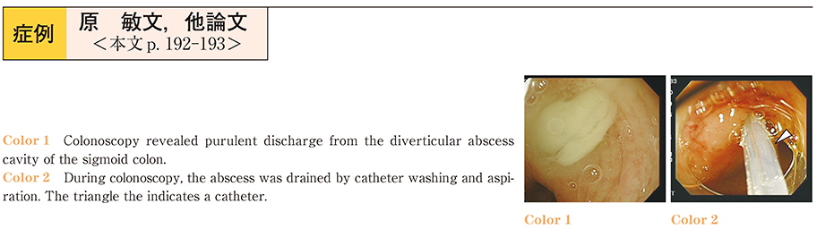
    2015Volume 86Issue 1 Pages 192-193
    Published: June 18, 2015
    Released on J-STAGE: June 23, 2015
    JOURNAL FREE ACCESS
    We describe a patient presenting diverticulitis with an abscess successfully treated by endoscopic drainage. A 59-year-old woman with left lower abdominal pain and fever was admitted to our hospital. Abdominal computed tomography revealed diverticulitis with abscess formation. Colonoscopy revealed colonic inflammation with abscess caused by diverticulitis. The abscess was drained by washing and aspiration during colonoscopy. After the purulent discharge from the abscess cavity was removed, abdominal pain and fever decreased, as expected. Endoscopic drainage, which is minimally invasive, has the potential to avoid emergency surgery in patients with many complications.
    Download PDF (835K)
  • Yoshihisa Saida, Tomotaka Ishii, Toshiyuki Enomoto, Kazuhiro Takabayas ...
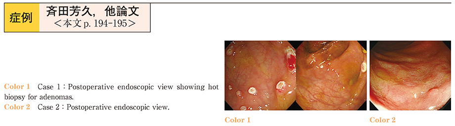
    2015Volume 86Issue 1 Pages 194-195
    Published: June 18, 2015
    Released on J-STAGE: June 23, 2015
    JOURNAL FREE ACCESS
    Usually, we conduct total coloproctectomy for familial adenomatous polyposis (FAP) . However, a problem regarding quality of life (QOL) of patients includes high stool frequency. Also, total coloproctectomy is relatively highly invasive surgery. We performed laparoscopic subtotal colectomy with ileo-sigmoid anastomosis following postoperative intensive endoscopic polypectomy for minimally invasive treatment of FAP.
    Case 1 ; A 39-year-old man. His stool frequency was about 4-5 times a day postoperatively. Colonoscopy was undergone every 3-6 months.
    Case 2 ; A 35-year-old man with sigmoid colon cancer and FAP. His stool frequency was about 3 times a day postoperatively. No recurrence of cancer in 4 years follow up was observed.
    Conclusion : Our procedures deliver high QOL for patients, and is relatively simple for surgeons and endoscopists to perform.
    Download PDF (1308K)
  • Mayo Tsuboi, Makoto Okamoto, Sachiko Kanai, Kazuma Sekiba, Azusa Matsu ...
    2015Volume 86Issue 1 Pages 196-197
    Published: June 18, 2015
    Released on J-STAGE: June 23, 2015
    JOURNAL FREE ACCESS
    Diverticular colitis is a segmental colitis, developing most commonly in the sigmoid colon of patients with diverticula. Its endoscopic features resemble ulcerative colitis or Crohn's disease, but by definition it does not involve the rectum. It is unknown if diverticular colitis is related to diverticula or inflammatory bowel disease.
    A 75-year-old male, who had previously undergone yearly follow-up colonoscopy to monitor adenoma and nonspecific inflammation without subjective symptoms, presented with lower abdominal pain and tenderness. Abdominal computed tomography (CT) revealed multiple diverticula and segmental inflammation involving the transverse and sigmoid colon. Colonoscopy showed stenosis near the splenic flexure, and segmental inflammation of the mucosa. The rectum was not involved. The case was diagnosed as diverticular colitis.
    The patient did not respond to antibiotics ; therefore, mesalazine therapy was initiated. Although abdominal pain resolved, laboratory results and CT findings worsened. Corticosteroids were added to the treatment regimen and follow up is ongoing.
    Download PDF (737K)
  • Yuki Shibata, Naoyuki Uragami, Haruo Ikeda, Syunsuke Omotaka, Genki Ts ...
    2015Volume 86Issue 1 Pages 198-199
    Published: June 18, 2015
    Released on J-STAGE: June 23, 2015
    JOURNAL FREE ACCESS
    We describe a case of sporadic cancer in a patient with Ulcerative Colitis in which detailed endoscopic examination did not show any signs of advanced neoplasia.
    This was a 50-year-old women that presented for surveillance colonoscopy. During colonoscopy under white light examination a flat (Paris 0-IIa) 10mm rectal lesion was detected. With magnifying endoscopy with narrow band imaging (M-NBI) , we recognized regular vessels as described with the Showa classification. Magnifying chromoendoscopy reveled type IIIL and type-IV were the dominant pit patterns. On endocytoscopy examination EC3a classification was seen indicating intramucosal carcinoma. The lesion was resected with EMR. A pathological diagnosis was well differentiated tubular adenocarcinoma (tub1) , pTis (M) , ly0, v0. Histologically, the lesion was growing down from the surface epithelium (top-down type) , which is not typical of dysplasia associated cancers.
    Download PDF (1050K)
  • Hiromichi Tsunashima, Shinpei Doi, Noritaka Ozawa, Masatoshi Mabuchi, ...
    2015Volume 86Issue 1 Pages 200-201
    Published: June 18, 2015
    Released on J-STAGE: June 23, 2015
    JOURNAL FREE ACCESS
    A 68-year-old man came to our hospital because he got edema in his left leg. Abdominal CT scan revealed multiple liver metastasis and pelvic CT scan showed marked thickening of the rectal wall and the border with the bladder became indistinct. Colonoscopic examination revealed stenosis of the rectum without mucosal abnormality. Histological examination of boring biopsies from this lesion led to the suspicion adenocarcinoma. His condition turned worse and died 1 month later. From the autopsy findings we diagnosed advanced bladder cancer with direct rectal invasion. Because this metastasis form is very rare, we report on this case along with a review of the literature.
    Download PDF (1053K)
  • Motoshi Miyagi, Yoshiro Tamegai, Kim Myungchul, Akiko Chino, Teruhito ...
    2015Volume 86Issue 1 Pages 202-203
    Published: June 18, 2015
    Released on J-STAGE: June 23, 2015
    JOURNAL FREE ACCESS
    A 67-year-old man was diagnosed by colonoscope to have superficial elevated type lesion at rectum. This lesion measuring about 15 mm in diameter, magnifying chromoendoscopy revealed a VI low irregularity Kudo's pit pattern system. Magnifying endoscopy with narrow band imaging revealed capillary pattern (CP) type IIIA. We diagnosed this lesion to adenocarcinoma invading until slight submucosal layer (SM1) , planed endoscopic submucosal dissection (ESD) . During maneuver, string blood vessel, fibrosis and retraction of proper muscle existed in submucosal layer. This findings shows that a carcinoma is invading submucosal layer around 1,000 μm. Pathological examination of the resected specimen revealed moderately differentiated tubular adenocarcinoma invading submucosal layer (1,100 μm, SM2) , with no vascular and lymphatic vessel involvement and budding Grade1. The surgical margin was negative for cancer (pHM0 pVM0) . This case report highlights the importance of this submucosal finding, it suggests that the cancer invade submucosal layer.
    Download PDF (1058K)
  • Makiko Mori, Koichi Koizumi, Go Kuwata, Taku Tabata, Susumu Iwasaki, K ...
    2015Volume 86Issue 1 Pages 204-205
    Published: June 18, 2015
    Released on J-STAGE: June 23, 2015
    JOURNAL FREE ACCESS
    An asymptomatic 80-years-old woman with an angiosarcoma of upper arm was incidentally found on PET to have a hot spot in the lower abdomen. She had been prescribed Calcium Polystyrene Sulfonate (CPS) .
    Colonoscopy revealed polypoid lesion about 30 mm in size in recto sigmoid, which endoscopic diagnosis was submucosal invasive carcinoma, biopsy specimen shows moderately differentiated adenocarcinoma. A year later, she complained bloody stool and lower abdominal pain. Second colonoscopy showed a white coated large polypoid lesion occupying a rectal lumen.
    Anterior resection was performed, and resected specimen showed 80×50 mm tumor with deep ulcer, pathological finding shows poorly differentiated with moderately differentiated component penetrating to right ovary, lymph node metastasis was positive for cancer. Many CPS crystals were deposited in the ulcer bed. CPS is known as mucosal toxic agents. Our case suggested CPS might be the cause of ulceration and rapid progression of the tumor.
    Download PDF (1179K)
  • Toyotaka Kasai, Koichi Kawabe, Seiji Muramatsu, Takuro Yamada, Yosuke ...
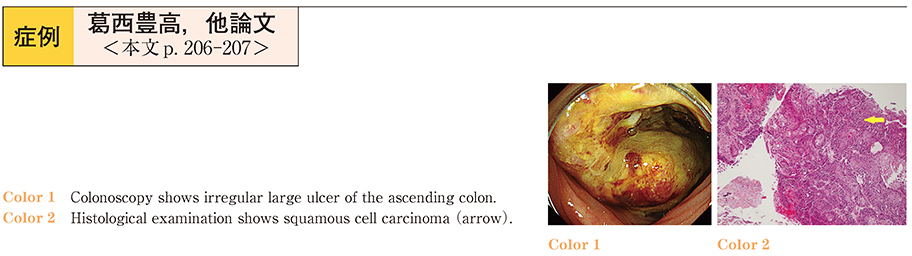
    2015Volume 86Issue 1 Pages 206-207
    Published: June 18, 2015
    Released on J-STAGE: June 23, 2015
    JOURNAL FREE ACCESS
    A 51-year-old man was admitted to our hospital with right lower quadrant abdominal pain, diarrhea, and fever. We diagnosed him with squamous cell carcinoma of the ascending colon after performing a colonoscopy. The upper gastrointestinal endoscopy revealed no carcinoma of the esophagus, and computed tomography revealed no lung cancer. Therefore, squamous cell carcinoma from the ascending colon was our final diagnosis. The positron emission tomography-computed tomography identified liver and bone metastases, and the patient died a month after admission. Squamous cell carcinoma of the colon is very rare. Squamous metaplasia of an adenocarcinoma is speculated to be the occurrence mechanism. It is very difficult to treat squamous cell carcinoma of the colon because it is often discovered at a highly advanced stage.
    Download PDF (682K)
  • Katsuyoshi Ando, Hiroki Tanabe, Eiichiro Ichiishi, Kiichi Sato
    2015Volume 86Issue 1 Pages 208-209
    Published: June 18, 2015
    Released on J-STAGE: June 23, 2015
    JOURNAL FREE ACCESS
    A 60-year-old-man who had been diagnosed with Lemmel’s syndrome and treated by endoscopic sphincterotomy (EST) three years ago, was presented with a developing fever and epigastralgia. Laboratory data revealed elevated serum bilirubin, white blood cell count and C-reactive protein levels. A computed tomography (CT) scan and magnetic resonance cholangiopancreatography (MRCP) revealed that the lower part of the common bile duct was displaced by a parapapillary diverticulum and that the upstream was dilated, but choledocholithiasis was not evident. We therefore diagnosed a recurrence of Lemmel's syndrome. Despite conservative therapy, serum bilirubin and biliary enzyme levels were continuously elevated for ten days from the time of admission. Accordingly, we performed an additional EST for the purpose of improving jaundice and bile duct congestion. After the EST, serum bilirubin and biliary enzyme levels decreased to within normal range. The patient is presently doing well and has been free from a relapse for nine months.
    An additional EST was effective against recurrent Lemmel’s syndrome.
    Download PDF (1061K)
  • Shigeto Ikeda, Sawako Kuruma, Makiko Mori, Susumu Iwasaki, Kazuro Chib ...
    2015Volume 86Issue 1 Pages 210-211
    Published: June 18, 2015
    Released on J-STAGE: June 23, 2015
    JOURNAL FREE ACCESS
    A 40-years-old woman came to our hospital for further evaluation of the dilatation of the common bile duct. Endoscopic retrograde cholangiopancreatography showed cystic dilatation of the common bile duct and pancreaticobiliary maljunction (PBM) with a 16-mm long common channel. Two narrow-branches were derived from the long common channel. The patient was diagnosed as congenital dilatation of the bile duct (Totani ClassificationIa) , and underwent laparoscopic bile duct resection and bilioenteric anastomosis. Histologically, there were no neoplastic lesions. Pathogenesis of PBM is not be fully elucidated. Side branches are derived from the long common channel in some PBM cases, which suggests that the long common channel in PBM is derived from the pancreatic duct.
    Download PDF (883K)
  • Yoriko Kuboi, Kazuya Sugimori, Shinpei Kondo, Atsuyoshi Kubo, Yuniba I ...
    2015Volume 86Issue 1 Pages 212-213
    Published: June 18, 2015
    Released on J-STAGE: June 23, 2015
    JOURNAL FREE ACCESS
    A 77-year-old woman underwent pancreaticoduodenectomy and Child's reconstruction for advanced duodenal cancer in 2012. During the 18 months following surgery, the patient was admitted six times with repeated cholangitis. Computed tomography showed intrahepatic bile duct dilation due to stones in the left hepatic duct. Using a colonoscope and double-balloon scope, we were not able to detect the choledochojejunostomy. After performing percutaneous biliary drainage, we successfully used the rendezvous method for cannulation of the bile duct. We attempted to extend the stenosis of the choledochojejunostomy using a biliary balloon dilatation catheter ; however, we were not able to achieve sufficient dilatation through the severe stenosis. Therefore, we deployed a covered metallic stent (CMS) , to dilate the stenosis of the choledochojejunostomy. After achieving sufficient dilatation of the stenosis with the CMS extension, the CMS was removed. This procedure sufficiently dilated the severe stenosis of the choledochojejunostomy to allow for safe removal of the intrahepatic bile duct stones. Cholangitis did not recur for six months after the procedure.
    Download PDF (889K)
  • Kohei Ono, Jun Ushio, Kensuke Yokoyama, Norikatsu Numao, Hisashi Hatan ...
    2015Volume 86Issue 1 Pages 214-215
    Published: June 18, 2015
    Released on J-STAGE: June 23, 2015
    JOURNAL FREE ACCESS
    A 70-year-old man was present with fever, chills and jaundice. Abdominal enhanced computed tomography (CT) showed hepatolithiasis of right hepatic lobe and common bile duct (CBD) stones. On endoscopic retrograde cholangiography, we successfully extracted CBD stones. However, intrahepatic stones of the posterior segment branches (B6) were left since conventional basket catheter could not pass. After several days, we succeeded to advance COAXIS deeply and extract residual stones of B6 without complications. Conventional basket catheter has the sideslit for the guidewire at the outside of the catheter. On the other hand, COAXIS has it at the center of the catheter, which achieves high followability to the guidewire. COAXIS might be useful for all the peripheral bile ducts that have been recognized difficult to be approached with the conventional basket catheters.
    Download PDF (892K)
  • Yuniba Ishii, Kazuya Sugimori, Atsuyoshi Kubo, Haruo Miwa, Eri Kameta, ...
    2015Volume 86Issue 1 Pages 216-217
    Published: June 18, 2015
    Released on J-STAGE: June 23, 2015
    JOURNAL FREE ACCESS
    A 66-year-old woman was referred to our hospital for further examination of gallbladder cancer. Contrast-enhanced computed tomography (CT) showed a ring formed by an enhanced mass measuring 10 mm in diameter in segment II of the liver, accompanied by the gallbladder cancer. Hepatic metastasis from the gallbladder cancer was suspected based on the findings of several imaging examinations. An adequate sample from the small lesion could not be obtained using abdominal ultrasound-guided biopsy because it was located deep under the skin. However, the lesion could be clearly visualized on endoscopic ultrasound because of its location near the stomach. Therefore, we performed ultrasound-guided fine needle aspiration (EUS-FNA) , and the lesion was diagnosed as an inflammatory mass. Surgery was selected as treatment for gallbladder carcinoma. According to a report by John DeWitt et al, the sensitivity for diagnosis of a malignant hepatic lesion by EUS-FNA was 82-94%. Our findings suggest that EUS-FNA may be useful for the histological diagnosis of hepatic lesions located near the stomach.
    Download PDF (764K)
  • Norimasa Matsushita, Yasuhiro Sudo, Takeshi Kubota, Tatsuo Inoue
    2015Volume 86Issue 1 Pages 218-219
    Published: June 18, 2015
    Released on J-STAGE: June 23, 2015
    JOURNAL FREE ACCESS
    We describe a case with a giant hepatic cyst and associated symptoms that was treated laparoscopically. A 63-year-old man who developed right hypochondrium pain in 2006 was previously diagnosed with cholecystitis. The pain recurred in February 2013, and he visited to our hospital. Computed tomography indicated a 20-cm hepatic cyst in the right liver lobe, which exerted pressure on the gallbladder and adjacent organs. A laparoscope and forceps were introduced following a single umbilical incision. Another port was established on the right flank for drain insertion. Deroofing of the cyst was performed, and the greater omentum was inserted into the cyst to prevent re-accumulation of cystic fluid. As hepatic cysts are benign, invasive procedures should be avoided. Thus, we performed laparoscopic surgery for a hepatic cyst as a radical treatment with low invasiveness, by ensuring that all ports, excluding the drainage hole, were restricted to the umbilical portion.
    Download PDF (1018K)
  • Kazuki Nomoto, Makoto Nishimura, Harutaka Kambayashi, Kenichiro Nakaji ...
    2015Volume 86Issue 1 Pages 220-221
    Published: June 18, 2015
    Released on J-STAGE: June 23, 2015
    JOURNAL FREE ACCESS
    A 75 year-old male with a history of diabetes was referred to our hospital to confirm the diagnosis for a hepatic mass in the caudate lobe, pointed out incidentally on a CT scan.
    The patient was admitted and underwent endoscopic ultrasound. From the stomach, the caudate lobe could be visualized, and endoscopic ultrasound-guided aspiration (EUS-FNA) was successfully performed. Histopathology results showed hepatocellular carcinoma (HCC) .
    EUS-FNA is widely used as a reliable method to make correct diagnoses for pancreatic tumor, and retroperitoneal / mediastinal lymphadenopathy. However, there have only been a few reports on EUS-FNA for the liver, particularly on the caudate lobe. In this case, we report that EUS-FNA for hepatic tumor is feasible, and technically easy, even for the caudate lobe.
    We conclude that EUS-FNA for hepatic tumor is feasible, and possibly useful as a complementally approach to be used in place of percutaneous liver biopsy.
    Download PDF (673K)
  • Taijiro Wake, Kei Tanaka, Hitoshi Kurata, Naoaki Dan
    2015Volume 86Issue 1 Pages 222-223
    Published: June 18, 2015
    Released on J-STAGE: June 23, 2015
    JOURNAL FREE ACCESS
    A 64-year-old male was admitted to our hospital with the chief complaint of severe abdominal distension. Abdominal CT findings showed massive ascites due to a non-malignant pancreatic disease. Amylase and protein in ascitic fluid were high level.
    From the above results, pancreatic ascites was suspected and he was treated with conservative therapy. On day 16, endoscopic retrograde pancreatography revealed transudation of contrast medium from the pancreatic duct. A pancreatic duct stent was placed, but he was complicated with bacterial peritonitis on day 23. The peritonitis was gradually improved by antibiotics and ascites drainage. The ascites disappeared in the CT before discharge. Thereafter we removed a pancreatic duct stent discharge 40 days later, he has now been in good health without a recurrence.
    Download PDF (1164K)
  • Fumisato Otaka, Mitsuhiro Kida, Kosuke Okuwaki, Hiroshi Yamauchi, Shir ...
    2015Volume 86Issue 1 Pages 224-225
    Published: June 18, 2015
    Released on J-STAGE: June 23, 2015
    JOURNAL FREE ACCESS
    A 15-year-old boy presented with low back pain. Abdominal radiography performed by a local physician showed calcification in the left upper abdomen. Abdominal computed tomography (CT) revealed a mass, 60 mm in diameter, arising in the tail of the pancreas, and the patient was referred to our hospital. Contrast-enhanced CT, contrast-enhanced magnetic resonance imaging, and endoscopic retrograde pancreatography suggested a solid pseudopapillary neoplasm (SPN) . Laparoscopic distal pancreatectomy was performed. The tumor was a well-demarcated cystic lesion, measuring 7×7×5.5 cm. The cut surface of the tumor had a solid, hemorrhagic, and cystic appearance. Histopathological examination revealed that the tumor components were surrounded by a cystic wall showing hyaline fibrosis. The tumor cells showed funicular or nest-like structures, and the nuclei were round or oval. Calcification and cholesterin crystals were found in the stroma. Immunohistochemically, the tumor cells were positive for CD10, CD56, vimentin, and α1-antitrypsin. An SPN was thus diagnosed. We describe our experience with a boy who had an SPN with extremely marked calcification.
    Download PDF (825K)
  • Ayako Matsui, Masashi Ijima, Eishin Kurihara, Yuta Watanuki, Yasumori ...
    2015Volume 86Issue 1 Pages 226-227
    Published: June 18, 2015
    Released on J-STAGE: June 23, 2015
    JOURNAL FREE ACCESS
    A 32-year-old woman with choriocarcinoma in 2008 has had a complete response to chemotherapy. Abdominal CT revealed a tumor at pancreatic body in April 2014. We suspected metastatic pancreatic cancer of choriocarcinoma, because the serum level of human chorionic gonadotropin (hCG) were elevated. So, we performed endoscopic ultrasound-guided fine needle aspiration (EUS-FNA) to exclude primary pancreatic cancer.
    However, EUS-FNA indicated adenocarcinoma. Finally, she was diagnosed as primary pancreatic cancer.
    She underwent distal pancreatectomy. Histopathological examination of the surgically resected specimen revealed tubular adenocarcinoma, pT2N0M0 Stage II in the pancreas.
    After surgery, serum hCG level was normal level.
    At this case, EUS-FNA can be useful for the preoperative diagnosis.
    Download PDF (831K)
  • Madoka Kataoka, Sawako Kuruma, Susumu Iwasaki, Makiko Mori, Kazuro Chi ...
    2015Volume 86Issue 1 Pages 228-229
    Published: June 18, 2015
    Released on J-STAGE: June 23, 2015
    JOURNAL FREE ACCESS
    A 70-year-old man complaining of epigastric discomfort and jaundice was admitted to our hospital in November 2013. Enhanced abdominal CT scan revealed a 2.5×3.0 cm-sized mass and enlargement of abdominal lymph nodes and left adrenal gland. MRCP showed obstruction of the dorsal pancreatic duct in the pancreatic head and distal bile duct. There was no visualization of the ventral pancreatic duct. Endoscopic pancreatography via the major duodenal papilla showed a short pancreatic duct with partial acinar filling, and pancreatography via the minor duodenal papilla revealed a stenosis in the dorsal pancreatic duct. There was no communication between the ventral and dorsal pancreatic ducts. Brushing cytology of the bile duct was adenocarcinoma.
    The diagnosis was pancreatic cancer (T4N1M1 Stage IVb) in the dorsal pancreas of pancreas divisum, and the patient underwent chemotherapy.
    Of our 69 cases of pancreas divisum, 6 cases (9%) had pancreatic cancer, and the cancer occurred from the dorsal pancreas in all cases. As longstanding stasis of pancreatic juice in the dorsal pancreatic duct caused by the relative stenosis of the minor duodenal papilla might be a factor that promotes pancreatic carcinoma, we should pay heed to changes of the dorsal pancreatic duct in individuals with pancreas divisum.
    Download PDF (706K)
feedback
Top