Progress of Digestive Endoscopy
Online ISSN : 2187-4999
Print ISSN : 1348-9844
ISSN-L : 1348-9844
Volume 62, Issue 2
Displaying 1-50 of 52 articles from this issue
Clinical study
  • Yoshiya Kumagai, Tsutomu Asaoka
    2003Volume 62Issue 2 Pages 26-30
    Published: May 31, 2003
    Released on J-STAGE: April 03, 2014
    JOURNAL FREE ACCESS
    In the disinfecting process of endoscopes with electrolyzed acid water and glutaraldehyde, we have found the deteriorations with rough texture on the outer coated surface of endoscopes after about 2,000 uses. We looked into the cause, analyzing the rugged surface, as follows. (1) The surface was observed by a digital microscope in 50 magnifications, and compared with the surface of new endoscopes. (2) The surface was analyzed and compared by a Fourier transform infrared spectrophotometer. (3) The rugged surfaces were cut off from the endoscopes in question to analyze the deteriorated material, and observed by a scanning electron microscope in 50,500 and 5,000 magnifications. (4) In use of sodium hypochlorite we found suitable to dissolve the deteriorated material, we transformed the dissolved material to powder through neutralization, salting out and freeze-drying. We made the analysis in the powder form.
    Conclusions : (1) The material dissolved in sodium hypochlorite was quantitated to 16 amino acids. We inferred that the deteriorated surface in hard accumulated layers was a substance polymerized and cross-linked of small amounts of protein, which was left after the endoscopes were washed, with glutaraldehyde. It was not known how electrolyzed acid water affected this. (2) For removal of this hard rugged substance, we found sodium hypochlorite useful and possible to recover the glossy smooth surface. We concluded that endoscopes should be thoroughly cleaned with enzymatic or neutral detergents before disinfecting process, without using glutaraldehyde.
    Download PDF (1981K)
  • Takeshi Matsuhisa, Nobutaka Yamada, Miki Suzuki, Mariko Sonoda, Toshiy ...
    2003Volume 62Issue 2 Pages 31-35
    Published: May 31, 2003
    Released on J-STAGE: April 03, 2014
    JOURNAL FREE ACCESS
    Difference of gastroduodenal disease between patients with hemodialysis and control group (non-hemodialisys patients) was studied by the match of age and gender. Helicobacter pylori (Hp) infection between two groups was also compared by the match of age, gender and endoscopic diagnosis.
    1) We found many gastritis cases (36.6%) , which means redness, erosion, hemorrhagic erosion and edema, and a few peptic ulcer cases (7.3%) in hemodialysis group. In control group, there were many peptic ulcer cases (42.7%) and a few gastritis cases (11.0%) . There were differences in these two groups.
    2) A prevalence of Hp infection in hemodialysis group was significantly low compare with control group (hemodialysis group : 28.6%, control group : 71.4%, P<0.001) . A prevalence of Hp infection in shorter than 3 months hemodialysis group showed 31.3% and it became lower percentage as hemodialysis period became longer.
    3) An average of chronic inflammation score in Hp negative hemodialysis group was higher than that of Hp negative control group (0.81 and 0.30, respectively, P=0.001) . A prevalence of serum anti-HpIgG antibody was also high in Hp negative hemodialysis group compare with Hp negative control group (hemodialysis group : 58.3%, control group : 20.0%, P<0.05) . According to these results, there was a possibility of natural eradication cases in patients with chronic renal failure.
    Download PDF (699K)
  • Fumiki Toriumi, Yoshiro Saikawa, Koichiro Kumai, Masahiko Aoki, Nobuna ...
    2003Volume 62Issue 2 Pages 36-40
    Published: May 31, 2003
    Released on J-STAGE: April 03, 2014
    JOURNAL FREE ACCESS
    <BACKGROUND>Endoscopic mucosal resection (EMR) or laparoscopic wedge resection have been performed for patients with mucosal gastric cancer, resulting in preservation of their stomach, keeping QOL. However indication of minimal surgery remains to be discussed, especially for undifferentiated type of gastric cancer, since undifferentiated type is known to invade into deep layer of gastric wall and lymph nodes. <PURPOSE>Clinico-pathological features of undifferentiated type of gastric cancer were considered, retrospectively, objecting early gastric cancer diagnosed by pre-operative examination. <PATIENTS AND METHODS>We retrospectively investigated 293 patients who were diagnosed as early gastric cancer before surgery, and performed gastrectomy between January, 1994 and December, 1997. Two subgroups were divided into either differentiated type (pap., tub1., tub2 ; 202 cases) or undifferentiated type (por 1., por 2., sig ; 91 cases) , and accuracy rates of pre-operative diagnosis for cancer invasion depth, incidence of mis-diagnosis of cancer margin, and distribution of lymph node metastasis were evaluated. <RESULTS>According to accuracy rates of pre-operative diagnosis for cancer invasion depth, differentiated and undifferentiated type resulted in 94.6% and 91.2%, respectively. We also showed 3 undifferentiated type which were histologically diagnosed as positive marginal of cancer after surgery, while we experienced no such case in differentiated type. In addition, positive rate of lymph nodes in undifferentiated mucosal cancer was statistically higher than in differentiated mucosal cancer (P=0.02) . <CONCLUSIONS>While small surgical procedure is a promising technique to preserve patients' QOL cancer therapy must be cautious to eliminate undesirable outcome like recurrence or cancer death caused by minimal surgery. In the study, we concluded that indication of minimal surgery should be considered properly, based on the risks of the difficulties of diagnosis for cancer invasion depth, surgical margin and lymph node metastasis in undifferentiated type of gastric cancer.
    Download PDF (1079K)
  • Masakazu Kamihira, Yukio Yoshida, Hiroyuki Miyatani, Ryuichi Hirakawa, ...
    2003Volume 62Issue 2 Pages 41-44
    Published: May 31, 2003
    Released on J-STAGE: April 03, 2014
    JOURNAL FREE ACCESS
    Angiectasia may be responsible for up to 1.2% to 8% all episodes of upper gastrointestinal bleeding. Among 170 patients with angiectasia of the stomach at Omiya Medical Center, Jichi Medical School from April 1990 to April 2000, 10 cases (5.9%) experienced episodes of bleeding. Four cases with active bleeding were managed by endoscopic therapy (clippig in 2 cases, EMR in 1 case, heater probe coagulation in 1 case) , and all four cases were successfully controlled. Long-term observation was possible in two cases. Case 2 : there has been no recurrence of angiectasia for 7 years since endoscopic mucosal resection (EMR) . Case 4 : there was an angiectasia appeared at the same place of site in the stomach 2 years after clipping, and a blood test showed anemia. Clipping is easier and safer than EMR, but might permit recurrences of an angiectasia.
    Endoscopic therapy is now the most widely used method for treating bleeding angiectasia. The use of ethanol injection, endoscopic ligation, clipping, microwave coagulation, heater probe coagulation, and EMR have been reported. Some of the authors reported no recurrence in brief follow-up periods from 1 month to 5 months. However, treatments should not be judged as recurrence-free in periods of less than one year, at minimum, and continuation endoscopic examination is necessary.
    Download PDF (461K)
  • Masato Ai, Taketo Yamaguchi, Takeo Odaka, Kanae Mitsuhashi, Tadayuki S ...
    2003Volume 62Issue 2 Pages 45-49
    Published: May 31, 2003
    Released on J-STAGE: April 03, 2014
    JOURNAL FREE ACCESS
    Background : Cholinergic blocking agents are regularly used to reduce colonic spasm in colonoscopy. However, these agents are thought to be contraindication for heart disease, prostate hypertrophy, or glaucoma. The aim of this study is to evaluate the effect of intracolonic spraying of Herbal Medicine, Shakuyaku-kanzo-to (TJ-68) on the suppression of colonic spasm during colonoscopy.
    Subject and methods : Twenty-six patients underwent colonoscopy without premedication. An endoscope was inserted from anal canal about to 25 cm of sigmoid colon. Then, identifying a certain colonic spastic region and keeping the distance between the spastic region and the tip of the colonoscope, the endoscopic view was recorded on digital videotape for 3 minutes before and after spraying of TJ-68 solution (0.5g of TJ-68 dissolved in 50 ml of lukewarm water) through a working channel. The 12 spot images recorded were selected every 3 minutes for the assessment of luminal change. The intraluminal area of the spastic region was measured with the computer image analyzer (Adobe Photoshop 7.0) and expressed as pixel counts. The areas under the curve (AUC) were calculated from the pixel curve obtained before and after the spraying of TJ-68.
    Results : The mean AUC before and after the TJ-68 spraying was 41,057 pixels and 98,348 pixels, respectively. Thus, AUC significantly increased after the TJ-68 spraying.
    Conclusion : A direct spraying of the TJ-68 on the colonic mucosa proved to suppress the colonic spasm. TJ-68 was suggested to be useful as a premedication of colonoscopy when the patients are not amenable to cholinergic blocking agent.
    Download PDF (1765K)
  • Tarou Hibiki, Naoyuki Uragami, Koichi Koizumi, Akiko Chino, Tomohiro T ...
    2003Volume 62Issue 2 Pages 50-54
    Published: May 31, 2003
    Released on J-STAGE: April 03, 2014
    JOURNAL FREE ACCESS
    Objectives : A complete resection of the lesion is the most important factor to determine the efficacy of the endoscopic therapy. This method was attempted to make up for weak point of piecemeal resection such as recurrence of lesion. A management of a rectal lesion should be more careful than that of a colon, because complications of rectal surgery are more frequent and risky. To avoid over-treatment, excision biopsy of rectal lesion is important, and the specimen should be good information to decide additional surgical treatment.
    Methods : Indication of this method is only in the rectum. Because the wall of rectum is thicker than that of colon, and there is less complication of perforation. Initially, saline with epinephrine solution was injected into submucosal layer around the lesion to lift it off the muscular layer. A needle knife was used for cutting around the lesion. Then insulation-tipped electro surgical (IT) knife was used for exfoliating the lesion from submucosal layer.
    Results : 4 patients were performed with this method. The lesions were completely resected with one piece. The mean diameter of resected tumors was 35 mm. All the lesions were consisted carcinoma with adenoma component. Immediate minor bleeding without transfusion was recognized in 2 patients, and was controlled with hemoclip. There were no perforations.
    Case 1 : 59 y/o F 45 mm, depth m, ly (-) , v (-) .
    Case 2 : 63 y/o M 20 mm, depth sm2, ly (+) , v (-) , addition surgical resection.
    Case 3 : 70 y/o M 40 mm, depth sm2, ly (-) , v (-) .
    Case 4 : 60 y/o M 30 mm, depth m, ly (-) , v (-) .
    Conclusion : This new method is able to remove the lesion with large one piece without serious complication. These specimens gave us good information for management of rectal tumor.
    Download PDF (1846K)
  • Tsunao Imamura, Hitoshi Yoshida, Katsuya Kitamura, Tatsurou Yanagawa, ...
    2003Volume 62Issue 2 Pages 55-59
    Published: May 31, 2003
    Released on J-STAGE: April 03, 2014
    JOURNAL FREE ACCESS
    Pancreatic carcinoma derived from pancreatic duct epithelium (pancreatic ductal carcinoma : PD Ca) is one of the most fatal cancers. Since it is still extremely difficult to detect early PD Ca, many patients with PD Ca will be considered to be unsuitable for surgery when they are diagnosed. At endoscopic retrograde cholangiopancrectography (ERCP) , exfoliated cytologic specimens are obtained from the main pancreatic duct (MPD) via the duodenal papilla by abrasion and aspiration with or without intravenous administration of secretin in Japan. However, the cytologic examination using the previously described techniques yields moderate sensitivity (30-79%) because desquamated ductal epithelial cells are presumably too small in number and too impaired to be accurately diagnosed. In order to obtain a large number of exfoliated cells without alteration from the pancreatic duct, we developed a novel sampling method,“pancreatic duct lavage fluid (PDLF) ”, by devising bronchoalveolar lavage fluid for pulmonary carcinoma. In the present study, cytologic examination using PDLF for diagnosis of PD Ca was evaluated for its usefulness.
    We examined 5 patients with PD Ca detected by abdominal CT scan. After brush cytology from the MPD having irregularities, stenosis or obstruction detected by ERCP, PDLF was collected with a catheter. The catheter consists of three parallel lumens for ballooning, injection and a guidewire. Isotonic saline was injected into the injection lumen following ballooning at the MPD, and“PDLF”was simultaneously aspirated from the guidewire lumen.
    Cytologic examination using PDLF was sensitive for diagnosis of malignancy compatible with pancreatic adenocarcinoma in all cases. Many exfoliated cells were collected without impairment from PDLF compared to those using brush or aspiration cytology methods. This examination was safely performed without shock, pancreatitis or cholangitis.
    Cytologic examination using PDLF is very sensitive for detection of PD Ca and available at any institution equipped with ERCP and cytologic. examination. In the present study, the novel examination.“PDLF”was evaluated as useful, sensitive, safe and not complicated. PDLF could be widely popularized for diagnosis of PD Ca.
    Download PDF (1971K)
Case report
  • Tsutomu Nomura, Takashi Tajiri, Masao Miyashita, Hiroshi Makino, Hiros ...
    2003Volume 62Issue 2 Pages 60-62
    Published: May 31, 2003
    Released on J-STAGE: April 03, 2014
    JOURNAL FREE ACCESS
    Since January 2001, the patients with a superficial esophageal cancer who could not undergo the endoscopic mucosal resection (EMR) have been treated by Argon Plasma Coagulation (APC) in our institute. A recurrence after the APC was observed in two cases.
    Case 1 : A 65-year-old man. He was pointed out to have a 0-IIc type of esophageal cancer at the middle thoracic esophagus in 2000. Firstly, radiation was selected because he had liver cirrhosis with esophageal varices. A recurrence was observed after the radiation, APC was performed in August 2001. In January 2002, APC was repeated because the recurrence after the ablation was observed.
    Case 2 : A 69-year-old man.Endoscopic examination revealed a 0-IIb type of esophageal cancer at the middle thoracic esophagus. EMR was avoided because of his bleeding tendency due to the medication for cerebral infarction and angina pectoris, then instead, APC was performed in January 2002. But the recurrence was observed about 6 months after the ablation and he underwent APC again.
    Because the depth of esophageal tissue degeneration by APC is thought to be limited to the submucosal layer, it is enough for superficial cancer of esophagus to be treated. EMR is thought to be the first primary method. However, APC may be useful in cases patients have complication which make EMR difficult.
    Download PDF (293K)
  • Shoji Maruyama, Masayuki Ando, Fumie Kobayashi, Ikuo Sakuma, Shin Han ...
    2003Volume 62Issue 2 Pages 64-65
    Published: May 31, 2003
    Released on J-STAGE: April 03, 2014
    JOURNAL FREE ACCESS
    We describe the successful endoscopic removal of a foreign body in the stomach by means of a snare technique. An 19-years-old woman ingested a table spoon and came to our hospital. She was under psychotherapy for overeating. An aout 18cm long spoon was noted by the X-ray photograph. We used the two-channel video endoscope and two polypectomy snare loops. The spoon was tied with two snare devices and located parallel to the esophagus. This device is simple to use, versatile, and effective and advances the safe endoscopic removal of a variety of gastroesophageal foreign bodies.
    Download PDF (588K)
  • Ryouhei Futami, Takashi Tajiri, Tsutomu Nomura, Hiroshi Makino, Koji S ...
    2003Volume 62Issue 2 Pages 66-67
    Published: May 31, 2003
    Released on J-STAGE: April 03, 2014
    JOURNAL FREE ACCESS
    A 61 years old male with esophageal carcinoma underwent right transthoracic esophagectomy and 3-field (cervix, mediastinum, abdomen) lymph node dissection. Protease inhibitor and a H2-Blocker were prescribed, but postoperative endoscopic findings and a biopsy specimen revealed Grade C esophagitis and patches of Barrett's mucosa at the oral side of the anastomosis.
    Patients undergoing esophagectomy and gastric tube reconstruction may develop Barrett's mucosa, because these patients are exposed to the loss of the mechanism to prevent reflux of tbe gastric contents. We report on a patient whose remnant esophagus was periodically followed up with endoscopy and biopsy for 7 years after surgical resection of esophageal carcinoma, and in whom patches of Barrett's mucosa developed within the remnant esophagus.
    Download PDF (450K)
  • Takeshi Shioya, Katsuyuki Ietomi, Kyoya Sakimura, Koichi Nakajima, Yui ...
    2003Volume 62Issue 2 Pages 68-69
    Published: May 31, 2003
    Released on J-STAGE: April 03, 2014
    JOURNAL FREE ACCESS
    A 73-year-old woman had been made a diagnosis that she had gotten reflux esophagitis and Long-Segment Barrett's Esophagus (LSBE) . However, it turned out to be the cancer due to the re-examination of endoscope after a year and two months from the first diagnosis. Although it had been thought to be mucosal cancer because there had been no unevenness of mucous membrane, histological examination after operation revealed that it was multifocal minimal cancers arised from Barrett's esophagus. Recently, the theory of dysplasia-cancer sequence is worldwidely accepted, in this case it is interesting that we could find out the initial appearance of carcinogenesis in the LSBE. We must take the contrast method with indigocarmine to find out very small cancer.
    Download PDF (867K)
  • Kenji Oomura, Kaori Matsuda, Mari Saito, Akira Shirai
    2003Volume 62Issue 2 Pages 70-71
    Published: May 31, 2003
    Released on J-STAGE: April 03, 2014
    JOURNAL FREE ACCESS
    We report a case of a 32-year-old man with HIV infection who has an esophageal perforation of an idiopathic ulcer. He had been treated by conservative therapy, and an endoscopic therapy was performed for the closure of the perforation. The HIV infection was well controlled by anti-HIV drugs administration and the ulcer became scared.
    In January 2002, he felt difficulty to swallow and had a high fever. He was admitted and computed tomography (CT) of the chest showed right pyothorax. Drainage of the pyothorax cavity and antibacterial therapy was performed. His blood examination indicated HIV infection.
    Esophagogram showed an esophageal perforation. Endoscopic examination showed giant ulcer that was considered as idiopathic one because cytomegalovirus antigen was not detected in his blood and it was cured without anti-herpes-virus drugs. He was conducted enteral nutrition and medication of anti-HIV drugs through gastoric tube. HIV infection was well controlled and the ulcer got healed progressively, but the perforation remained. So we performed an endoscopic therapy which was a brushing technique with irregular epithelium on inner face of perforation cavity by a pancreatic duct biopsy brush. Successfully the perforation was closed and he could regain to take orally soon. He discharged 152 days after admission and his HIV-infection is controlled as an outpatient.
    Download PDF (1166K)
  • Toshiyuki Mashimo, Jyunko Iwasaki, Hideyuki Suzuki, Shinya Saruya, Yuu ...
    2003Volume 62Issue 2 Pages 72-73
    Published: May 31, 2003
    Released on J-STAGE: April 03, 2014
    JOURNAL FREE ACCESS
    A 76-year-old woman suddenly had epigastralgia and dyspnea after vomiting of polyethylene glycol electrolyte lavage solution in the bowel preparation for colonoscopy. Emergency upper endoscopy showed the laceration at the gastro-esophageal junction and chest CT scan revealed the existence of mediastinal emphysema and bilateral pleural effusions. Consequently, she was diagnosed as having spontaneous esophageal rupture and admitted to our hospital. Conservative therapy was given to her because she had little mediastinal pollution and none of complications such as mediastinal abscess, pneumothrax, and emphysema. After the conservative therapy, the disappearance of the laceration, mediastinal emphysema, and pleural effusions were demonstrated by gastroesophageal fluorography and chest CT scan.
    Here we present this case because spontaneous esophageal rupture due to vomiting is rare. We suggest that conservative therapy may be useful for a patient with spontaneous esophageal rupture if little mediastinal pollution, only slight symptom and inflammation are observed.
    Download PDF (612K)
  • Toshitake Mitsuhashi, Nobuo Murata, Tsuyoshi Suzuki, Hirofumi Yamada, ...
    2003Volume 62Issue 2 Pages 74-75
    Published: May 31, 2003
    Released on J-STAGE: April 03, 2014
    JOURNAL FREE ACCESS
    A 55-year-old male began to feel precordial pain after meals in the end of April, 2001. He was in hospital to receive the steroid therapy for dermatomyositis since December 2000.
    Endoscopic examination showed a large ulcer and mucosal bridges resulting from laceration of the submucosal layer, which produced a false channel throughout the middle and distal esophagus. The patient was first managed conservatively to refrain from any drinks and meals for two weeks. In the middle of May, esophagoscopy showed retention of fluid and food in the false channel of the esophagus. On 28 of May, the mucosal bridge of the esophagus was cut through with a diathermy knife under endoscopic control. The false channel was abolished. The patient was able to drink water 20 days after the endoscopic treatment and to eat meals 26 days after it. Endoscopic examination after 5 months showed almost perfect epitherization of the esophageal mucosal membrane. The endoscopic treatment to open the false channel by cutting the mucosal bridge shortened the admission period. It is recommended that the mucosal bridge should be cut through endoscopy as soon as possible.
    Download PDF (930K)
  • Yuta Nemoto, Hitoshi Shimao, Masaki Morise, Natsuya Katada, Shin-ichi ...
    2003Volume 62Issue 2 Pages 76-77
    Published: May 31, 2003
    Released on J-STAGE: April 03, 2014
    JOURNAL FREE ACCESS
    A 68 years old male was admitted for esophageal carcinoma with tracheal invasion. After 2 course of chemotherapy, esophago-tracheal fistula was defined.
    He was received a double stents implantation for trachea (Tracheal Ultraflex coverd 18mm, 4cm) and esophagus (Esophageal Ultraflex covered 17mm, 10cm) . After implantation he could ingest a diet and discharge at home. Although 5 courses of chemotherapy were performed, were evaluated as NC (no change) . After 339 days, he was died of esophageal carcinoma. Due to the development of esophageal stent, long survival cases were reported in literature. But, for esophageal carcinoma with esophago-tracheal fistula, the reports of long survival were rare. Mean survivals of those ranged from 11 to 119 days by the literature. We experienced a long survivals case and conclude that a double stents for esophago-tracheal fistula can be effective therapy for a quality of life.
    Download PDF (900K)
  • Masaaki Hanai, Yoichi Hoshino, Moriya Machida, Ichiro Ohki, Terumasa K ...
    2003Volume 62Issue 2 Pages 78-79
    Published: May 31, 2003
    Released on J-STAGE: April 03, 2014
    JOURNAL FREE ACCESS
    A 58-year-old female was referred to our hospital for endoscopic gatric polypectomy in January, 2002. Endoscopic examination revealed Yamada type IV polyp, 4.5cm in length, on the posterior wall of the middle gastric body. Histological findings of the polypectomied specimen suggested a hyperplastic polyp with cells of papillary adenocarcinoma and signet ring cell carcinoma, respectively, in the polyp. The phenotype of the carcinoma in the polyp was evaluated by immunohistochemicaly with CD10, MUC2 and human gastric mucin (HGM) . The carcinoma and the hyperplastic foveolar epithelium were only positive for HGM. Therefore the carcinoma was classified as gastric type. It is said that this type changed more commonly to undifferentiated type during the course of invasion of the mucosa and/or the submucosa or deeper. This case was thought to be very rare in that signet ring cell carcinoma and papillary adenocarcinoma were developing in a hyperplastic polyp in the stomach.
    Download PDF (692K)
  • Shingo Sato, Shigeaki Nagao, Keisuke Okudaira, Hitomi Tajima, Kazutosh ...
    2003Volume 62Issue 2 Pages 80-81
    Published: May 31, 2003
    Released on J-STAGE: April 03, 2014
    JOURNAL FREE ACCESS
    An 83-year-old male was diagnosed as high-grade MALT lymphoma of the stomach both endoscopically and pathologically. Eradication of Helicobacter pylori with 40mg of omeprazole, 1,500mg of amoxicillin, 400mg of clarithromycin a day for 10 consecutive days at first and 3 courses of anthracyline-based chemotherapy (CHOP ; 650mg of cyclophosphamide, 40mg of doxorubicine hydrochloride, 1.2mg of vincristine sulfate per a course and 50mg of predoninn a day during a course) were done secondly and 40Gy of ratiation therapy was added to them. 2 months after whole therapies done, endoscopic examination was performed to evaluated the effect of whole therapies. A type of IIa + IIc, an early gastric adenocarcinoma, was newly revealed to be located on the lesser curvature of the middle body of the stomach. Endoscopic therapy with semiconductor lazer was performed due to the patient's request instead of surgery. The effect of H. pylori infection on the pathogenesis of gastric neoplasia has not been cleared yet. This case suggested the some carcinogen from H. pylori might cause both MALT lymphoma and adenocarcinoma of the stomach.
    Download PDF (231K)
  • Mamiko Nagashima, Yasuhiro Onozato, Masaaki Aiba, Mitsuo Toyoda, Atsus ...
    2003Volume 62Issue 2 Pages 82-83
    Published: May 31, 2003
    Released on J-STAGE: April 03, 2014
    JOURNAL FREE ACCESS
    The presented case is a 78-year-old woman who had three IIa lesions adjacent to each other at the lesser curvature of the gastric anglus. These lesions were all diagnosed as a well-differentiated adenocarcinoma by histopathological examination of the biopsy specimen. The depth of invasion was predicted to be within the mucosal layer by an endoscopic ultrasonography. They could be excised in a lump by endoscopic mucosal resection, following the marginated cutting and exfoliating method using an insulated-tip electrosurgical knife. The resected specimen was measured 45×56 mm in size ; each size of three IIa lesions was 19×11×3 mm, 20×14×4 mm, and 9×8×4 mm, respectively. All three lesions were histopathologically diagnosed as a well-differentiated adenocarcinoma localized within the mucosal layer. The patient had an uneventful postoperative course without development of any complications nor new lesions. As with this case, synchronous multiple early gastric cancers adjacent to each other could be resected in a lump by the marginated cutting and exfoliating method using an insulated-tip electrosurgical knife and this procedure could provide a complete specimen for the histopathological examination.
    Download PDF (746K)
  • Masahiko Aoki, Yoshiro Saikawa, Yoshihide Otani, Fumiki Toriumi, Nobun ...
    2003Volume 62Issue 2 Pages 84-85
    Published: May 31, 2003
    Released on J-STAGE: April 03, 2014
    JOURNAL FREE ACCESS
    Seventy-four-year-old male underwent endoscopic mucosal resection (EMR) for double lesions of early gastric cancers. Another elevated lesion at the cardia was pointed out at the time. While strict observation with periodically repeated endoscopic biopsies did not demonstrate malignant change in gastric mucosa in the lesion. Group V was diagnosed by gastric biopsy in the lesion at 8 months after initial EMR. Since endoscopic and radiological examinations suggested invasion into the submucosal layer, whichi increase possibility of lymph node metastasis, proximal gastrectomy with D1+α lymph node dissection was performed, preserving gastric function. Histopathological findings revealed advanced cancer with invasion into the muscularis propria. One year and two months after the surgery, EMR was performed again against early gastric cancer. We concluded that strict follow-up for metachronously developed gastric cancer will be important, in order to detect cancer at early stage, which will enable to preserve stomach, maintaining QOL, even for quadruple gastric cancers.
    Download PDF (245K)
  • Takayuki Sakurai, Akira Nakamura, Yukako Yoshikumi, Ei Itobayashi, Ken ...
    2003Volume 62Issue 2 Pages 86-87
    Published: May 31, 2003
    Released on J-STAGE: April 03, 2014
    JOURNAL FREE ACCESS
    A 61-year-old male was admitted to our hospital because of hematemesis. Emergent gastroendoscopy and CT was performed at once, and they had detected a 4 cm of splenic arterial aneurysm which perforated into his stomach. We performed emergent abdominal operation but couldn't reach the aneurysm because of hard adhesion.
    So we performed Angiography and TAE twice, stuffed aneurysm with metallic coil, and finally success with stop the hemorrhage. 6 months later, gastroendoscopy showed that metallic coil had penetrated into the stomach, but there were no bleeding.
    Download PDF (907K)
  • Masayuki Mizuno, Takashi Kawai, Kohei Kawakami, Shinichi Takagaki, Mas ...
    2003Volume 62Issue 2 Pages 88-89
    Published: May 31, 2003
    Released on J-STAGE: April 03, 2014
    JOURNAL FREE ACCESS
    A 65-year old women visited our hospital for further examination of gastric ulcer lesion. Upper gastrointestinal endoscopy revealed multiple irregular ulcer lesions in the lesser curvature of anglus. Histological examination of biopsy specimens demonstrated diffuse infiltration centrocyte-like cells and formed lymphoepithelial lesions. Helicobacter pylori (H. pylori) was demonstrated by rapid urease test, culture and histological examination. Low grade mucosa-assiciated lymphoid tissue (MALT) lymphoma (stageI) with H. pylori was diagnosis by further examination of endoscopic ultrasonography, Computer tomography and 67Ga scintigraphy. One week eradication therapy (lansoprazole 60mg/day + Amoxicillin (AMPC) 1,500mg/day + clarithromycin (CAM) 800mg/day) unsuccessfully carried out and MALT lymphoma cells were remained. H. pylori strain separated from gastric mucosa after the first line eradication was reveled CAM-resistant strain by susceptibility test. Second line eradication (rabeprazole 20mg/day + AMPC 1,500mg/day + metronidazole 750mg/day 1wk) was carried out successfully and the lesion was regressed endoscopically and histologically.
    Download PDF (685K)
  • Atsushi Yoshioka, Yuka Miyasaka, Keiichi Ono, Yosuke Adachi, Shigeru K ...
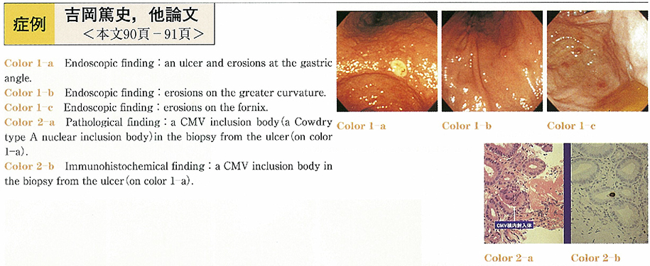
    2003Volume 62Issue 2 Pages 90-91
    Published: May 31, 2003
    Released on J-STAGE: April 03, 2014
    JOURNAL FREE ACCESS
    A patients, 51 years old female had been treated with prednisolone (7.5mg/day) and azathioprin (50mg/day) for autoimmune hepatitis. She had been also treated for duodenal ulcer with famotidine. High grade fever continued for 20 days without the evidence of bacterial infection and blood examination showed high titer of cytomegarovirus (CMV) IgG antibody and CMV antigenemia. After the discontinuation of azathioprine, her body temperature become normal and cytomegarovirus antigenemia disappeared.
    Endoscopic examination revealed an ulcer at the gastric angle, and gastric erosions on the fornix and the greater curvature. CMV inclusion bodies were recognized in the biopsies from the ulcer edge. CMV was not detected from former gastric biopsy samples which were taken for the examination of Helicobactor pylori. CMV disappeared in biopsy samples with the improvement of gastric ulcer. The clinical course and histopathological findings suggested gastric ulcer was induced by CMV infection.
    Download PDF (254K)
  • Hiroshi Shiozaki, Toshiyuki Tahara, Yoshiyuki Yamagishi, Kou Nakada, K ...
    2003Volume 62Issue 2 Pages 92-93
    Published: May 31, 2003
    Released on J-STAGE: April 03, 2014
    JOURNAL FREE ACCESS
    A 50-year-old female was pointed out to have abnormalities by a barium meal for medical checkup and visited our hospital. At the age of 40, she had been pointed out to have mediastinal lymphadenopathy by a chest X ray for medical checkup. At another hospital, biopsy of the lymphnode had been done and the diagnosis of sarcoidosis had been made.
    She was free of gastrointestinal symptoms and her fecal occult blood test was negative. Gastroscopy showed multiple protruding lesions in the fornix, body and angulus. Center of the lesion was yellowish and slightly depressed. Some of the lesions were also accompanied with converging folds. Endoscopic ultrasonography of the lesion showed a thickened hypoechoic area in the second inner layer.
    Biopsy specimens taken from the lesions showed non-caseous epithelioid granulomas with Langhans' giant cells in the lamina propria microscopically.
    Neither tubercle bacillus nor fungus was found. Serological tests of syphilis were also negative. These findings led to the diagnosis of gastric sarcoidosis.
    An ulcer scar with Helicobacter pylori infection was also observed in the stomach, then eradication of H. pylori was preformed. Her gastroscopic finding has not changed during the follow-up period (about 1 year) .
    Download PDF (763K)
  • Naoko Okamoto, Masao Tani, Tomotaka Kaisan, Ichiro Saeki, Naoya Saito, ...
    2003Volume 62Issue 2 Pages 94-95
    Published: May 31, 2003
    Released on J-STAGE: April 03, 2014
    JOURNAL FREE ACCESS
    The case was a 64-year-old male who was noted to have a submucosal tumor. 6 mm in size, at the duodenal balb. Biopsy taken by endoscopy showed the presence of carcinoid tumor. But he had no symptoms of carcinoid syndrome.
    The tumor was resected successfully with no complication by endoscopic mucosal resection using a cap-fitted panendscope (EMRC) . Histopathologically, there was no carcinoid invasion to the resected margins and to the vessels.
    We suggested that EMRC is a safe and effective procedure for treatment of such small lesions in the duodenum.
    Download PDF (1136K)
  • Shigeru Iwase, Hideaki Anan, Rie Matueda, Osamu Akasaka, Makoto Ueno, ...
    2003Volume 62Issue 2 Pages 96-97
    Published: May 31, 2003
    Released on J-STAGE: April 03, 2014
    JOURNAL FREE ACCESS
    Case was a 63-year-old female. She was admitted to hospital because of hematoemesis. Laboratory data showed slight anemia, increasing data of eosinophil and IgG. No pathological findings were recognized by chest plain X-ray films and abdominal CT scan, At first, we suspected rupture of esophago-gastric varices, portal hypertensive gastropathy and so on, because she attended our hospital for Primary biliary cirrhosis. Upper GI endoscopy performed in an emergency showed oozing from multiple erosions in the stomach.
    She was hospitalized by the diagnosis of acute gastric mucosal lesion. We performed 2nd upper GI endoscopy on 3 hospitalized days for checking the state of affairs. Endoscopic pictures showed a white roundworm in 2nd portion of the duodenum. This worm could be removed by grasping forceps led to the diagnosis of Ascaris lumbricoides. And then, She was cured of this disease by taking Pyrantel pamoate.
    In recent years, the Incidence of Ascaris lumbricoides has been decreasing because of improved public hygiene and living standards in Japan. However, there is a slight increasing in number of cases due to the natural foods boom and increasing contact with foreign countries. In this case, the origin was natural foods. It is necessary for clinicians to be able to take parasitosis into consideration.
    Download PDF (1215K)
  • Ken Sumiyoshi, Hiroyuki Komoriyama, Naotaka Tobe, Masahiro Hamaya, Hid ...
    2003Volume 62Issue 2 Pages 98-99
    Published: May 31, 2003
    Released on J-STAGE: April 03, 2014
    JOURNAL FREE ACCESS
    A case of a 35-year-old woman who was hospitalized at near clinic because of fervescence after abroad traveling. Laboratory data showed slight liver dysfunction and a high level of CRP. We suspected that she had a common cold and administrated her with antibiotics but no defervescence was observed. The results of tests denied the possibilities of collagen disease, meningitis, and imported infection. Abdominal CT showed mural tylosis of pars descendens duodeni, and the superior endoscopic image showed ulcers and multiple polyps dotted at pars descendens duodeni. She was moved to our hospital because of suspicion of duodenum Crohn's disease and aim of exact examination of fever unaccounted for. Five days after removal, sudden hematemesis induced Hb5.0 and hemorrhagic shock. We had to operate coil embolization in superior pancreaticoduodenal artery for hemostasis because she didn't response to blood transfusion. Multiple polyps dotted at duodenum tended to be cured but existed, but pathological observation suggested inflammatory reproductive polyp and denied Crohn's disease. We will report our experience of duodenal lesion unaccounted for with a discussion about the literatures.
    Download PDF (750K)
  • Mikitaka Iguchi, Naohisa Yahagi, Shintaro Kondo, Naomi Kakushima, Kats ...
    2003Volume 62Issue 2 Pages 100-101
    Published: May 31, 2003
    Released on J-STAGE: April 03, 2014
    JOURNAL FREE ACCESS
    A 51-year-old male was admitted to our hospital for the treatment of a laterally spreading tumor in the rectum. A protrusion with bridging folds, which suggested a SMT was found in the second part of the duodenum by screening endoscopy for preoperative check up. The lesion had slightly reddish and erosive surface, and several biopsy specimens were taken from the eroded area. The diagnosis of carcinoid tumor was made, since these specimens demonstrated positive reactions to chromogranin A, synaptophysin and Grimelius stain. Endoscopic ultrasography revealed that the tumor was mainly located in the submucosal layer, and the tumor size was 15mm in diameter. It is reported that the risk of metastasis depends on its size, and since large tumors more than 10mm are judged to be high risk, the patient underwent laparotomy. Operative findings showed no obvious metastatic lesions in the liver, no lymph node swelling or disseminated lesions in the peritoneum, therefore partial duodenectomy was performed. The resected specimen revealed that the tumor was originated from the submucosa and the surgical margin was negative for carcinoid.
    Download PDF (962K)
  • Takefumi Kouro, Maya Watanabe, Hidehiko Utsuki, Kumiko Tahara, Masao A ...
    2003Volume 62Issue 2 Pages 102-103
    Published: May 31, 2003
    Released on J-STAGE: April 03, 2014
    JOURNAL FREE ACCESS
    A 68 years old woman was admitted to our hospital with chief complaint of tarry stool. She was administrated the anti coagulant for hypertension and atrial fibrillation. The gastrofiberscopy was done to examine with tarry stool on the day of hospitalization. We did not found bleeding point by the gastrofiberscopy and colonoscopy, except for a gastric ulcer scar. However we found the dark red stool when we inserted to the terminal ileum. She had tarry stool at the 4th hospitalized day once again. We performed the small intestine fiberscopy and inserted to the jejunum. The submucousal tumor that had the ulcer with the bleeding from the visible vessel was recognized. We injected 1/10,000 epinephrine for hemostasis. We judged that the possibility of bleeding is high, and then an emergent operation was carried out. The histological examination reviewed small intestine GIST. We experienced the case of small intestine GIST that was able to find out a lesion by the small intestine fiberscopy. As long as we had reviewed in the literature, only four cases had been reported.
    Download PDF (459K)
  • Akiko Hashimoto, Hironori Yamamoto, Tomonori Yano, Noriko Hashimoto, H ...
    2003Volume 62Issue 2 Pages 104-105
    Published: May 31, 2003
    Released on J-STAGE: April 03, 2014
    JOURNAL FREE ACCESS
    A 69 year-old woman was admitted to our hospital because of a tarry stool and severe anemia. She was diagnosed with small bowel bleeding due to malignant lymphoma distributed from the distal portion of the duodenum to the ileum by using double-balloon enteroscopy. Then, she became hypovolemic shock due to large blood loss from the gastrointestinal tract. 99mTcO4-scintigraphy indicated bleeding from the small intestine. In order to control bleeding from the small intestine, double-balloon enteroscopy was carried out in emergency and the electrocoagulation therapy was performed after the identification of the bleeding lesion. While the technique of endoscopy usually provides visualization of the most proximal and distal end of the gut. The small bowel is among the most difficult part of the gastrointestinal tract to access. Both small bowel radiograophy and capsule endoscopy are useful for the diagnosis of the small intestine, but interventional capabilities are totally absent in these methods. Conventional push enteroscopy provides tremendous discomfort to the patient, and cannot access to the entire small bowel as well. The double-balloon method is a new technique capable of accessing entire small intestine with intervention capabilities. We have demonstrated a case in which double-balloon enteroscope was very helpful not only for the diagnosis of small bowel tumors but also for the control of bleeding from the tumor in the small intestine.
    Download PDF (494K)
  • Eishin Nonaka, Koji Shinmura, Naoki Negami, Masahiko Satou, Takeshi Oh ...
    2003Volume 62Issue 2 Pages 106-107
    Published: May 31, 2003
    Released on J-STAGE: April 03, 2014
    JOURNAL FREE ACCESS
    The case involved a 64-year-old woman referred to our department with a chief complaint of uncomfortable feeling in the right lower quadrant. Abdominal CT examination showed a mass-like lesion with a laminated appearance in the ileocecal area. Barium enema findings include a filling defect, size 2×1cm, and a sharply marginated mass within the cecum with no visible appendix. At colonoscopy, a steeply rising and subpedunculated mass with smooth surface was noted in the cecum. These examination findings diagnosed the patient with appendiceal intussusception and laparoscopic appendectomy involving removal of part of the cecal wall was subsequently carried out. In the excised specimen, the appendix protruded and inverted into the cecum.
    We experienced a case of appendiceal intussusception, a rare disease found in the ileocecal area, and here report it.
    Download PDF (821K)
  • Kazuhiro Endo, Hisanaga Horie, Kazutomo Togashi, Masaki Okada, Hideo N ...
    2003Volume 62Issue 2 Pages 108-109
    Published: May 31, 2003
    Released on J-STAGE: April 03, 2014
    JOURNAL FREE ACCESS
    Mucinous cystadenocarcinoma of the appendix is rare. Some cases of early carcinoma were reported, but many of them were diagnosed incidentally. In this case, We report a unique picture of the vermiform appendix supposed to be early lesion. A 89-year-old man visited Tochigi Cancer Center complaining of lower abdominal pain. Colonoscopic examination showed the elevated lesion and circular fold around the opening of the vermiform appendix with discharge of mucin. At that time, pathologic examination of the biopsy specimen revealed adenoma. He refused to undergo a surgical resection. Three years later, he was admitted to Jichi Medical School Hospital with the diagnosis of cecal tumor. Colonoscopic examination showed the large tumor covered with mucin and blood in the cecum. He underwent ileocecal resection, and the pathological diagnosis was mucinous cystadenocarcinoma of the appendix. We supposed that such colonoscopic findings as the elevated lesion and circular fold around the opening of the vermiform appendix with discharge of mucin showed early stage of mucinous cystadenocarcinoma of the appendix.
    Download PDF (1261K)
  • Kenichi Iizuka, Tomohiro Kudoh, Toshiyuki Mashimo, Keiichirou Yuasa, S ...
    2003Volume 62Issue 2 Pages 110-111
    Published: May 31, 2003
    Released on J-STAGE: April 03, 2014
    JOURNAL FREE ACCESS
    We presented the case of 73-year-old man with the chief complaint of the constipation and lower abdominal pain. Colonoscopic examination showed the slight elevated lesion at the bottom of the cecum without the appendical orifice. Magnifying endoscopic examination revealed type VN pit pattern surrounded by the lesion with type I pit pattern. Histological findings of specimen from the lesion with type VN pit pattern was mucinous adenocarcinoma. It is difficult to make a definite diagnosis of the carcinoma of the appendix, which is very rare. before surgery. It is useful to observe the cecum in detail.
    Download PDF (760K)
  • Makoto Takase, Yoshihisa Saida, Yoshinobu Sumiyama, Jiro Nagao, Youich ...
    2003Volume 62Issue 2 Pages 112-113
    Published: May 31, 2003
    Released on J-STAGE: April 03, 2014
    JOURNAL FREE ACCESS
    Recently. polypectomy using colonoscopy has been becoming general procedure. But colonoscopy has some complications of bleeding and perforation. We report a case of iatrogenic colon penetration recovering by conservative treatment. A 61-year-old male was admitted to our hospital for treatment of transverse colon tumor. Lower GI series, colonoscopy and Endoscopic ultrasoundsonography showed a giant large intestinal lipoma in Transverse colon. The spherical lesion was 50mm in diameter with a stalk. Polypectomy using colonoscopy was performed. Penetration was suspected by abdominal X-ray immediately after polypectomy. But abdominal pain was localized and the inflammatory change was not increased rapidly. Therefore penetration was treated conservatively. The extra colon gas was existed in the mesenterium and retroperitoneum, but fluid collection was not observed in the abdomen by computed tomography. The change of the extra colon gas observating by abdominal X-ray and computed tomography was decrease from 2nd days after polypectomy using colonoscopy. The inflammatory change was in the normal range after 4th postpolypectomy day, and the patient could begin drinking after 6th postpolypectomy day and eating the foods after 8th postpolypectmy day. After that, the course was uneventful, the patient was discharged from hospital on 12th postplypectomy day. The penetration was successfully treated conservatively. We considered that treatment of penetration in colon was not always surgical operation.
    Download PDF (957K)
  • Tetsuya Sato, Tomoyuki Kushida, Yukio Fujino, Gohichi Hayashi, Kohki M ...
    2003Volume 62Issue 2 Pages 114-115
    Published: May 31, 2003
    Released on J-STAGE: April 03, 2014
    JOURNAL FREE ACCESS
    A 42-year-old woman was consulted to our hospital due to abdominal pain, diarrhea, and hematochezia. Laboratory tests showed leukocytosis, elevated CRP level, and Escheria coli (serum type O-166) was detected. First we diagnosed infectious colitis, so prescribed antibiotics. But her symptoms still continued. Colonoscopy and barium enema study revealed the total colitis type of ulcerative colitis, and it was comfirmed pathologically. We continued medical therapy with total parenteral nutrition, salazosulfapyridine, and prednisolone. After these treatment the patient condition improved. Follow up study by colonoscopy showed that the erosion, and pseudopolyposis remained, but inflammatory findings were improved markedly.
    The special type of Escheria coli caused infectious enterocolitis including diarrhea and gastroenteritis.
    It is concluded that emergent colonoscopy and stool culture are important for the diagnosis and treatment of ulceraive colitis.
    Download PDF (468K)
  • Takamichi Yamane, Soichi Hotta, Toshiro Kamoshida, Shinji Hirai, Yuji ...
    2003Volume 62Issue 2 Pages 116-117
    Published: May 31, 2003
    Released on J-STAGE: April 03, 2014
    JOURNAL FREE ACCESS
    A 64-year-old man visited us to undergo further detailed examination for the sigmoid colon tumor which had been revealed by endoscopic examination in Otashiro Clinic. Colonoscopic examination revealed a 11mm-sized flat elevated lesion which was well demarcated and the type VI pit pattern was observed by magnifying endoscopic examination. This lesion was also observed positive non-lifting sign and the histopathological diagnosis of the biopsied specimen was well differentiated adenocarcinoma. Double contrast barium enema showed semilunar deformity in the lateral view of the lesion. Our diagnosis was sigmoid colon cancer invading deep submucosal layer or muscularis propria.
    Laparoscopic assisted sigmoidectomy was carried out. Macroscopic examination revealed a well demarcated flat elevated lesion which size was 11×8×2mm. Stereomicroscopic examination of the lesion revealed type VI pit pattern. Histopathological diagnosis was well differentiated adenocarcioma, mp3, ly1, v0, n0.
    According to the previous reports, a small advanced colorectal cancer of flat elevated type (IIa-like) is considered to be rare and that with type VI pit pattern is considered to be very rare.
    Download PDF (1106K)
  • Nobukazu Takahashi, Masaru Morimoto, Yoshiya Kaneshiro
    2003Volume 62Issue 2 Pages 118-119
    Published: May 31, 2003
    Released on J-STAGE: April 03, 2014
    JOURNAL FREE ACCESS
    Endoscopic colorectal polypectomies were performed for multiple polyp of colon in the 59 years old male. However, pneumoretroperitoneum occurred after multiple clippings for hemostasis and defect closure after EMR of Isp type polyp near the SD junction. It is considered that muscle layer was injured by clipping procedure. Therefore, careful procedure should be requied in clipping. Fortunately, post EMR mucosal defect was finally closed and recovered by conservative therapies in this case.
    Download PDF (967K)
  • Chihiro Ono, Kanji Yaegashi
    2003Volume 62Issue 2 Pages 120-121
    Published: May 31, 2003
    Released on J-STAGE: April 03, 2014
    JOURNAL FREE ACCESS
    A 51-year-old female referred to our hospital because of a swelling of the right lower extremity on April in 2002. She underwent endoscopic mucosal resection for lower rectal cancer measuring 14mm 4 years and 8 months ago. Though the resected specimen revealed well differentiated adenocarcinoma invading massively into the submucosal layer, no lymphvascular invasion was detected. Surgical resection was not performed and she was followed up for 3 years and 5 months. After admission, wide spreading lymph node metastasis was disclosed by abdominal and pelvic CT scan and MRI, which caused the extrinsic compression of the right common and an external iliacvein. Lymphadenectomy and postoperative combined chemoradiotherapy was performed. Patients with submucosal invasive carcinoma having adverse prognostic factors should be treated with colorectal resection and lymphnode dissection, but otherwise, meticulous follow up is necessary for long period.
    Download PDF (683K)
  • Yasunori Ishido, Shigeru Shirota, Yousuke Uchida, Hiroyuki Iwase, Moto ...
    2003Volume 62Issue 2 Pages 122-123
    Published: May 31, 2003
    Released on J-STAGE: April 03, 2014
    JOURNAL FREE ACCESS
    A 68-year-old man was seen at the hospital because of right abdominal palpable mass. A barium enema study showed an apple-core-sign of the ascending colon, and an abdominal CT showed multiple liver metastases and mass of right lower abdomen. Due to appearing of ileus, right hemicolectomy was performed with a diagnosis of the ascending colon cancer with multiple liver metastases. Histlogically, the tumor consisted of polygonal-shaped cells and grew in a solid pattern without grand formation. Immnohistochemically, tumor cells were positive for Chromogranine A and NSE. Electron-microscopically, these cells contained abundant neuroendocrine granules in cytoplasm, and diagnosis of endocrine carcinoma was made. Histologically, in addition to operation, intestive treatment with chemotherapy and radiotherapy are necessary for patients with colorectal endocrine cell carcinoma.
    Download PDF (762K)
  • Sachio Takekawa, Hideki Satou, Kazushige Nirei, Yoshikazu Iwasaki, Hit ...
    2003Volume 62Issue 2 Pages 124-125
    Published: May 31, 2003
    Released on J-STAGE: April 03, 2014
    JOURNAL FREE ACCESS
    We report here in a rare case of Cronkhite Canada Syndorome associated with early stage of colon cancer. Historical and genetic background of polyposis was moleculary investigated in terms of mutations in APC, K-ras and p53 genes.
    Download PDF (602K)
  • Kyoko Iwase, Tomoharu Yajima, Hiroshi Serizawa, Satoshi Tsunematsu, No ...
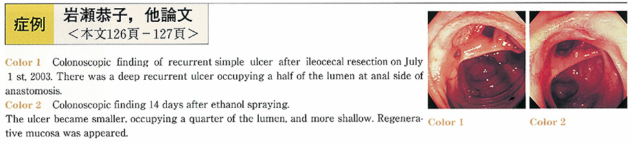
    2003Volume 62Issue 2 Pages 126-127
    Published: May 31, 2003
    Released on J-STAGE: April 03, 2014
    JOURNAL FREE ACCESS
    A 55-years-old male with simple ulcer resistant to sulfasalazopyrine and steroid therapy admitted to our hospital for lower abdominal pain. Ileocecal resection had been performed because of ileal perforation 9 years before. His symptom became a little better once after increase of prednisolone up to 40mg/day and total parenteral nutrition, however, oral diet induced worsening of abdominal pain again. Colonoscopy showed a deep and geographical ulcer at anastomotic site. Absolute ethanol spraying through endoscope at 20ml directly for the anastomotic ulcer was performed and pain was relieved a few days later. Colonoscopy after two weeks revealed that the size of the ulcer was remarkably reduced, and absolute ethanol spraying was repeated at 20ml. Lower abdominal pain has been relieved even after starting a meal. Absolute ethanol spraying through endoscope is easy to be performed and should be considered in the case of simple ulcer which is resistant to medical therapy.
    Download PDF (263K)
  • Yuji Nagashima, Yuji Okihama, Takeshi Matsuda, Yoshihiro Hiramoto, Sho ...
    2003Volume 62Issue 2 Pages 128-129
    Published: May 31, 2003
    Released on J-STAGE: April 03, 2014
    JOURNAL FREE ACCESS
    A Case Report of solitary ulcer syndrome of rectum to differentiate from rectal cancer by colonscopy is reported. The patient was 40 years-old, male. He visited our hospital with complaint of occult blood of stool in 1997. Colonscopy was performed. It shows solitary ulcer in rectum. Biopsy is no malignancy. 4 years after (2001) he visited again with same complaint. Colonscopy shows tumor (like type 2 rectal carcinoma) with ulcer in rectum. Biopsy is no malignancy, but we highly suspected rectal cancer. We performed rectal biopsy 2 times, and huge specimen by snear. These show no malignancy in this rectal tumor. Then, we diagnose this case as solitary ulcer syndrome of rectum.
    It is important to perform biopsys frequently, suspected solitary ulcer syndrome of rectum.
    Download PDF (560K)
  • Keizo Yoneda, Tatehiko Wada, Nobuaki Sakamoto, Jirou Ogata, Koichiro K ...
    2003Volume 62Issue 2 Pages 130-131
    Published: May 31, 2003
    Released on J-STAGE: April 03, 2014
    JOURNAL FREE ACCESS
    A 87-year-old man was admitted with right chest pain and an abnormal chest density on the chest X-ray examination or chest CT examination. A malignant tumor was diagnosed by CT guided needle biopsy. Radiotherapy was given for the lung tumor. Twelve days after radiotherapy, colonoscopy was performed and an ulcer in the rectum was recognized, performed. Because hemorrhage did not continue, he was treated with IVH with no sustenance by mouth. After about 2 weeks, colonoscopic findings showed extensive erosion occupying the entire lumen in the lower rectum. Microscopic findings of biopsy specimens obtained from the ulcers showed cytomegalic inclusion bodies, and we obtained positive stains by a immunohistochemical technique using a monoclonal antibody to CMV. The bloody discharge disappeared following treatment with ganciclovir for two weeks, but he died of respiratory failure due to his original illness after one month.
    The lung tumor was diagnosed as a malignant lymphoma and the rectal ulcer had disappeared on autopsy. This was a case of CMV enterocolitis associated with malignant lymphoma. CMV colitis should be included in the differential diagnosis of colitis of unknown etiology in immunocompromised hosts.
    Download PDF (983K)
  • Shuta Nishinakagawa, Masakatsu Hukuzawa, Ayako Hiraide, Motoki Okitsu, ...
    2003Volume 62Issue 2 Pages 132-133
    Published: May 31, 2003
    Released on J-STAGE: April 03, 2014
    JOURNAL FREE ACCESS
    A 71-year-old female patient underwent an abdominal simple panhysterectomy with bilateral adnexectomy and received radiation therapy for carcinoma of the corpus uteri in January 2000. She began having repeated alternate episodes of diarrhea and constipation six months after the operation, and pain in the lower quadrants of the abdomen also developed. As the symptoms were persistent the patient was examined by colonoscopy in March 2002. The examination disclosed in the sigmoid colon the presence of a net-like foreign body partly embedded in the intestinal mucosa and unexcisable endoscopically. The foreign body was considered to be a Marlex mesh that had been applied to an area of peritoneal defect during a previous operation and probably perforated into the intestine. After two months, the foreign body was spontaneously excreted on bowel evacuation. While endoceliac foreign bodies, such as retained surgical gauze, sometimes form granulomas and are subjected to surgical extirpation, reports of spontaneous excretion of a foreign body perrectum have been as few as 3 cases in Japan, including the one documented herein. The underlying mechanism is assumed to be that the segment of intestine adhering to the periphery of the mesh became necrotized due to inflammation caused by radiation therapy, so that the mesh perforated into the colon and was gradually dragged into the intestinal lumen by peristalsis.
    Download PDF (687K)
  • Yukio Fujino, Tomoyuki Mochizuki, Tetsuya Sato, Tomoyuki Kushida, Kouj ...
    2003Volume 62Issue 2 Pages 134-135
    Published: May 31, 2003
    Released on J-STAGE: April 03, 2014
    JOURNAL FREE ACCESS
    In recent years, diagnostic laparoscopy has widely been used in gastroenterological surgery, gynecology, urology and pediatric surgery. In this present study, we experienced a case of hepatic tumor which was difficult to make preoperative diagnosis due to iodine allergy and the history of surgery but finally diagnosed by laparoscopy. In this case, hand-assisted laparoscopic surgery (hereinafter abbreviated as HALS) was useful for resection.
    The case was a 30-year-old woman whose tumor was palpated in a left epigastric region in a standing position and a sitting position. In a right lateral position, the tumor exceeded a median line and moved to the right side. No definite diagnosis was made by gastroscopy, abdominal ultrasonography and abdominal CT scan, and the tumor was laparoscopically diagnosed as exophytic one originating from the lateral segment of the liver. The tumor was resected using the HALS by inducing it to the outside of abdominal cavity. The tumor was pathologically diagnosed as focal nodular hyperplasia.
    Download PDF (619K)
  • Hidehiko Utsuki, Maya Watanabe, Masao Araki, Fumitake Kouro, Kumiko Ta ...
    2003Volume 62Issue 2 Pages 136-137
    Published: May 31, 2003
    Released on J-STAGE: April 03, 2014
    JOURNAL FREE ACCESS
    A 53-year-old female complaining with epigastralgia was raferred to our hospital. She was recognized hyperglycemia and a rise of pancreatic enzyme in peripheral blood examination. Abdominal US showed the solid and cystic tumor which was size of 41×37×32mm in pancreas head and the obvious dilatation of MPD which was 22mm of in diameter. Abdominal CT showed the pancreas head tumor with enhancement. EUS revealed the obvious dilatation of MPD and papillary tumor inside of MPD at pancreas head. ERCP showed wide opening of the MPD with mucous exudates and the dilatation of MPD. Cytological diagnosis of pancreatic juice was definite malignancy (ClassV) . The patient was underwent surgical operation. Pathological diagnosis for resected specimen was IPMT, borderline malignancy. We reported a case of an intraductal papillary mucinous tumor, with summary of IPMT literature in Japan.
    Download PDF (512K)
  • Terumi Kamisawa, Naoto Egawa, Hitoshi Nakajima, Kozue Amemiya, Kumiko ...
    2003Volume 62Issue 2 Pages 138-139
    Published: May 31, 2003
    Released on J-STAGE: April 03, 2014
    JOURNAL FREE ACCESS
    A 71-year-old female was admitted to our hospital because of advanced cholangiocellular carcinoma. She underwent endoscopic retrograde cholangiopancreatography for evaluation of the tumor. Endoscopically, the longitudinal fold of the major duodenal papilla was slightly swelling but readily compressible with a smooth appearance before cannulating. Cholangiography in the initial phase showed no abnormal findings in the terminal common bile duct. The mucosa proximal to the orifice bulged endoscopically into the duodenum, during contrast injection from the cannulating catheter situated in the terminal common bile duct near the orifice. Cholangiography demonstrated a round contrast-filled structure adjacent to the termination of the common bile duct.
    In conclusion, choledochocele could be diagnosed by duodenoscopic and cholangiographic findings during ERCP. Ballooning of the papilla during contrast injection is a clue to the presence of a choledochocele. It might be a sign which should alert the endoscopist to the presence of a choledochocele.
    Download PDF (763K)
  • Hitoshi Ono, Tsunao Imamura, Tatsurou Yanagawa, Kazuo Konishi, Katsuhi ...
    2003Volume 62Issue 2 Pages 140-141
    Published: May 31, 2003
    Released on J-STAGE: April 03, 2014
    JOURNAL FREE ACCESS
    A 65-year-old male had been previously diagnosed as having alcoholic chronic pancreatitis with pancreatic stones at 40 year-old. In April 2002, he was admitted to another hospital for relapse of chronic pancreatitis. He was initially treated conservatively, however he was consulted our hospital for complication with a 15-cm giant pancreatic pseudocyst. Abdominal computed tomography (CT) and ultrasonography showed formation of multiple pseudocysts, and severe dilatation of the main pancreatic duct. Endoscopic retrograde pancreatography visualized 1-cm-stenosis in length of the main pancreatic duct at the head and severe dilatation of the main pancreatic duct at the tail. We judged that the stenosis was caused by chronic pancreatitis, and performed endoscopic naso-pancreatic drainage. After treatment, abdominal CT scan showed reduction of size of pseudocysts, then we exchanged the drainage to a 10 Fr 7-cm pancreatic stent. On his discharge, pancreatic pseudocysts vanished and dilatation of the main pancreatic duct improved. Our experience suggests that endoscopic transpapillary pancreatic drainage is one of the effective treatments for pancreatic pseudocyst.
    Download PDF (1213K)
  • Jun Inoue, Junya Kashimura, Katsuya Endo, Katsuhisa Sato
    2003Volume 62Issue 2 Pages 142-145
    Published: May 31, 2003
    Released on J-STAGE: April 03, 2014
    JOURNAL FREE ACCESS
    A 53-year-old man admitted to our hospital with a complaint of epigastralgia. Ultrasonography showed some small stones in the gallbladder. Magnetic resonance cholangiopancreatography (MRCP) revealed two stones, about 5mm in diameter, in the common bile duct (CBD) . Endoscopic examination showed two separated papillae which had each orifice, and the oral papilla was located inside the diverticulum. Endoscopic retrograde cholangiopancreatography (ERCP) revealed two stones, 5mm in diameter, in CBD and couldn't prove the existence of choledocoduodenal fistura. From these findings, we diagnosed as CBD stones with the completely separation of CBD and the main pancreatic duct (MPD) . Endoscopic papillary balloon dilatation (EPBD) was performed and CBD stones were completely removed by a balloon catheter endoscopically. After that, laparoscopic cholecystectomy was performed for cholecystolithiasis.
    The completely separated orifices of CBD and MPD is rare, reported to be lower than 4% in Japan. EPBD is thought to be safe and useful for the treatment of CBD stones in the patient with the complete separation of CBD and MPD, especially the orifice of CBD was located inside the diverticulum.
    Download PDF (997K)
  • Tatsuro Yanagawa, Tsunao Imamura, Hitoshi Yoshida, Hitoshi Ono, Katsuh ...
    2003Volume 62Issue 2 Pages 144-145
    Published: May 31, 2003
    Released on J-STAGE: April 03, 2014
    JOURNAL FREE ACCESS
    A 59 year-old man was diagnosed as having primary sclerosing cholangitis (PSC) according to elevated serum ALP and IgG levels, biliary stricture and irregularity evaluated by endoscopic retrograde cholangiopaucreatography (ERCP) , inflammatory cell infiltration and fibrosis not only surrounding interlobular bile ducts but within periportal parenchyma on histologic features 10 months before admission to our hospital. Although he had been treated well with oral administration of 600mg/day ursodeoxycholic acid (UDCA) , he suffered from cholangitis on admission March 2002 due to dominant stricture of the common bile ducts (CBD) . Since bile ducts carcinoma and syndromic autoimmune pancreatitis were ruled out by biliary exofoliative cytology, ERCP and CT, we safely repeated endoscopic balloon dilation of dominant stricture in the CBD 3 times at 6 atm for 60 seconds and administered 900mg/day UDCA and bezafibrate for peripheral biliary stenosis. After the treatment, appearance of dominant stricture of the CBD diminished and serum ALP and IgG levels decreased. Endoscopic treatment and medication presumably render him free of cholangitis symptom for 10 months.
    Our successful experience indicated that endoscopic balloon dilation is an effective and noninvasive treatment for dominant strictures in the extrahepatic bile ducts with PSC.
    Download PDF (745K)
feedback
Top