Progress of Digestive Endoscopy
Online ISSN : 2187-4999
Print ISSN : 1348-9844
ISSN-L : 1348-9844
Volume 90, Issue 1
Displaying 1-50 of 60 articles from this issue
Clinical study
  • Rie Kure, Ayako Kobayashi, Yukiko Himukai, Hirotaka Yamamoto, Maiko Ki ...
    2017 Volume 90 Issue 1 Pages 46-50
    Published: June 09, 2017
    Released on J-STAGE: July 19, 2017
    JOURNAL FREE ACCESS

    Background : The clinical features of acute esophageal mucosal lesion (AEML) have not been fully investigated because of its relative rarity, and almost all cases recover rapidly according to endoscopic findings. This study aimed to clarify the clinical features of AEML.
    Methods : AEML was diagnosed in 13 cases who were admitted to our hospital between January 2008 and April 2016. We investigated clinical features and endoscopic findings. Six black esophagitis cases were compared with 7 non-black esophagitis cases.
    Result : Various comorbidities had been found in AEML cases, the most common being malignancy. Six cases (46%) used PPI (proton pump inhibitor) before AEML attack. Eleven cases rapidly improved, but 2 died. In the black esophagitis group, there were significantly more smokers than in the non-black group. The 2 dead cases had black esophagitis.
    Conclusion : There are several participating factors in the onset of AEML. Our findings suggest that smoking might be a risk factor for AEML, and it was difficult to prevent AEML with PPI. Further, black esophagitis may represent more severe underlying conditions.

    Download PDF (722K)
  • Masao Kusano, Daisuke Komazawa, Hiromichi Ito, Masaki Tosa, Kaname Uno ...
    2017 Volume 90 Issue 1 Pages 51-54
    Published: June 09, 2017
    Released on J-STAGE: July 19, 2017
    JOURNAL FREE ACCESS

    We have been able to eradicate chronic atrophic gastritis related to Helicobacter pylori (H. pylori) in Japan since February 2013. We observed the appearance of many gastric black dots in patients after H. pylori eradication. We here describe typical cases of gastric black dots in three patients. In case 1, a 68-year-old man who received esophagogastroduodenoscopy (EGD) was diagnosed as having H. pylori-related gastritis. After 7 months of H. pylori eradication, EGD revealed many black dots in the gastric body. Histopathological examination revealed black granules in the cystically dilated fundic gland. In case 2, a 62-year-old man who received EGD was diagnosed as having H. pylori-related gastritis. After 6 months of H. pylori eradication, EGD revealed many black dots in the gastric body and fornix. Histopathological examination revealed tiny black granules in the cystically dilated fundic gland. In case 3, a 74-year-old man who received EGD was diagnosed as having H. pylori-related gastritis. After 10 months of H. pylori eradication, EGD revealed many black dots in the gastric body and fornix. Gastric black dots may be a new EGD finding after H. pylori eradication.

    Download PDF (1040K)
  • Koji Takahashi, Toshio Tsuyuguchi, Harutoshi Sugiyama, Junichiro Kumag ...
    2017 Volume 90 Issue 1 Pages 55-58
    Published: June 09, 2017
    Released on J-STAGE: July 19, 2017
    JOURNAL FREE ACCESS

    Study Objective : We examined the efficacy and safety of rendezvous ERCP using percutaneous biliary drainage (PBD) route to salvage previously unsuccessful ERCP cases. Patients and Methods : Patients who underwent rendezvous ERCP using PBD route were retrospectively examined from November 2012-April 2016. Results : The subject of the study was 14 patients. Therapeutic interventions were successful in 13 out of 14 patients (93%) . Twenty-nine percent underwent reconstructed gastrointestinal anatomy ; ERCP was successful for all. Bile leak occurred in one patient. At first endoscopy, major papilla was not detected in 4 patients ; the rendezvous procedure was successful in 3 out of 4 patients. Conclusion : Rendezvous ERCP using PBD route to salvage unsuccessful ERCP cases showed a high success rate with few complications.

    Download PDF (901K)
Case report
  • Yoshikazu Yakami, Hiroki Yazama, Tomoki Bando
    2017 Volume 90 Issue 1 Pages 59-61
    Published: June 09, 2017
    Released on J-STAGE: July 19, 2017
    JOURNAL FREE ACCESS

    A 33-year-old man presented to our hospital with the chief complaint of epigastric pain. Abdominal CT revealed localized wall thickening in the gastric antrum. In the same area, upper gastrointestinal endoscopy revealed multiple shallow ulcers fused into a geographical pattern. Based on the endoscopic findings and the patient’s social history (history of sexual activity with many and unspecified persons) , gastric syphilis was initially suspected. However, a serological test for syphilis turned out negative, and immunostaining of a tissue sample from the lesion demonstrated the absence of treponema. In a culture of a gastric mucosal tissue specimen, Streptococcus viridans was detected, which led to the diagnosis of phlegmonous gastritis. He started receiving conservative treatment with antibiotics (AMPC) and recovered. Patients with acute abdomen should be diagnosed and treated with care in consideration of phlegmonous gastritis or gastric syphilis as differential diagnosis.

    Download PDF (1070K)
  • Kotaro Matsumoto, Masatoshi Mabuchi, Takako Adachi, Hanae Kajiyama, Ta ...
    2017 Volume 90 Issue 1 Pages 62-65
    Published: June 09, 2017
    Released on J-STAGE: July 19, 2017
    JOURNAL FREE ACCESS

    A 71-year-old woman receiving long-term oral steroids for treatment of Vogt-Koyanagi-Harada disease was admitted to our hospital with symptoms of fever, vomiting, diarrhea, and lower abdominal pain. A diagnosis of peritonitis caused by infectious gastroenteritis was made. Following treatment with fasting, antibiotics, and intravenous fluids, her symptoms, physical findings, and laboratory results improved. However, after cessation of antibiotic treatment, her fever returned and laboratory results indicated increased inflammation. A pelvic abscess was observed using computed tomography (CT) . It was located adjacent to the rectum as observed using endoscopic ultrasonography. Endoscopic ultrasound-guided fine-needle aspiration was performed through the rectum wall and two internal stents were inserted. Her symptoms subsequently improved and a follow-up CT scan showed the abscess had significantly decreased in size.

    Download PDF (1173K)
  • Haruko Furuya, Takahiro Kawamura, Jun Ohara, Mizuki Nagai, Takehito As ...
    2017 Volume 90 Issue 1 Pages 66-69
    Published: June 09, 2017
    Released on J-STAGE: July 19, 2017
    JOURNAL FREE ACCESS

    A 74-year-old man was admitted due to the persistent blood per rectum and dizziness. Emergency upper endoscopy showed no sign of hemorrhage in stomach but a string of coagulated blood attached to the papilla of Vater was noticed. Intermittent flow of blood along with yellow and translucent bile was observed. The enhanced abdominal computed tomography was unable to detect active bleeding in gastrointestinal tract, but showed atrophy and multiple calcification of pancreas with a gastroduodenal artery aneurysm suggesting the previous condition of chronic pancreatitis which is one of the main causes of HP. He was successfully treated with angiography and transcatheter embolization of the aneurysm and his hemoglobin stabilized without requiring further treatment. HP is a rare cause of gastrointestinal bleeding associated with pancreatitis. While rarity of the condition and elusive bleeding pattern pose a challenge to an experienced gastroenterologist, HP should be considered in a case of occult bleeding with a history or an evidence of preceding pancreatitis.

    Download PDF (961K)
  • Masanori Odaira, Ryohei Miyata
    2017 Volume 90 Issue 1 Pages 70-71
    Published: June 09, 2017
    Released on J-STAGE: July 19, 2017
    JOURNAL FREE ACCESS

    We report two cases of accidental ingestion of a press-through package (PTP) . Case 1 was a 57-year-old woman who visited our hospital and stated that she possibly swallowed a PTP. Abdominal CT did not detect a foreign body. During upper gastrointestinal endoscopy, the PTP was identified directly in the esophagus and removed endoscopically. Case 2 was a 75-year-old woman who visited our hospital because of ingestion of a PTP. Abdominal CT revealed an object with high-density opacity, which was suspected of being a PTP, in the esophagus. Under the diagnosis of ingestion of a PTP, upper gastrointestinal endoscopy was performed and the PTP was removed.
    Abdominal CT imaging is generally useful for correct diagnosis of ingestion of a PTP, although CT sometimes cannot detect a PTP depending on the materials it is made of. It is noteworthy that the materials that make up a PTP affect various CT imaging results.

    Download PDF (874K)
  • Masanori Ochi, Dai Kubota, Nozomu Miyajima, Takahiro Seimiya, Maki Mat ...
    2017 Volume 90 Issue 1 Pages 72-73
    Published: June 09, 2017
    Released on J-STAGE: July 19, 2017
    JOURNAL FREE ACCESS

    The patient was a 77-year-old female. She was brought to our hospital by ambulance with hematemesis. Upper gastrointestinal endoscopy revealed a submucosal hematoma in the thoracic/upper esophageal regions. A proton pump inhibitor was administered, but vomiting frequently occurred thereafter, showing exacerbation. Acute heart failure developed, and a catecholamine preparation was introduced, but there was no improvement, leading to a fatal outcome. Esophageal biopsy at the periphery of the submucosal hematoma showed amyloid deposition. Pathologically, AL-type amyloidosis was suggested. A urine sample was positive for Bence Jones protein, and cephalic CT revealed a punched-out lesion, leading to a diagnosis of multiple myeloma. Although symptoms of multiple myeloma include low back pain, anemia, and renal dysfunction, a submucosal hematoma may also become a diagnostic criterion.

    Download PDF (805K)
  • Akira Shimomura, Chizu Yokoi, Toru Igari, Koh Imbe, Kazuhiro Watanabe, ...
    2017 Volume 90 Issue 1 Pages 74-75
    Published: June 09, 2017
    Released on J-STAGE: July 19, 2017
    JOURNAL FREE ACCESS

    An 80-year-old man presented emergently with acute melena and hematemesis. Blood tests revealed anemia, with prolonged activated partial thromboplastin time and normal prothrombin time. Esophagogastroduodenoscopy showed severe esophagitis with hemorrhage ; and he required 10 units of blood transfusion. The intractable esophagitis improved gradually with acid suppression medication. However, 21 days after discharge, he developed a subcutaneous hematoma in his neck. This led to a diagnosis of acquired hemophilia A. We think that the patient had developed acquired hemophilia A during the first admission. Here we report this rare case of repeated hemorrhagic esophagitis caused by acquired hemophilia A.

    Download PDF (633K)
  • Momoko Hayashi, Youko Ootsuka, Teruhiko Sakamoto, Yoshihiko Naritaka, ...
    2017 Volume 90 Issue 1 Pages 76-77
    Published: June 09, 2017
    Released on J-STAGE: July 19, 2017
    JOURNAL FREE ACCESS

    An 83-year-old man presented with high blood pressure, type 2 diabetes, and a renal function disorder. He complained mainly of chest pressure and dyspnea and was hospitalized on suspicion of colon cancer ileus, based on thickening and directly visualized intestinal tract expansion of the intestinal wall by CT examination. Lower gastrointestinal tract endoscopy was also performed, indicating rectal cancer of the Borrman2 type. And We identified acute necrotizing esophagitis from the esophagogastric junction to the middle esophagus by upper gastrointestinal tract endoscopy.
    Surgical intervention was performed in acknowledgment of the rapid improvement with PPI, followed by a supplementary liquid.
    With necrotic esophagitis, possibility of a contribution to the onset by hypoalimentation due to the underlying malignant tumor with a microcirculatory disorder as the background factor. Acute necrotic esophagitis is rare and this case is presented with a discussion of the relevant literature on malignant tumors.

    Download PDF (591K)
  • Masashi Yokota, Jun Koike, Masaya Sano, Toshiki Kodama, Ryutaro Fujimo ...
    2017 Volume 90 Issue 1 Pages 78-79
    Published: June 09, 2017
    Released on J-STAGE: July 19, 2017
    JOURNAL FREE ACCESS

    An elderly man with dysphagia was referred to our hospital due to a type 3 lesion in the middle thoracic esophagus. A computed tomography showed no metastasis but a partial infiltration to the adjacent aorta. Histology of the biopsy specimen showed sheet-like proliferation of rather light-colored rounded cells in HE staining and synaptophysin and CD56 expression by immunostaining. He was diagnosed as neuroendocrine carcinoma (small cell carcinoma) in cStage IVa and planned to receive chemoradiotherapy (CRT) . As for chemotherapy, combination of carboplatin and etoposide was used according to the regimen of lung small cell carcinoma, but the doses were reduced to 70 % considering his age. Due to granulocytopenia, the chemotherapy was quitted after only one course, however radiotherapy could be completed. Lesions markedly decreased in size and the tumor markers also decreased drastically by the treatment and we evaluated its response as PR.

    Download PDF (775K)
  • Ryutaro Fujimoto, Hirokazu Shiozawa, Reiko Nishina, Jun Nakamura, Jun ...
    2017 Volume 90 Issue 1 Pages 80-81
    Published: June 09, 2017
    Released on J-STAGE: July 19, 2017
    JOURNAL FREE ACCESS

    A 66-year-old woman was hospitalized with alcoholic liver cirrhosis classified as Child-Pugh A. Esophagogastroduodenoscopy revealed three columns of esophageal varices (LmF1CbRC (-) Lg-C (+) ) and a superficial depressed lesion of 15 mm in diameter located about 30cm from the incisors as a brownish area by NBI (narrow band imaging) . Magnifying endoscopy with NBI showed many inhomogeneous loop-like vessels varying in caliber with small avascular areas (B1 vessels with AVA-small) in the lesion. The lesion was unstained by Lugol’s solution. It was, therefore, diagnosed as an early esophageal carcinoma of depth EP/LPM and endoscopic submucosal dissection (ESD) was indicated. Due to the risk of bleeding from the varices near the lesion during the procedure, the varices were treated by endoscopic variceal ligation before ESD. After confirming the disappearance of the varices, ESD was performed by the double channel method with EEMR-tube without complications such as active bleeding. Pathologically, the tumor was diagnosed as squamous cell carcinoma, 0-IIc, pT1a-EP, ly0, v0, pHM0, pVM0. Pretreatment of EVL may be useful for avoiding the risk of active bleeding during ESD for superficial esophageal cancer near esophageal varices.

    Download PDF (896K)
  • Nana Mamuro, Hideo Shimada, Takayuki Tajima, Takayuki Nishi, Junnya Og ...
    2017 Volume 90 Issue 1 Pages 82-83
    Published: June 09, 2017
    Released on J-STAGE: July 19, 2017
    JOURNAL FREE ACCESS

    A 68-year-old male was referred to our hospital with the suspicion of early esophageal cancer. Endoscopy-revealed a shallow erythematous depression on a mucosal elevation of 1cm in diameter at 27cm from the incisors. Histopathologic examination of biopsy specimen confirmed squamous cell carcinoma,type0-IIc. The mucosal elevation was pulsatile. Contrast-enhanced computed tomography-revealed that the elevation was the meandering left bronchial artery arising from the aorta at the Th10 level. Endoscopic submucosal dissection (ESD) was performed with sufficient submucosal injection for mucosal incision, and submucosal dissection involved countertraction to assure better visualization. Various etiologies including submucosal tumors, the aorta, varices, hemangioma, and a vertebra cause esophageal elevations. It is imperative to identify the cause of a mucosal elevation when endoscopically treating lesions on top of such elevation.

    Download PDF (943K)
  • Takako Endo, Akitake Uno, Syuhei Arima, Tetsuro Fujii, Kotoyo Ouchi, A ...
    2017 Volume 90 Issue 1 Pages 84-85
    Published: June 09, 2017
    Released on J-STAGE: July 19, 2017
    JOURNAL FREE ACCESS

    Treatment of esophageal cancer and recent advances in the diagnosis have improved patient survival after esophagectomy. We experienced a case of ESD for GTC, which was occurenced on the suture line of a reconstructed gastric tube (GT) after esophagectomy. The patient was a 73-year old man who had received esophagectomy with reconstruction of the GT for esophageal squamous cell carcinoma. Three years later, endoscopy showed a slightly depressed mucosal lesion (0-IIc) with red-colored mucosa was found on the suture line of the GT. It was diagnosed as a well differentiated tubular adenocarcinoma, and we performed ESD without complication. ESD for GTC after esophagectomy is a technically difficult because of the limited working space and the presence of gastric fibrosis with staples under the suture line, but considered a useful treatment.

    Download PDF (838K)
  • Takahiro Matsunaga, Yukishige Okamura, Mari Akasaka, Kazuhiro Togasaki ...
    2017 Volume 90 Issue 1 Pages 86-87
    Published: June 09, 2017
    Released on J-STAGE: July 19, 2017
    JOURNAL FREE ACCESS

    Patient was a 50-year-old man with anemia and high CEA. Esophagogastroduodenoscopy (EGD) showed a 0-IIb lesion in the middle thoracic esophagus. Narrow band imaging (NBI) revealed a brownish area, and magnifying endoscopy with NBI showed an intrapapillary capillary loop with mild dilation. After iodine spraying, the lesion showed partial circularity. Whole-body contrast CT showed no metastasis. Lesion was removed by ESD on post admission day (PAD) 1. 100mg triamcinolone acetonide (TA) was injected to the resected site to prevent postoperative stricture. Oral prednisolone (PSL) 20mg/day was started from PAD 2 and decreased to 10mg/day within 2 weeks after ESD. EGD was done bi-weekly, with confirmation of healing on PAD 68. PSL was finally stopped on PAD 89. No postoperative stricture was observed during follow up.

    Download PDF (586K)
  • Koji Takahashi, Kazuki Kato, Shingo Kasamatsu, Mari Aminaka, Shin Tsuc ...
    2017 Volume 90 Issue 1 Pages 88-89
    Published: June 09, 2017
    Released on J-STAGE: July 19, 2017
    JOURNAL FREE ACCESS

    A 82-year-old man was referred to our hospital for epigastric pain. Computed tomography revealed a linear high-density structure that perforated the stomach and reached near the neck of the gallbladder. A few hours before he felt pain, he had consumed a sea bream. We diagnosed his condition as a fish bone perforation of the stomach. An upper gastrointestinal endoscopic examination revealed that a fish bone had perforated the antrum of the stomach. The bone was extracted using forceps, and the hole was closed with clip. The bone was 35 mm long and 3 mm wide. After placing a nasogastric tube, a proton pump inhibitor and antibiotics were administered. The patient’s clinical symptoms subsequently improved, and the clinical course was uncomplicated. He was discharged 5 days after the endoscopic treatment. Thus, our results reveal that an endoscopic treatment may be an appropriate choice for treating fish bone perforation without peritonitis.

    Download PDF (792K)
  • Yurika Ikegami, Tomoyuki Yada, Koichi Ito, Yoshiyuki Itakura, Naomi Ue ...
    2017 Volume 90 Issue 1 Pages 90-91
    Published: June 09, 2017
    Released on J-STAGE: July 19, 2017
    JOURNAL FREE ACCESS

    A 69-year-old woman was referred to us because of abnormally high serum pepsinogen (PG) levels in the ABC method (blood test for gastric cancer screening) done in April 2016, which resulted in group A. EGD performed in May 2016 revealed diffuse redness and mucosal swelling, indicating HP-associated gastritis, although the endoscopic findings in 2014 had indicated a HP-negative gastric mucosa. A rapid urease test was positive. From April to June 2016, serum PGI levels decreased, and serum HP antibody levels increased and changed from negative to positive. Thus, the initial HP infection was found by abnormally high PG levels and then converted to chronic persistent infection. The ABC method should be evaluated not only by the groups but also by serum PG and HP antibody levels, as well as endoscopic findings.

    Download PDF (631K)
  • Ayako Watanabe, Dai Inoue, Gen Maeda, Naoya Tsugawa, Shiori Itou, Nats ...
    2017 Volume 90 Issue 1 Pages 92-93
    Published: June 09, 2017
    Released on J-STAGE: July 19, 2017
    JOURNAL FREE ACCESS

    A 52-year-old man had been experiencing black stools and and admitted to hospital. On emergent endoscopy, he was found to have a 40mm submucosal tumor with hemorrhagic ulcer in the gastric antrum. The hemorrhagic points were stopped bleeding by clipping. On next day, he had been changing our hospital. We found mucosal surface was appeared from submucosal tumor in endoscopic examination. Endoscopic ultrasound fine needle aspiration (EUS-FNA) showed a high echoic mass from the third and fourth layer. Gastric lipoma had been diagnosed from tissue collection by EUS-FNA. The laparoscopic partial gastrectomy was performed. When gastric lipoma is larger than 4cm, it has high risk of bleeding. The risk of bleeding is involved with the size of the gastric lipoma. Surgery is enforced in many cases. But, in the case of small size ,the endoscopic resection is enforced.

    Download PDF (801K)
  • Fumi Sakuma, Mari Iwasaki, Ken Kashima, Ko Fukushi, Takeshi Sugaya, Ka ...
    2017 Volume 90 Issue 1 Pages 94-95
    Published: June 09, 2017
    Released on J-STAGE: July 19, 2017
    JOURNAL FREE ACCESS

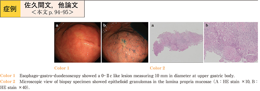
    A 59 year old female complained of vomiting and hematemesis, was referred to our hospital for close examination of 0-IIc like lesion of the stomach.Esophago-gastro-duodenoscopy (EGD) showed a discolored IIc like lesion, 10 mm in size, at the greater curvature of the upper corpus.
    The results of the biopsy demonstrated epithelioid cell granulomas in the lamina proprea, leading to the diagnosis of gastric sarcoidosis. Because the administration of proton pump inhibitor relieved her symptoms. She has been followed up regularly without steroids. Sarcoidosis is a rare systemic disease characterized by non-creating granulomas in affected organs. Involvement of gastrointestinal tract is documented in 1 % of the patients, but most frequently found in the stomach. The endoscopic finding may vary, without any specific characteristic. Because western report indicate causal relationship between gastric sarcoidosis and malignant lymphomas, surveillance of the gastric sarcoidosis will be required.

    Download PDF (698K)
  • Shiori Ito, Dai Inoue, Shun Fujiwara, Daiki Yamada, Natsuki Miura, Kou ...
    2017 Volume 90 Issue 1 Pages 96-97
    Published: June 09, 2017
    Released on J-STAGE: July 19, 2017
    JOURNAL FREE ACCESS

    A 71-year-old man was admitted to our hospital complaining of appetite loss. An esophagogastroduodenoscopy (EGDS) revealed poor distensibility of the antrum wall, and an abdominal CT showed a markedly thickened stomach wall. A biopsy of the gastric wall was performed to test for scirrhous gastric cancer but no malignancy was found. One month later, the patient’s appetite loss worsened, and a second EGDS was performed, revealing multiple shallow ulcers on the antrum. Eight days later, he passed massive bloody stools and went into shock. CT revealed a remarkably decreased contrast effect on the wall of the small intestine. Because there was no sign of intraperitoneal artery obstruction, non-occlusive mesenteric ischemia (NOMI) was diagnosed. The patient died on the next day. Based on the autopsy findings of amyloid-A protein deposition on the stomach, the diagnosis of AL amyloidosis followed by NOMI was finally made. In retrospect, the direct fast scarlet staining of the biopsy specimen on the first EGDS had demonstrated amyloid-A protein deposition on gastric mucosa. Our findings suggest that amyloidosis should be suspected whenever unidentified digestive symptoms are encountered.

    Download PDF (853K)
  • Yoshihiro Sasaki, Yukihiro Kiya, Takeshi Kamijyo, Yusuke Shimada, Masa ...
    2017 Volume 90 Issue 1 Pages 98-99
    Published: June 09, 2017
    Released on J-STAGE: July 19, 2017
    JOURNAL FREE ACCESS

    A 50 year-old woman was found to have gastric submucosal tumor with central ulceration, was observed in the greater curvature by upper gastrointestinal endoscopy. Biopsy was done but pathological diagnosis was not obtained.
    A hypoechoic, partially non-echoic mass was observed by EUS and EUS-FNA was performed, but pathological diagnosis could not be made. Gastrointestinal stromal tumor was suspected, and Laparoscopy and Endoscopy Cooperative Surgery was done. Pathological examination was observed dense growth of spindle-shaped cells. Immunohistochemical staining were S100 (+) , CD117 (-) , CD34 (-) , desmin (-) , so it was diagnosed gastric schwannoma.
    We present a rare case of gastric schwannoma, whose diagnosis was established histologically after surgery.

    Download PDF (781K)
  • Yohei Furumoto, Tomoyo Machida, Yukito Okura, Mana Matsuoka, Takahito ...
    2017 Volume 90 Issue 1 Pages 100-101
    Published: June 09, 2017
    Released on J-STAGE: July 19, 2017
    JOURNAL FREE ACCESS

    An 18-year-old woman was referred to our department for examination of a large gastric polyp. She had one pigmented spot on her upper lip. She did not have a family history of Peutz-Jeghers syndrome. Esophagogastroduodenoscopy revealed a large semipedunculated polyp measuring 50mm in diameter at the greater curvature of the upper gastric body. Pathological examination of a biopsy specimen showed hyperplasia of glandular epithelium and no evidence of malignancy (Group 1) . Endoscopic submucosal dissection (ESD) was performed. Pathological examination of the resected specimen showed smooth muscle bundles with an arborizing appearance throughout the lamina propria with a tiny focus of well-differentiated adenocarcinoma. We reported a case of adenocarcinoma associated with Peutz-Jeghers syndrome-type gastric polyp that was curatively resected by ESD.

    Download PDF (748K)
  • Taihei Iwaya, Tomohiro Kawamura, Tomohiko Matsuda, Masahiro Okada, Kaz ...
    2017 Volume 90 Issue 1 Pages 102-103
    Published: June 09, 2017
    Released on J-STAGE: July 19, 2017
    JOURNAL FREE ACCESS

    An 84-year-old woman with history of carcinoma of the papilla of Vater, in which percutaneous transhepatic stent was placed, was admitted to our hospital with a complaint of fever. The patient was diagnosed as cholangitis.
    Immediate endoscopy examination revealed that the stent was projecting long and distal side of the stent had been embedded into the duodenal wall which located opposite to the papilla of Vater. Because of tumor in-growth, removal of the stent was impossible. In the literature, as well-known, endoscopic section of migrated stent using argon plasma coagulation (APC) is useful. However, the risk of the duodenal mucosa injury was the matter because the stent was embedded. We removed the distal side stent which is embedded to the duodenal wall using biopsy forceps without any complication.
    Afterwards we put covered stent into the original metallic stent with stent-in-stent manner. We conclude that biopsy forceps is safe and useful to dissect an embedded uncovered biliary stent.

    Download PDF (581K)
  • Naoki Konishi, Junichi Iwamoto, Jun Asaoka, Tadakuni Monma, Masashi Mu ...
    2017 Volume 90 Issue 1 Pages 104-105
    Published: June 09, 2017
    Released on J-STAGE: July 19, 2017
    JOURNAL FREE ACCESS

    A 83-year-old woman with hematemesis was admitted to our hospital . Emergency endoscopic examination showed multiple ulcerations with bleeding in the duodenum and the proximal jejunum. Enhanced abdominal CT scan showing wall thickness of proximal jejunum. Histopathological studies showed proliferation of atypical lymphocytes. Immunohistohemical staining of the tumor cells revealed CD3 (+) , CD4 (-) , CD8 (+) , CD20 (-) , and CD56 (+) . From these findings, enteropathy associated T-cell lymphoma (EATL) was diagnosed. Because she had dementia and poor ADL, she did not receive an active treatment including chemotherapy and died on the 59th hospital day. We report a rare case of primary T-cell lymphoma of the duodenum and the proximal jejunum.

    Download PDF (866K)
  • Takahiro Kobayashi, Yuichi Takano, Kenta Iwahashi, Akihiro Tabuchi, Na ...
    2017 Volume 90 Issue 1 Pages 106-107
    Published: June 09, 2017
    Released on J-STAGE: July 19, 2017
    JOURNAL FREE ACCESS

    A 40-year-old female patient visited the hospital because of a stomachache and vomiting 3 days after barium gastrography. The abdominal computed tomography scan indicated a diverticulum around the third part of the duodenum, high-density area inside the diverticulum, and fluid collection and increased fat tissue concentration around the retroperitoneum. The patient was diagnosed as having a retroperitoneal abscess due to a perforated duodenal diverticulum, and she was treated conservatively because the extent of the abscess was limited and her general condition was not poor.
    In many similar cases, surgical therapy is selected, but this patient achieved complete recovery with conservative treatment. We experienced a rare case of a perforated duodenal diverticulum after barium gastrography.

    Download PDF (709K)
  • Haremaru Kubo, Shinji Morishita, Kou Haraguchi, Genki Asai, Asuka Okaz ...
    2017 Volume 90 Issue 1 Pages 108-109
    Published: June 09, 2017
    Released on J-STAGE: July 19, 2017
    JOURNAL FREE ACCESS

    A 38-year-old woman and a 29-year-old man were referred to our hospital for abdominal pain. In both cases, gastroendoscopy revealed a duodenal ulcer. H. pylori and NSAIDs (Non-Steroidal Anti-Inflammatory Drugs) intake history were negative in both cases. These duodenal ulcers were refractory even with PPI (Proton Pump Inhibitor) treatment for a few months. Colonoscopies were performed for further evaluation, and they showed vascular pattern loss and fine granular mucosal pattern. From colonoscopy and histopathological examination of the colon, we diagnosed them as ulcerative colitis. Mesalazine therapy was started, and mucosal inflammation of the duodenum and colon gradually subsided. Histopathological examinations of duodenal biopsy showed basal plasmacytosis and crypt distorsion, which is characteristic in gastroduodenitis associated with ulcerative colitis. Therefore, we diagnosed these duodenal ulcers as gastroduodenitis associated with ulcerative colitis. When we see a refractory duodenal ulcer without H. pylori infection and NSAIDs intake, we should consider the coexistence of ulcerative colitis.

    Download PDF (1092K)
  • Ayako Kimura, Hideki Ohno, Takuya Ono, Mana Hosoda, Yuko Okabe, Mineyo ...
    2017 Volume 90 Issue 1 Pages 110-111
    Published: June 09, 2017
    Released on J-STAGE: July 19, 2017
    JOURNAL FREE ACCESS

    A 67-year-old man was referred to our department because of tumor marker outliers pointed out by his nearby physician, whom he had visited for epigastric pain. Upper gastrointestinal endoscopy revealed an irregular mucosa on the posterior wall of duodenal bulb and was diagnosed as a primary duodenal cancer (adenocarcinoma) by biopsy. However, a contrast-enhanced computed tomography (CT) showed many small nodules considered compatible with liver metastasis that limited effectiveness of surgery. And then, he was admitted to this department for his tarry stool and rapidly progressive anemia. A CT scan showed a rapid progression of both primary tumor and liver metastasis compared to a month before. On the 9th hospital day he had a large amount of melena considered intractable to endoscopic hemostasis or interventional radiology. Therefore, octreotide was administered for his symptomatic relief, and then melena and the need of blood transfusion were reduced. However, he gradually developed conscious disturbance and tumor fever, and died on the 26th hospital day. Octreotide reduced the bleeding from the terminal duodenal cancer and provided effective symptomatic relief of acute hemorrhage.

    Download PDF (811K)
  • Eriko Nakagomi, Yuri Enomoto, Yusuke Nishikawa, Tomoko Umakoshi, Mitsu ...
    2017 Volume 90 Issue 1 Pages 112-113
    Published: June 09, 2017
    Released on J-STAGE: July 19, 2017
    JOURNAL FREE ACCESS

    A 75-year-old woman was admitted to our hospital for persistent edema. She had been previously admitted to another hospital for 40 days, where she had presented with diarrhea, vomiting, and stomach pain, and had undergone blood tests, computed tomography (CT) , and other tests. However, all tests showed normal findings. Treatment with mesalazine was started for suspected Crohn’s disease. The patient was discharged with a diagnosis of anorexia. After discharge, she was unable to perform routine daily activities because of edema ; therefore, she visited our hospital. Laboratory tests showed hypoalbuminemia. CT showed wall thickening in the small intestine. We then performed protein-losing scintigraphy. Protein loss was noted in the same region showing abnormality on CT. Therefore, we diagnosed protein-losing gastroenteropathy, and steroid therapy was initiated. The therapy was effective. Here we report this case of protein-losing gastroenteropathy without an underlying primary disease and review the literature.

    Download PDF (805K)
  • Yukito Okura, Tomoyo Machida, Emi Aonuma, Mana Matsuoka, Takahito Noza ...
    2017 Volume 90 Issue 1 Pages 114-115
    Published: June 09, 2017
    Released on J-STAGE: July 19, 2017
    JOURNAL FREE ACCESS

    A 21-year-old male visited emergency room in our hospital with right abdominal pain. He had a fever and symptoms of upper respiratory infection. There were tenderness in the right abdominal region, but no signs of peritoneal irritation. Abdominal CT scan revealed an invagination of the ileocecal intestinal tract into the ascending colon, and intussusception was diagnosed. We attempted repositioning by colonoscopy. During colonoscopy, we confirmed that a reddened ileocecal region had invaginated into the ascending colon. The invaginated intestinal tract was gradually repositioned by air insufflation. The symptoms did not relapse, and the patient was discharged. One month after discharge, we found no obvious problems in colonoscopy. A majority of adult-onset intussusception cases are accompanied by neoplastic lesions ; however, in this case, no obvious problems were noted. Therefore, we report this patient as a rare case.

    Download PDF (772K)
  • Soichiro Yamamoto, Hirohito Miyako, Takahiro Kenmochi, Yuu Kitahara, K ...
    2017 Volume 90 Issue 1 Pages 116-117
    Published: June 09, 2017
    Released on J-STAGE: July 19, 2017
    JOURNAL FREE ACCESS

    A 60s woman with frequent watery diarrhea who was urgently hospitalized with renal dysfunction. Previously she had been prescribed olmesarton and rosuvastatin by a local doctor beause her medical history included hypertension and hyperlipidemia.
    Colonoscopic examination revealed edematous mucosa of the left colon and an indistinct vascular pattern. Biopsy of the same region revealed abundant lymphocytic infiltration in the mucosa, and the likely diagnosis was lymphocytic colitis. Two weeks after cessation of these medications, the diarrhea had improved. Then approximately three months after discontinuation of the drugs, colonoscopic examination showed improvement in the mucosal edema and the vascular pattern.
    Examination of a biopsy specimen revealed that the pathological findings of lymphocytic colitis had also disappeared. Thereafter, treatment with olmesartan was resumed temporarily. However, it was again discontinued because the diarrhea had recurred. Subsequently these symptoms resolved. Thus, the patient was diagnosed as having lymphocytic colitis associated with olmesartan.

    Download PDF (842K)
  • Tomoaki Moriyama, Minoru Shimizu, Tomoaki Fujikawa, Kohei Kato, Naoko ...
    2017 Volume 90 Issue 1 Pages 118-119
    Published: June 09, 2017
    Released on J-STAGE: July 19, 2017
    JOURNAL FREE ACCESS

    A 69-year-old woman was referred to the hospital because of a positive fecal occult blood test. She had taken kamisyoyosan continuously for twenty-five years due to menopausal syndrome and anxiety disorder. Colonoscopic examinations showed dark purple mucosa with irregular vascular structure from the cecum to the descending colon and a deep ulcer in the ascending colon. Plain abdominal radiograph showed calcification along the ascending colon.Abdominal CT revealed thickening of the colonic wall and calcification of the mesenteric vein. Histopathological findings disclosed fibrous thickening of the veins in the submucosa and collagen fiber deposits in the lamina propria. A diagnosis of mesenteric phlebosclerosis was made, then kamisyoyosan was discontinued. Colonoscopy after about a half year showed healed ulcer in the ascending colon.
    As mesenteric phlebosclerosis may be discovered incidentally, endoscopic and radiographic characteristic images of the disease need be kept in mind. In patients with a long-term history of taking Chinese herbal medicines containing gardenia fruit colonoscopy or abdominal CT is thought usually necessary.

    Download PDF (943K)
  • Eiko Takeichi, Kosei Hamano, Osamu Noguchi
    2017 Volume 90 Issue 1 Pages 120-121
    Published: June 09, 2017
    Released on J-STAGE: July 19, 2017
    JOURNAL FREE ACCESS

    A 25-year-old female with an endometriotic cyst on her left ovary came to our hospital, claiming she had bloody stool, fever, and abdominal pain. We diagnosed her with an endometriotic cyst infection and gave her antibiotics, which eased the condition. We also performed a colonoscopy to examine the bloody stool, in which we discovered a submucosal tumor and a pine cone-like elevation in her rectosigmoid colon. Based on the biopsy of the lesion, we diagnosed it as intestinal endometriosis because of ER (+) , PgR (+) , and CDX - 2 (-) . After the episode, the patient continued to have blood stool at each menstruation. Therefore, we put her on an ultra low dose birth-control pill, and the symptom has receded.
    It is hard to diagnose intestinal endometriosis by biopsy because of the location of the main lesion and the poor pathological characteristics in normal staining. However, in this case, by suspecting it from clinical course and communicating it to the pathologist, we could diagnose it from biopsy by adding immunostaining, enabling us to successfully treat it conservatively.

    Download PDF (774K)
  • Noboru Yatagai, Kazuki Ohta, Osamu Kobayashi, Daichi Watanabe, Ryuta K ...
    2017 Volume 90 Issue 1 Pages 122-123
    Published: June 09, 2017
    Released on J-STAGE: July 19, 2017
    JOURNAL FREE ACCESS

    A 66-year-old female with ulcerative colitis (UC) has been treated with salazosulfapyridine, steroids and infliximab for the past 24 years since the initial diagnosis of UC. She had a good clinical course. Follow-up colonoscopy revealed multiple round, elevated lesions with a smooth surface along the ascending colon and transverse colon. The patient was diagnosed as having pneumatosis cystoides intestinalis (PCI) . There are only 40 reported cases of PCI associated with UC in Japan, including our case. We report the findings of our case with a literature review.

    Download PDF (871K)
  • Fumiko Sunada, Hirotsugu Sakamoto, Mamiko Tsukui, Hidekazu Kurata
    2017 Volume 90 Issue 1 Pages 124-125
    Published: June 09, 2017
    Released on J-STAGE: July 19, 2017
    JOURNAL FREE ACCESS

    In 1994 Fujinuma et al. reported that mucosal polyp consist of normal submucosa tissue with normal membrane. So far, 16 cases were reported. A 73 year-old man took total colonoscopy. He had multiple diverticulosis and a polyp with normal membrane in the sigmoid colon. We recommended that the polyp would be followed up. Total colonoscopy was performed after 14 months. The polyp did not change in size and shape. We performed endoscopic mucosal resection of the polyp depending on his request. The pathological finding of the polyp was normal membrane without muscularis mucosae. We diagnosed the polyp as mucosal polyp in colon. There was only case report about a mucosal polyp in colon pathologically without muscularis mucosae. This is the first report to follow up on mucosal polyp.

    Download PDF (906K)
  • Jun Ohara, Yuta Sugiyama, Takehito Asakawa, Minami Endo, Yasuhiro Iizu ...
    2017 Volume 90 Issue 1 Pages 126-127
    Published: June 09, 2017
    Released on J-STAGE: July 19, 2017
    JOURNAL FREE ACCESS

    A 62-year-old woman presented to our hospital with watery diarrhea for 5 months since esomeprazole (EMZ) was first administered. On colonoscopy, linear ulcers and aphtha were noted in the sigmoid colon and cecum. Multiple biopsy specimens showed thickened collagen bands in the sub-epithelial layer. She was diagnosed with collagenous colitis (CC) . To dissolve doubts of EMZ-associated CC, she was administered famotidine. After 2 weeks, she showed an improvement in diarrhea. In Japan, the most common cause of drug-induced CC is lansoprazole. We report our experience with a case of EMZ-associated CC, which is rare in drug-induced CC.

    Download PDF (640K)
  • Yoshiyuki Itakura, Tomoyuki Yada, Yurika Ikegami, Koichi Ito, Hitohiko ...
    2017 Volume 90 Issue 1 Pages 128-129
    Published: June 09, 2017
    Released on J-STAGE: July 19, 2017
    JOURNAL FREE ACCESS

    An 85-year-old woman visited our hospital complaining of sustained watery diarrhea after starting a course of lansoprazole for treatment of gastroesophageal reflux disease. Routine laboratory studies and stool cultures were normal. A colonoscopy was performed during which it was technically easy to reach the cecum, however two mucosal lacerations were identified in the transverse colon on withdrawal of the scope. An abdominal CT scan taken immediately after colonoscopy showed free air. The patient was stable and successfully managed using a conservative course of antibiotics. Biopsy specimens of every colonic segment resulted in a diagnosis of collagenous colitis. A postulated mechanism for the mucosal lacerations might be the rigid character of the collagen containing subepithelium. Once collagenous colitis is suspected, colonoscopy should be performed more carefully, moreover, if mucosal lacerations are seen during colonoscopy, colonic perforation should be considered.

    Download PDF (651K)
  • Aya Yamashita, Chiemi Nakayama, Jun Tashiro, Jun Miwa
    2017 Volume 90 Issue 1 Pages 130-131
    Published: June 09, 2017
    Released on J-STAGE: July 19, 2017
    JOURNAL FREE ACCESS

    A 30-years-old woman was admitted due to chronic diarrhea and foot ulcer. Her clinical grounds and colonoscopic findings were compatible with ulcerative colitis (UC) . The foot ulcer was aseptic, and histopathological examination of the ulcer base showed nonspecific inflammation with neutrophil infiltration and pseudovasculitis ; pyoderma gangrenosum (PG) was hence diagnosed as an extraintestinal complication of UC. We initiated treatment with 5-aminosalicylate (5-ASA) 4,000 mg/day and granulocyte and monocyte adsorption apheresis (GMA) . The response to the treatment was favorable and rapid-acting. After 10 GMA sessions she achieved remission from UC and PG was completely epithelized. We reduced 5-ASA to 3,000 mg/day and continued observation. This report demonstrates the efficacy of GMA for PG in line with recent studies.

    Download PDF (1197K)
  • Iichiro Ohtsu, Ryo Chinzei, Masanori Takahashi, Kei Funada, Taira Sato ...
    2017 Volume 90 Issue 1 Pages 132-133
    Published: June 09, 2017
    Released on J-STAGE: July 19, 2017
    JOURNAL FREE ACCESS

    A 66-year-old man, who received surgical resection for advanced colorectal cancer of ascending colon 5 years ago, visited our hospital for surveillance colonoscopy. A colonoscopic examination revealed a IIa + IIc type lesion (10mm in diameter) in the sigmoid colon and a conventional image showed redness in the depressed area and fold convergence around the lesion. In narrow-band imaging (NBI) magnified images, a Japan NBI Expert Team (JNET) type 3 classification tumor was observed in the depressed area. A VI high-grade pit pattern was observed in magnified images after crystal-violet staining. We diagnosed the lesion as submucosal massively invasive cancer and surgical resection was performed with D2 lymph node dissection. Histological examination revealed well-to-moderately differentiated adenocarcinoma invading into the muscularis propria as well as a #241 lymph node metastasis. Although advanced colorectal cancers less than 10mm in diameter are relatively rare, we should mind possibility and importance of such a small lesion as shown in this case.

    Download PDF (933K)
  • Yusuke Nishikawa, Eriko Nakagomi, Takahisa Matsuno, Tomoko Umakoshi, M ...
    2017 Volume 90 Issue 1 Pages 134-135
    Published: June 09, 2017
    Released on J-STAGE: July 19, 2017
    JOURNAL FREE ACCESS

    A 73-year-old woman with a history of perforated diverticulitis in the sigmoid colon and chronic kidney disease was brought to our hospital with acute left lower abdominal pain. Abdominal computed tomography showed descending colitis. Contrast enema and colonoscopy revealed that the entire circumference of the descending colon was constricted. Abdominal angiography showed obstruction at the root of the inferior mesenteric artery. The patient was diagnosed with chronic ischemic changes combined with chronic diverticulitis. Proctosigmoidectomy (Hartmann procedure) was performed, and the postoperative course was uneventful. The patient has experienced no recurrence or complications to date.

    Download PDF (1003K)
  • Keisuke Koroki, Nobuyuki Sugiura, Masaaki Saito
    2017 Volume 90 Issue 1 Pages 136-137
    Published: June 09, 2017
    Released on J-STAGE: July 19, 2017
    JOURNAL FREE ACCESS

    An 83-year-old man was admitted to our hospital complaining of left lower abdominal pain for a month. Abdominal ultrasonography and computed tomography showed descending and sigmoid colon wall thickening, left hydronephrosis and a small amount of ascites. Colonoscopy showed edematous mucosa and stenosis of sigmoid colon, so the scope could not pass the lesion. Histological examination of biopsy specimens from the colonoscopy showed non-neoplastic cells. Malignant cells were detected by cytological examination of ascitic fluid. The immunostaining pattern (CK7-, CK20+, CDX2+) suggested colorectal cancer. After palliative care, the patient died. A pathological autopsy showed type 4 colorectal cancer with peritoneal dissemination. Type 4 colorectal cancer is rare and accounts for only 0.5-1.3% of all colorectal cancer. Diagnosis of type 4 colorectal cancer is difficult because the detection rate by biopsy is low. The cytodiagnosis of ascites combined with immunostaining is useful for diagnosis of type4 colorectal cancer with malignant ascites.

    Download PDF (1407K)
  • Umi Tahara, Naoto Kurihara, Hideshi Matsuda, Yasuhiro Sasaki, Yuko Kim ...
    2017 Volume 90 Issue 1 Pages 138-139
    Published: June 09, 2017
    Released on J-STAGE: July 19, 2017
    JOURNAL FREE ACCESS

    An 81-year-old man who had been diagnosed as having situs inversus totalis and had suffered from repeated episodes of sigmoid volvulus was admitted with a history of right upper quadrant abdominal pain. Physical examination showed no evidence of peritoneal irritation. A plain radiograph of the abdomen showed a markedly dilated sigmoid colon with an inverted U-shaped appearance. Abdominal CT showed situs inversus totalis, no free air, no ascites, and a whorled appearance of the sigmoid mesentery, with dilated bowel loops. Based on these findings, the patient was diagnosed as having recurrence of sigmoid volvulus. Colonoscopy performed for repositioning showed converging mucosa signifying the distal point of the torsional obstruction, and a dilated section of the bowel with gas and feces proximal to the obstruction in the sigmoid colon. After endoscopic decompression, colonoscopy showed no evidence of mucosal ischemia. We treated this case successfully as we would have a case of sigmoid volvulus without situs inversus.

    Download PDF (760K)
  • Kohei Tajima, Hiroshi Miyakita, Takashi Ogimi, Tomoki Nakamura, Takayu ...
    2017 Volume 90 Issue 1 Pages 140-141
    Published: June 09, 2017
    Released on J-STAGE: July 19, 2017
    JOURNAL FREE ACCESS

    A 75-year-old man complained of lower abdominal pain for a week. Obstructive colorectal cancer was suspected by abdominal CT, and the patient was hospitalized immediately. A transanal ileus tube was inserted during emergency surgery to avoid colostomy. All circumference stenosis was observed, and the inflow of contrast medium to the oral side was not confirmed. Adenocarcinoma was reported in the biopsy results. Depression was insufficient, and a self-expandable metallic stent (SEMS) was placed on the 3rd day of hospitalization. After placing the SEMS (Wall FlexTM 90 mm) , the patient was scheduled to receive elective surgery and left our hospital. A temporary radical operation (laparoscopic low anterior resection and D3 lymph node dissection) was performed on the 24th day after hospitalization. Observations of the resected specimen showed that the SEMS covered the lesion without perforation, and that the SEMS expansion was sufficient. In conclusion, we experienced a case of obstructive colorectal cancer applicable to the temporary radical operation and avoided emergency surgery by inserting a SEMS. We report this case with some literature review.

    Download PDF (759K)
  • Yorinari Ochiai, Toshifumi Mitani, Yugo Suzuki, Yumiko Wada, Masami Ta ...
    2017 Volume 90 Issue 1 Pages 142-143
    Published: June 09, 2017
    Released on J-STAGE: July 19, 2017
    JOURNAL FREE ACCESS

    We report on two cases of villous neoplasm in the rectum that were resected by ESD.
    Case 1 : A 57-year-old man had copious mucous stool ; colonoscopy demonstrated villous neoplasm occupying three-fourths of the circumference of the rectosigmoid portion. We performed ESD in 515 minutes and enabled en bloc resection. A pathological examination showed a 148×142 mm cancer in the villous adenoma, sm 4,000 µm, and additional surgery was performed (no remaining tumor and no lymph node metastasis) .
    Case 2 : A 64-year-old-man had severe diarrhea and electrolyte depletion syndrome (EDS) . Colonoscopy demonstrated villous neoplasm that occupied the entire circumference of the lower rectum. We performed en bloc resection in 865 minutes by ESD. A pathological examination showed a 192×180 mm, villous adenoma with high-grade dysplasia. The patient needed steroid injection and balloon dilation to prevent stenosis after treatment. This patient has currently no stenosis in the lower rectum.

    Download PDF (680K)
  • Kouichi Tabei, Soutarou Tokunaga, Takahiro Yatagai, Hirotaka Oota, Gen ...
    2017 Volume 90 Issue 1 Pages 144-145
    Published: June 09, 2017
    Released on J-STAGE: July 19, 2017
    JOURNAL FREE ACCESS

    A 75-year-old man visited our hospital because of constipation and bloody stool. The inspection dayof colonoscopy, he was admitted due to a subileus condition. On colonoscopy Type 2 tumor was found in the rectum. On gastric endoscopy, three small gastric elevated leisions ware noted. The lesions appeared to be submucosal tumors with erosion on the top.
    Microscopic findings of biopsy specimens of elevated leisionshowed signet ring cell carcinoma, similar to the primary rectum cancer. He was diagnosed with gastric cancer metastasized from rectal signet ring cell carcinoma from immunohistochemical staining. He underwent systemic chemotherapy but died of cancer progression 6 months. Immunohistochemical staining are useful tools to distinguish metastatic gastric cancer from primary gastric cancer.

    Download PDF (1077K)
  • Naoki Miyao, Yuichi Takano, Kenta Iwahashi, Akihiro Tabuchi, Misako To ...
    2017 Volume 90 Issue 1 Pages 146-147
    Published: June 09, 2017
    Released on J-STAGE: July 19, 2017
    JOURNAL FREE ACCESS

    Currently, histopathological diagnosis of liver tumors is mainly obtained by percutaneous needle biopsy in Japan. We report a case of a small metastatic liver tumor diagnosed with endoscopic ultrasonography-guided fine needle aspiration (EUS-FNA) , in whom the lesions were located in unapproachable area by percutaneous needle biopsy.
    A 67-year-old woman with a previous history of sigmoid colon cancer and pancreatic neuroendocrine tumor was suspected to have metastatic liver tumors of < 10 mm on computed abdominal tomography and magnetic resonance image. She had Chilaiditi’s syndrome and the tumors could not be visualized on external abdominal ultrasonography. Therefore, EUS-FNA was attempted to the lesion. EUS-FNA was successfully performed to the lesion in the right hepatic lobe and it was diagnosed with NET G2 from the histopathologically findings of the biopsied specimen.
    We believe that EUS-FNA is feasible and useful for liver tumors in cases that are difficult to visualize on external abdominal ultrasonography.

    Download PDF (903K)
  • Akitoshi Kobayashi, Hideaki Mizumoto
    2017 Volume 90 Issue 1 Pages 148-149
    Published: June 09, 2017
    Released on J-STAGE: July 19, 2017
    JOURNAL FREE ACCESS

    The patient was a 100-year-old woman. She was admitted because of cholangitis by common bile duct stones (CBDs) at the age of 93 years. Endoscopic retrograde cholangiopancreatography (ERCP) revealed multiple CBDs, and we placed a plastic biliary stent (PS) for CBD. She complained of lower abdominal pain a year after she discharged and computed tomography (CT) revealed PS in the sigmoid colon. We removed it by colonoscopy and placed PS again for CBD. She was admitted because of acute cholangitis 2 years later. ERCP showed migration of PS. We could not remove that by a basket catheter and balloon catheter but successfully removed it using grasping forceps. Finally, we placed two PSs for CBD, and they are under observation for more than 3 years. A long-term PS placement is useful in very elderly patients with choledocholithiasis, but it is necessary to pay attention to stent dislodgement and migration.

    Download PDF (784K)
  • Fukiko Kinoshita, Yukiko Takayama, Nao Okuno, Junichi Akao, Kenta Naga ...
    2017 Volume 90 Issue 1 Pages 150-151
    Published: June 09, 2017
    Released on J-STAGE: July 19, 2017
    JOURNAL FREE ACCESS

    The patient was a 71-year-old man with a chief complaint of fever. He was referred to our hospital because upper intrahepatic bile ducts extending into the porta hepatis and superior bile duct stricture were found in abdominal ultrasonography. Computed tomographic scan and magnetic resonance imaging showed the same findings. Endoscopic retrograde cholangiopancreatography showed a continuous bile duct stricture from 2 cm in the porta hepatis and superior bile ducts. The pathological evaluation did not produce a diagnosis. SpyGlassTM view revealed an irregular granular papillary stricture with white mucus. We performed biopsy with SpyBite from the tumor papilla. We diagnosed intraductal papillary neoplasm of the bile duct derived from a hepatic hilar cholangiocarcinoma by using SpyGlass.

    Download PDF (755K)
  • Kentaro Takagi, Kenichiro Imai, Ayako Oyama, Mari Kitamura, Akiko Kita ...
    2017 Volume 90 Issue 1 Pages 152-153
    Published: June 09, 2017
    Released on J-STAGE: July 19, 2017
    JOURNAL FREE ACCESS

    A 64-year-old man was referred to our hospital due to jaundice. Abdominal CT and MRI revealed a mass in the hepatic hilum and the intrahepatic bile ducts were dilated. We performed ERC following transpapillary biopsy. The pathological findings of biopsy sample suggested adenocarcinoma. The cholangiogram suggested the tumor extent to the left hepatic duct and lower bile duct. Therefore, we performed peroral cholangioscopy (POCS) and target biopsies under its direct vision to evaluate the exact tumor extent. As a result, we successfully determined the resection line before surgery.
    POCS was useful to diagnose the exact tumor extent of cholangiocarcinoma.

    Download PDF (1039K)
  • Takeshi Mine, Kiyofumi Ishii, Tatsuya Fujimoto, Yu Yoshida
    2017 Volume 90 Issue 1 Pages 154-155
    Published: June 09, 2017
    Released on J-STAGE: July 19, 2017
    JOURNAL FREE ACCESS

    A 71-year-old Japanese woman was admitted to investigate the cause of common bile duct dilatation. She had no previous history of pancreatic diseases. Endoscopic retrograde cholangiopancreatography (ERCP) was performed on the admission day. During ERCP, biliary cannulation was difficult and we used the pancreatic duct guidewire technique. To prevent post-ERCP pancreatitis, a pancreatic stent was placed into the main pancreatic duct, but was accidentally migrated into the duct. The stent could not be retrieved by various devices because of the narrow pancreatic duct.
    We performed ERCP again on hospital day 5 and successfully removed the stent using a 5-French Soehendra stent retriever (SSR-5) . The patient was discharged on hospital day 7 without any other adverse events. Pathological examination of biopsy specimens of the common bile duct revealed no malignancy. In cases of stent migration into the non-dilated pancreatic duct, SSR-5 may be useful for the stent removal.

    Download PDF (807K)
  • Yuichi Uchida, Naoyuki Hasegawa, Kazunori Ishige, Kuniaki Fukuda, Tosh ...
    2017 Volume 90 Issue 1 Pages 156-157
    Published: June 09, 2017
    Released on J-STAGE: July 19, 2017
    JOURNAL FREE ACCESS

    A 68-year-old man was admitted because of an infected pancreatic pseudocyst after alcoholic pancreatitis. Computed tomography revealed multiple pancreatic duct stones and multilocular pancreatic pseudocysts in his pancreatic tail. We performed endoscopic ultrasonography-guided transmural drainage of the pancreatic pseudocyst adjacent to the fundus of the stomach. It was effective for the punctured cyst, but not for the distant cysts. Therefore, we additionally performed transcutaneous drainage. Although the size of the cysts decreased, the drainage tube could not be removed because of continuous drainage of pancreatic juice. Then, we performed trans-papillary drainage by using a pancreatic duct stent. After that, the amount of pancreatic juice drained from the transcutaneous tube decreased and was successfully removed. Combined drainage from various routes is effective for treating multilocular pancreatic pseudocysts, not only when the cysts do not communicate with each other but also when they communicate with an obstructed pancreatic duct.

    Download PDF (877K)
feedback
Top