Progress of Digestive Endoscopy
Online ISSN : 2187-4999
Print ISSN : 1348-9844
ISSN-L : 1348-9844
Volume 87, Issue 1
Displaying 1-50 of 65 articles from this issue
Clinical study
  • Mikinori Koga, Sho Suzuki, Takemasa Sato, Chika Kusano, Masakatsu Fuku ...
    2015Volume 87Issue 1 Pages 40-44
    Published: December 12, 2015
    Released on J-STAGE: January 06, 2016
    JOURNAL FREE ACCESS
    ]Background/Objective] In our hospital, dental floss-clip traction (DFC) method has been used for gastric endoscopic submucosal dissection (gastric ESD) . We examined the safety and efficacy of the DFC method in gastric ESD. ]Methods] We retrospectively examined 288 epithelial tumor lesions of the stomach in which gastric ESD was performed during the period May 2012 to December 2014 in our hospital. We conducted a comparative study of the en bloc resection rate, complete resection rate, treatment time, and complication rate between 57 lesions which underwent gastric ESD using DFC method and 231 lesions which underwent gastric ESD without it. ]Results] The background data of patients in both groups were comparable. Only the mean treatment time of the DFC group (mean±standard deviation ]SD], 81.6±78.4 min) was significantly shorter than that of the non-DFC group (mean±SD, 97.3±68.1 min) (p=0.020) . ]Conclusion] DFC method is effective for improving the technical difficulty of gastric ESD.
    Download PDF (860K)
  • Kei Tanaka, Hitoshi Kurata, Naoaki Dan
    2015Volume 87Issue 1 Pages 45-48
    Published: December 12, 2015
    Released on J-STAGE: January 06, 2016
    JOURNAL FREE ACCESS
    Background : To reduce the number of missed gastric tumor lesions during endoscopy, we examined patients with multiple lesions and patients with missed lesions.
    Subjects and methods : The subjects were patients with gastric tumor lesions who underwent endoscopic submucosal dissection at our hospital from May 2013 to June 2015.
    Results : Of the 27 enrolled patients, 15 had solitary gastric tumor lesion (solitary group) and 12 had multiple gastric tumor lesions (multiple group) . Patients in the multiple group showed more severe atrophy of the background gastric mucosa than patients in the solitary group (p<0.001, Mann-Whitney U test) .Of the 11 enrolled patients in multiple group, 6 had no missed gastric tumor lesion (detected group) and 5 had missed gastric tumor lesion (undetected group) during the first endoscopy. The use of chromoendoscopy was more frequent in the detected group than in the undetected group (p=0.015, Fisher’s exact test) .
    Conclusion : In patients with severe gastric atrophy and one gastric tumor lesion, we should use chromoendoscopy during endoscopy.
    Download PDF (663K)
  • Masaaki Shiina, Shigehiro Kokubu
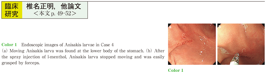
    2015Volume 87Issue 1 Pages 49-52
    Published: December 12, 2015
    Released on J-STAGE: January 06, 2016
    JOURNAL FREE ACCESS
    Alimentary canal anisakiasis is important among the differential diagnoses of acute abdomen. We experienced 10 cases of anisakiasis (7 males and 3 females) from August 2012 to April 2015, including 2 cases diagnosed through cancer screening. Anisakiasis was diagnosed throughout the year (4 cases diagnosed in February-March, 2 cases in April-June, 4 cases in September-November) and in patients of all ages. Epigastralgia was the most frequent symptom, and mackerel (raw or in vinegar) was the leading suspected food. Abdominal attack occurred 2 to 7 hours after ingestion, and upper gastrointestinal endoscopy was performed 8 to 72 hours later. Anisakis larvae were removed by using biopsy forceps in all ten cases, including 3 cases with additional use of l-menthol, a compound that inhibits gastrointestinal peristalsis. In Japan, complete prophylaxis against anisakiasis is difficult because of eating habits and food preferences. Since removal of larvae under endoscopy is the only definite treatment upon abdominal attack, we have to perform endoscopy as soon as possible when migration of Anisakis larvae is suspected by clinical findings and/or CT. In addition, spray injection of l-menthol to moving larvae might be helpful.
    Download PDF (743K)
  • Satoru Nonaka, Ichiro Oda, Seiichiro Abe, Haruhisa Suzuki, Shigetaka Y ...
    2015Volume 87Issue 1 Pages 53-57
    Published: December 12, 2015
    Released on J-STAGE: January 06, 2016
    JOURNAL FREE ACCESS
    Background and study aims : Duodenal endoscopic resection (ER) is the most challenging compared to any other site in the gastrointestinal tract. The aim of this study was to analyze the clinical outcomes of duodenal ER and assess their feasibility as a therapeutic procedure.
    Patients and methods : This study involved 127 patients with 134 nonampullary duodenal tumors who underwent ER included endoscopic submucosal dissections (ESD) and endoscopic mucosal resections (EMR) between January 2000 and March 2015.
    Results : The median tumor size was 12mm (range, 3-50) consisting of 80 adenocarcinomas (59%) and 54 adenomas (41%) . ER performed included 125 EMRs (93%) and 9 ESDs (7%) . En-bloc resections were achieved in 87 lesions (65%) and piecemeal resection in 46 lesions (35%) except with 1 discontinuation case because of perforation. There were 11 delayed bleeding (8%) , 2 perforation (1.5%) and 1 delayed perforation (0.7%) requiring emergency surgery. Of 96 patients who were followed for more than 1 year, none of the patients died from a primary duodenal neoplasm and there were 2 local recurrences during the 38-month median follow-up period (range, 12-181) .
    Conclusions : It can be acceptable to perform a piecemeal resection by EMR for small lesions based on the excellent long-term outcomes.
    Download PDF (712K)
  • Hiroki Osumi, Yoshiro Tamegai, Seita Kataoka, Shoichi Yoshimizu, Kenji ...
    2015Volume 87Issue 1 Pages 58-62
    Published: December 12, 2015
    Released on J-STAGE: January 06, 2016
    JOURNAL FREE ACCESS
    Background : It is important for us to evaluate the depth of submucosal cancer invasion because there have been reports of significantly increased risk factors for lymph node metastasis of early colorectal cancers in cases where the lesions invaded the deep submucosa (depth of invasion ≧ 1,000 µm : T1b) . The aim of this study is to evaluate diagnostic accuracy in estimating the depth of submucosal invasion of colorectal cancer using both pit pattern and narrow band imaging (NBI) classifications, and to extract endoscopic features for misdiagnosis patients.
    Patients and Methods : We enrolled the 522 patients who performed colorectal ESD from January 2,011 to December 2,014. Among them, 283 patients, 286 lesions (57.2%, 162 men, 42.8%, 121 women, Median age ±SD : 66±11.3 years) that we could confirm type VIVI pit pattern were enrolled for this study. We evaluated the ability of differential diagnosis between Tis-T1a (depth of invasion < 1,000 µm) and T1b cancer using type VI pit pattern and NBI, and extract endoscopic features for misdiagnosis patients.
    Results : In the relationship between type VI pit pattern and the depth of invasion, sensitivity, specificity, and accuracy were 96.0%, 33.3%, and 88.1%, respectively. On the other hand in the relationship NBI and the depth of invasion, sensitivity, specificity, and accuracy were 89.4%, 32.3%, and 82.5%, respectively. Endoscopic features of misdiagnosis patients of type VI mild were more than 30 mm in max diameter, depressed area, shrunk in size of villous component, constitutive of the focal fold, large nodule, expansiveness, and white spots. For the patients of type VI severe, 5 patients had loss of dyeing and this was the factor of over diagnosis. The remaining 5 patients high frequency of endoscopic features were severe redness, elevation, depressed component, and the area of type VI severe within 5 mm in diameter.
    Conclusion : We should utilize the findings of the lesions invaded with deep submucosa in the white light and chromo-endoscopic observation and evaluate submucosal fibrosis of the cancer when we perform ESD.
    Download PDF (1003K)
  • Kazuhiro Watanabe
    2015Volume 87Issue 1 Pages 63-67
    Published: December 12, 2015
    Released on J-STAGE: January 06, 2016
    JOURNAL FREE ACCESS
    Guidelines for colorectal cancer screening by the American College of Physicians do not recommend screening for patients aged 75 years or with a life expectancy of <10 years. In the rapidly aging Japanese population, and also the number of colonoscopy (CS) for elderly has been increasing. [Subjects and Methods] A retrospective study was conducted in 2013 for 78 patients aged 90 years who underwent CS at our hospital between 2005 and 2008. Questionnaire respondents were divided into asymptomatic, symptomatic (such as bloody stool) , or treatment groups (surgical or endoscopic treatment) . Median survival time (MST) for each group was calculated using Kaplan−Meier analysis to determine the effectiveness of CS in elderly patients. Patients in the control group with the same age and gender distribution as the respondents underwent upper endoscopy at our hospital during the same 4−year period. [Results] All 56 respondents or their family members were satisfied with CS. The initial mean patient age was 91.7±1.6 (90−98) years. The final mean patient age was 95.1±2.3 years. MST after screening was 3.7 years and not significantly different from that in the control group. Among the five groups (asymptomatic [n=26], symptomatic [n=30], treatment [n=25], all cases [n=56], and control [n=56]) , no significant differences in MST or survival rate were observed. [Conclusions] Life expectancy for patients 90 years was not extended significantly by CS. These symptomatic patients receiving screening and treatment were unlikely to have reduced life expectancy. And these patients and their family members were highly satisfied with CS. The upper age limit for CS screening in asymptomatic elderly patients must be re-evaluated in Japan.
    Download PDF (957K)
  • Toshifumi Mitani, Kosuke Nomura, Yasutaka Kuribayashi, Masami Tanaka, ...
    2015Volume 87Issue 1 Pages 68-71
    Published: December 12, 2015
    Released on J-STAGE: January 06, 2016
    JOURNAL FREE ACCESS
    Endoscopic submucosal dissection (ESD) can be performed en bloc for large superficial colorectal tumors that are difficult to resect, but requires considerable technical skill. Is safe, reliable treatment possible for large superficial rectal tumors >50 mm? We retrospectively analyzed 160 patients with superficial rectal tumors, divided into 2 groups by tumor diameter : >50 mm (L group : 44 lesions) and<50 mm (S group : 116 lesions) . The groups did not significantly differ by clinical background, en bloc resection rate and curative resection rate. Procedure duration was prolonged in the L group (146.4 : 52.3 min) , but performance speed was lower despite the large tumor size (38.7 ; 23.8 mm2/min) . Perforation occurred in 2 L group patients but was endoscopically managed. Postoperative hospitalization was longer in the L group (12.95 : 9.00 days) . Difficulty performing the procedure was greater for large tumors than small tumors. But ESD is safe, reliable, and possibly beneficial despite the large tumor size.
    Download PDF (718K)
Case report
  • Satoshi Adachi, Toshiharu Kakimoto, Hitoshi Kanda, Toshikazu Yamaguchi
    2015Volume 87Issue 1 Pages 72-75
    Published: December 12, 2015
    Released on J-STAGE: January 06, 2016
    JOURNAL FREE ACCESS
    In a 77-year-old male undergoing conservative treatment for suture failure following total gastrectomy, anastomotic stenosis and fistula expansion were observed, making it difficult to continue the treatment. Furthermore, postoperative cardiac hypofunction was diagnosed, making repeat surgery difficult ; therefore, the patient was referred to our department for endoscopic treatment on postoperative day 36. Upper gastrointestinal (GI) endoscopy revealed pinhole stenosis in the anastomotic region, and upper GI series indicated a 4-cm stenosis and contrast medium leakage from the fistula. After gradual dilation using an esophageal balloon, an Ultraflex esophageal covered stent was placed and the fistula was closed. Three days after stent placement, the patient was started on meals and discharged with no complications. Because stent blockages due to leftover food were observed twice, the stent was removed after 6 months using a two-channel endoscope for X-ray monitoring. Subsequently, the closure of the fistula was confirmed, and the patient is currently undergoing regular monitoring with no evidence of restenosis.
    Download PDF (1090K)
  • Eisaku Ito, Hironori Ohdaira, Keigo Nakashima, Norihiko Suzuki, Imakit ...
    2015Volume 87Issue 1 Pages 76-79
    Published: December 12, 2015
    Released on J-STAGE: January 06, 2016
    JOURNAL FREE ACCESS
    This report describes the case of a 67-year-old woman who developed refractory ileus of the small intestine. The patient had been taking steroidal medication and immunosuppressive agents for myasthenia gravis. She had previously undergone resection anastomosis for obstructive ascending colon cancer and had received a total of six surgeries for conditions such as dehiscence and anastomotic recurrence. The patient had repeatedly developed ileus of the small intestine, and a transnasal ileus tube had accordingly been placed. We diagnosed the patient with refractory ileus of the small intestine, and determined that repeat surgery would be difficult. We treated the condition successfully and painlessly through the placement of a percutaneous transgastric ileus tube and magnetic compression anastomosis of the small intestine and S-shaped colon (the Yamanouchi method) . Here, we report our observations along with a review of the literature.
    Download PDF (870K)
  • Eisaku Ito, Hironori Ohdaira, Keigo Nakashima, Norihiko Suzuki, Tomono ...
    2015Volume 87Issue 1 Pages 80-83
    Published: December 12, 2015
    Released on J-STAGE: January 06, 2016
    JOURNAL FREE ACCESS
    A 63-year-old man elected to undergo laparoscopic cholecystectomy for symptomatic cholecystolithiasis. Bile leakage from the gallbladder dissection surface was observed intraoperatively, but was able to be repaired with an additional clip, so surgery concluded in this state. On postoperative day 13, transection-type bile leakage from a subsegmental biliary branch of the anterio-superior area (B8) was observed. No organ was interposed with the transected bile ducts on the other side and the distance between bile ducts was o3 cm, several days had passed since surgery, and anastomosis by repeat laparotomy was considered excessively invasive ; therefore, creation of a B8-common bile duct fistula by magnetic compression anastomosis (Yamanouchi method) was considered. a daughter magnet via PTCD and a parent magnet via endoscopic retrograde cholangiography were placed so that mutual attraction created a B8-common bile duct fistula. No progress of jaundice was subsequently observed, nor was recurrent bile leakage identified, and the postoperative course was uneventful. Transection-type bile leakage can be challenging to treat, but this case was addressed in a minimally invasive manner, which we discuss with reference to the literature.
    Download PDF (975K)
  • Takuto Torimitsu, Yukishige Okamura, Kosuke Yoshida, Yuta Yoshino, Yuu ...
    2015Volume 87Issue 1 Pages 84-85
    Published: December 12, 2015
    Released on J-STAGE: January 06, 2016
    JOURNAL FREE ACCESS
    An 83-year-old-woman with dementia choked while having dinner, and visited our hospital with fever and dyspnea. The patient was diagnosed with aspiration pneumonia, and treated with antibiotics. Afterwards, the patient attempted oral intake but developed dysphagia resulting in the recurrence of aspiration pneumonia. We unsuccessfully attempted insertion of a nasogastric tube to introduce tube feeding. Subsequent endoscopic imaging showed the presence of an artificial tooth in the pyriform sinus ; the tooth was removed using a snare. The artificial tooth was confirmed by computed tomography (CT) on admission. In the event of accidental ingestion of an artificial tooth, CT is useful in addition to upper gastrointestinal endoscopy. Accidental ingestion of an artificial tooth should be considered when treating for aspiration pneumonia, particularly when patients with dementia repeat aspiration pneumonia.
    Download PDF (742K)
  • Yu Kigasawa, Rieko Nakamura, Tai Omori, Tsunehiro Takahashi, Norihito ...
    2015Volume 87Issue 1 Pages 86-87
    Published: December 12, 2015
    Released on J-STAGE: January 06, 2016
    JOURNAL FREE ACCESS
  • Seiji Sagara, Osamu Goto, Ai Fujimoto, Yasutoshi Ochiai, Toshio Uraoka ...
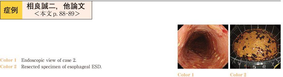
    2015Volume 87Issue 1 Pages 88-89
    Published: December 12, 2015
    Released on J-STAGE: January 06, 2016
    JOURNAL FREE ACCESS
    We experienced two esophageal ESD cases in which intraoperative introduction of dexmedetomidine (DEX) was useful for poor responders to conventional sedation mainly by flunitrazepam. The patients having a large, superficial esophageal cancer were referred to our hospital for endoscopic resection. We started ESD with flunitrazepam and pethidine hydrochloride which had been effective on preoperative endoscopy. During the procedure, they moved frequently, and additional injection of flunitrazepam, midazolam, and pethidine hydrochloride was ineffective. Therefore, we decided to switch the sedation to continuous infusion of DEX. After using DEX, favorable sedation was obtained and vital signs including respiratory function became stable. Finally, we could complete the procedure without complications.
    Download PDF (625K)
  • Shuichiro Iwasaki, Takafumi Yano, Kazuho Uehara, Teppei Nakame, Miyuki ...
    2015Volume 87Issue 1 Pages 90-91
    Published: December 12, 2015
    Released on J-STAGE: January 06, 2016
    JOURNAL FREE ACCESS
    We describe a 59-year-old woman who coughed and vomited a small amount of blood on the day of presentation. Upper endoscopy was performed on the same day. The examination revealed only fibrin clot-like coagula in the esophagus. Contrast-enhanced computed tomography of the chest and abdomen was performed to locate the source of bleeding. The patient was given a diagnosis of dissecting aortic rupture and esophageal perforation.
    A search of the database of the Japan Medical Abstracts Society for the period from 2001 through 2015 revealed only 23 reported cases of esophageal perforation associated with rupture of a dissecting aortic aneurysm as confirmed on upper endoscopy in Japan, including our patient. We describe our experience with this very rare case along with a discussion of the literature.
    Download PDF (1020K)
  • Tsunehiro Suzuki, Masakazu Nakano, Naoya Izawa, Kazuhiro Takenaka, Hid ...
    2015Volume 87Issue 1 Pages 92-93
    Published: December 12, 2015
    Released on J-STAGE: January 06, 2016
    JOURNAL FREE ACCESS
    This is a case of a male patient in his 30’s who underwent esophagogastroduodenoscopy (EGD) for epigastric pain. EGD revealed a longitudinal ulcer in the esophagus of unknown cause for which a mucosal protective agent and proton-pump inhibitor were prescribed. He presented again with a lower abdominal pain. Colonoscopy (CS) was also performed. It showed cobblestone appearance and longitudinal ulcer in the ileocecal region. He was diagnosed with Crohn’s disease (CD) and previous esophageal ulcers it was found to be an upper gastrointestional tract lessions of CD. He was admitted for further investigations and instigation of medical treatment. Treatment with azathioprine and infliximab was initiated. To this date, he has remained in remission, and has not had any relapses of esophagus ulcers.
    Download PDF (918K)
  • Naoya Izawa, Takeshi Sugaya, Akira Kanamori, Ruiko Muraoka, Hitoshi Ki ...
    2015Volume 87Issue 1 Pages 94-95
    Published: December 12, 2015
    Released on J-STAGE: January 06, 2016
    JOURNAL FREE ACCESS
    52-year-old woman with a left colitis type ulcerative colitis (UC) was referred to our hospital for the treatment of steroid-dependent UC. For the investigation of her swallowing pain on the chest, she underwent esophagogastroduodenoscopy (EGD) , which showed an ulcer in the middle esophagus. Colonoscopy also showed modelate inflammation in the left colon. PPI administration did not improve esophageal ulcer. Then, induction therapy for UC was started with steroid and immunomodulator, which improved bloody stool and also swallowing pain in a week. Healing of esophageal ulcer, was confirmed by EGD. She was diagnosed as a rare case of esophageal ulcer complicated with UC.
    Download PDF (612K)
  • Yoko Kubosawa, Kaoru Takabayashi, Yusaku Takatori, Keiichiro Abe, Yosh ...
    2015Volume 87Issue 1 Pages 96-97
    Published: December 12, 2015
    Released on J-STAGE: January 06, 2016
    JOURNAL FREE ACCESS
    A 60-year-old man suffered from persistent vomiting and was admitted to our hospital. Esophagogastroduodenoscopy showed type 3 lesions affecting 2/3 of the circumference of the esophagogastric junction and submucosal tumor-like elevated lesions with erosions in the upper part of the stomach. Flat submucosal tumor-like elevated lesions were found in the duodenum. PET-CT showed FDG accumulation throughout the body. The biopsy from all lesions demonstrated that melanin pigment was negative and HMB45 and Melan-A were positive. Thus, he was diagnosed with an amelanotic malignant melanoma. No primary lesion was noted on the skin. Endoscopically, the lesions in the esophagogastric junction showed features consistent with those of primary malignant melanoma in the gastrointestinal tract, and the lesions in the stomach and duodenum showed features consistent with those of metastatic tumor in the gastrointestinal tract. Melanocytes were noted in the squamous epithelial tissue of the esophagus. Thus, this is a rare case of amelanotic malignant melanoma derived from the melanocytes of the esophagogastric junction, which grew and progressed in the stomach, resulting in multiorgan metastasis.
    Download PDF (759K)
  • Masami Omae, Junko Fujisaki, Yusuke Horiuchi, Toshiyuki Yoshio, Akiyos ...
    2015Volume 87Issue 1 Pages 98-99
    Published: December 12, 2015
    Released on J-STAGE: January 06, 2016
    JOURNAL FREE ACCESS
    A 79-year-old male was diagnosed with Long segment Barrett’s esophagus in 2011. Three metachronous superficial Barrett’s esophageal adenocarcinomas (s-BEAs) were detected two years later. Two of the 3 cancers were detected by magnifying endoscopy with narrow band imaging (ME-NBI) . Endoscopic submucosal dissection was performed on all 3 lesions and the pathological findings were intramucosal cancers. In our case, ME-NBI was very useful for detecting s-BAE. s-BEA on ME-NBI was observed as a brownish area.
    Download PDF (1101K)
  • Yumiko Fukuma, Tsukasa Furuhata, Syu Hoteya, Kousuke Nomura, Yasutaka ...
    2015Volume 87Issue 1 Pages 100-101
    Published: December 12, 2015
    Released on J-STAGE: January 06, 2016
    JOURNAL FREE ACCESS
    A 73-year-old man with esophageal squamous cell carcinoma was referred to our hospital for the treatment. The white light endoscopy revealed that the invasion depth was m3-SM1, and NBI magnified endoscopy revealed it was EP-LPM. But Endoscopic ultrasonograhy (EUS) revealed it was SM2. Other evaluation method revealed no metastasis. All things considered, we diagnosed as cT1bN0M0, stageI. This lesion was thought to be outside the guideline for ESD. But the patient strongly requested the endoscopic resection, we decided to perform the ESD. The pathological diagnosis revealed it was SCC, pT1a (LPM) , ly0, v0. The depth of cancer resulted in LPM, but there were a lot of hyperplastic lymphoid follicles under the cancer. This case indicates that it is very difficult to differentiate the cancer from hyperplastic lymphoid follicles and the focal fibrosis by EUS.
    Download PDF (853K)
  • Hisafumi Yamagata, Masateru Hataji, Sae Nakayama, Atsuki Moroi, Isao A ...
    2015Volume 87Issue 1 Pages 102-103
    Published: December 12, 2015
    Released on J-STAGE: January 06, 2016
    JOURNAL FREE ACCESS
    A case of a 24-year-old man with schizophrenia. After ingesting a button battery (witnessed by others) , he visited the outpatient department of our hospital. Radiographic examination confirmed the presence of the battery in the stomach, leading to the diagnosis of ingestion of a foreign body with toxic contents. Emergency endoscopy was performed. The button battery found in the stomach was recovered with a retrieval net. On arrival, the patient was in an agitated state. Because our hospital’s psychiatrists were unavailable, sedation was provided by the same doctor who performed endoscopy. To examine patients in a state of psychomotor agitation, it is necessary that doctors who perform endoscopy be able to sedate them with haloperidol or levomepromazine when necessary.
    Download PDF (764K)
  • Kana Yamamoto, Maiko Kishino, Ayako Kobayashi, Hirotaka Yamamoto, Shin ...
    2015Volume 87Issue 1 Pages 104-105
    Published: December 12, 2015
    Released on J-STAGE: January 06, 2016
    JOURNAL FREE ACCESS
    We experienced five cases of gastric bezoars for 8 years from 2006 to 2014. They included two males and three female, mean age was 68 years. Main symptom were vomiting, dyspepsia, abdominal pain and tarry stool. All patients had a history of diabetes and three patients had operation history of distal gastrectomy. In two cases, endscopy revealed a gastric ulcer and one case showed small bowel obstruction. A size of bezoars were 2〜10cm, one case had two bezoars. And all components of bezoars were tannic acid. Two cases treated by endscopic fragmentation, one case treated by dissolution herapy with Cola and endscopic fragmentation, an one case treated by endscopic extraction. All cases could be achieved complete dissolution without any complications.
    Download PDF (747K)
  • Toshiya Akai, Tomohiko Setoguchi, Kenichi Sunayama, Toshiyuki Ori
    2015Volume 87Issue 1 Pages 106-107
    Published: December 12, 2015
    Released on J-STAGE: January 06, 2016
    JOURNAL FREE ACCESS
    A 53 year-old-man was admitted to our hospital because of upper gastrointestinal bleeding that occurred 3 weeks after eating simesaba. Emergency esophagogastroduodenoscopy (EGD) revealed an A1 stage hemorrhagic ulcer at the fornix. Endoscopic hemostasis was performed by local injection of hypertonic saline with epinephrine and argon plasma coagulation. However, due to saburra it was difficult to observe in detail. The next day, we performed EGD and found an Anisakis simplex at the base of the ulcer, and was removed during EGD with biopsy forceps. The patient was treated with proton pump inhibitor. His condition was improved to the H1 stage at 7days after the admission and on the 11th day he was discharged from the hospital. Gastric anisakiasis is usually occurred in association with stomach ache and diagnosed by the endoscopic findings of the larvae of the nematode Anisakis simplex, mucosal edema or erosion. We report a rear case of hemorrhagic gastric ulcer associated with anisakiasis.
    Download PDF (662K)
  • Jun Morikawa, Masaru Nakano, Taku Kobayashi, Satoko Umeda, Sayaka Shim ...
    2015Volume 87Issue 1 Pages 108-109
    Published: December 12, 2015
    Released on J-STAGE: January 06, 2016
    JOURNAL FREE ACCESS
    An 88-year-old woman was admitted to our hospital with general malaise. Upper gastrointestinal endoscopy revealed a submucosal tumor-like lesion in the body of stomach. Endoscopic ultrasonography (EUS) revealed the lesion to be in the submucosal layer with multiple cyst-like masses. As definitive diagnosis was difficult from these findings and adenocarcinoma could not be ruled out, we performed partial gastrectomy. Pathological examination revealed the mucosa invaginated into the submucosal layer. Based on these findings, we diagnosed the lesion as HIP. If one is able to suspect HIP, EUS becomes useful in its diagnosis. If it can be confirmed that the lesion is localized within the submucosal layer by EUS, minimally invasive treatments such as endoscopic submucosal dissection can be selected. HIP thus should be included in the differential diagnoses of protruding lesions in order to perform appropriate and accurate pre-operative diagnoses.
    Download PDF (895K)
  • Akifumi Tanaka, Kengo Tokunaga, Masao Toki, Hideaki Mori, Tadakazu His ...
    2015Volume 87Issue 1 Pages 110-111
    Published: December 12, 2015
    Released on J-STAGE: January 06, 2016
    JOURNAL FREE ACCESS
    Approximately 90% of patients with gastric mucosa-associated lymphoid tissue (MALT) lymphoma are infected with H. pylori. The eradication of H. pylori leads to complete remission (CR) of the disease in 70-80% of the gastric MALT lymphoma with H. pylori infection. We reported three cases of H. pylori negative gastric MALT lymphoma that were confirmed as CR treating by radiation therapy (RT) . All cases confirmed H. pylori negative by 4 or 5 H. pylori tests. In addition, H. heilmannii was negative in all cases. CR was confirmed at the time of 1 or 2 years after RT.
    In conclusion, RT is useful for treating H. pylori negative gastric MALT lymphoma.
    Download PDF (554K)
  • Takahito Minaguchi, Naoto Yoshitake, Hitoshi Kino, Yoshihito Kaneko, M ...
    2015Volume 87Issue 1 Pages 112-113
    Published: December 12, 2015
    Released on J-STAGE: January 06, 2016
    JOURNAL FREE ACCESS
    A 56-year-old woman received gastric cancer-risk screening (ABC method) , and was classified into group D.She underwent esophago-duodeno-gastroscopy (EGD) for the close examination of the stomach at a nearby clinic,and a superficially depressed lesion tubular adenocarcinoma measuring 18mm in diameter was found at the anterior wall of the antrum.
    She referred to our hospital for endoscopic treatment. Re-endoscopy revealed the lesion was limited to the mucosal layer without the metastasis, therefore it was resected by endoscopic submucosal dissection (ESD) . Pathologically, the lesion was curatively resected with ESD. EGD after three months indicated that gastric body had severe mucosal atrophy compared with the antrum. The histopathological examination demonstrated similar findings, and demonstrated chromogranin A-positive endocrine cell nest. The laboratory data showed hypergastrinemia, negative result for H. pylori antibody, and positive for gastric parietal cell antibody. From the above findings, she was diagnosed as a rare case of early gastric cancer associated with type A autoimmune gastritis
    Download PDF (1110K)
  • Tomoaki Moriyama, Naoko Nagasu, Tomoaki Fujikawa, Ichiro Kato, Hirokaz ...
    2015Volume 87Issue 1 Pages 114-115
    Published: December 12, 2015
    Released on J-STAGE: January 06, 2016
    JOURNAL FREE ACCESS
    An 84-year-old Japanese woman was referred to the hospital because of abdominal pain, gastric fullness, and vomiting. Plain abdominal roentgenogram showed dilation of the stomach and gastric outlet obstruction was suspected. Upper endoscopic examinations showed a protruding lesion in the gastric antrum prolapsing into the duodenum. The tumor was circumferential, endoscopically intractable, and an estimate of its nature and depth was difficult, then endoscopic resection was considered inappropriate. Barium x-ray study revealed the lobular lesion prolapsing into the duodenum. A diagnosis of gastric tumor presented with ball valve syndrome was made, laparoscopy-assisted distal gastrectomy was performed. Histopathological findings disclosed well differentiated tubular adenocarcinoma slightly invading the submucosa.
    An endoscopic treatment is thought to deserve considering when the early gastric cancer prolapsing into the duodenum is successfully reduced. In case of endoscopic intractability or contraindication laparoscopy-assisted distal gastrectomy is a good choice.
    Download PDF (921K)
  • Shuhei Tazaki, Hitoshi Yoshida
    2015Volume 87Issue 1 Pages 116-117
    Published: December 12, 2015
    Released on J-STAGE: January 06, 2016
    JOURNAL FREE ACCESS
    A 65-year-old man complaining of nausea underwent upper gastrointestinal endoscopy in June 2012 which showed chronic gastritis. Upon reexamination in October 2013, a gastric cancer of IIa+IIc on the greater curvature of the gastric antrum was detected. Preoperative endoscopy of 15 days later showed a steep rise of the lesion. Histopathologic findings of the resected specimen showed a well-differentiated adenocarcinomatous gastric cancer that invaded the submucosal layer without vascular invasion and lymph node metasis. Because this well-differentiated adenocarcinoma grew and changed in the short-term, we suspected this case was a special type of gastric cancer. Immunohistochemical analysis of AFP was positive in the gastric cancer resulting in the diagnosis of an AFP-producing gastric cancer. AFP-producing gastric cancer has an extremely poor prognosis, and readily metastasizes to the liver. This case has not recognized metastasis and increase of the serum AFP after operation.
    We report our experience with a rare case of AFP-producing early gastric cancer that grew in the short-term.
    Download PDF (1116K)
  • Masahiro Yura, Rieko Nakamura, Tsunehiro Takahashi, Norihito Wada, Hir ...
    2015Volume 87Issue 1 Pages 118-119
    Published: December 12, 2015
    Released on J-STAGE: January 06, 2016
    JOURNAL FREE ACCESS
    A flat elevated lesion with a central depression was observed in the lower gastric body of a 75-year-old man. The epithelium of the first biopsy specimen was atypical, and no significant change was apparent 3 weeks later. The epithelial surface was covered with a whitish substance rendering it difficult to observe the surface structure. Three months later, the lesion had grown. A biopsy revealed malignancy, and endoscopic submucosal dissection was performed. A mucinous carcinoma exhibiting submucosal invasion was histopathologically identified. Additional surgery was performed, but lymph node metastasis developed. Thus, an early-stage mucinous carcinoma may rapidly grow and metastasize. If a flat elevated lesion covered with a whitish substance (indicative of an early-stage mucinous carcinoma) is observed, diagnostic endoscopic submucosal dissection or strict follow-up is required.
    Download PDF (866K)
  • Yoshihiro Sasaki, Hajime Kamijyo, Yukihiro Kiya, Yusuke Shimada, Masat ...
    2015Volume 87Issue 1 Pages 120-121
    Published: December 12, 2015
    Released on J-STAGE: January 06, 2016
    JOURNAL FREE ACCESS
    A 50 year-old woman visited our hospital because of epigastric pain. Upper GI endoscopy revealed a gastric tumor resembled advanced type 3 gastric cancer. Histological examination of the biopsy specimens revealed poorly differentiated adenocarcinoma with small portion of signet ring cell carcinoma. There was no distant metastases by CT examination, we diagnosed this case as an advanced gastric cancer (T2, N0, M0 cStageIB) , then distal gastrectomy was performed.
    Pathological study of the resected specimen showed intramucosal gastric cancer (por2 (por2>sig) , pT1a (M) , ly0, v0, pPM0, pDM0, CY0, pR0, pN0, StageIA) ,
    The accuracy of the diagnosis of the depth of gastric cancer invasion in the undifferentiated adenocarcinoma and the differentiated carcinoma are reported to be about 70% and 90% respectively. EUS is useful for further accurate diagnosis, but sometimes it is difficult to make a correct diagnosis of cancer invasion, in particular in a case with a deep ulcer formation.
    Download PDF (1072K)
  • Minoru Tomizawa, Rumiko Hasegawa, Fuminobu Shinozaki, Yoshinori Shirai ...
    2015Volume 87Issue 1 Pages 122-123
    Published: December 12, 2015
    Released on J-STAGE: January 06, 2016
    JOURNAL FREE ACCESS
    A 69-year-old male visited our hospital with a complaint of low abdominal pain. He was diagnosed with acute colon diverticulitis and consequently admitted and treated with antibiotics. He was subjected to screening endoscopy. The endoscopy findings revealed a gastric ulcer, and he was administered a proton pump inhibitor. Moreover, a depressed area was also observed. On follow-up endoscopy after discharge, the depressed area was obvious, and its surface was irregular. Biopsy was performed, and the lesion was diagnosed as a well-differentiated adenocarcinoma. Upon endoscopic ultrasound, the tumor was found to be restricted within the mucous. Accordingly, we speculate that the gastric cancer might have become obvious after the administration of the proton pump inhibitor, as the gastritis was cured and the mucus was clear.
    Download PDF (991K)
  • Michiko Sato, Kaoru Takabayashi, Keiichiro Abe, Yoshiaki Takada, Tetsu ...
    2015Volume 87Issue 1 Pages 124-125
    Published: December 12, 2015
    Released on J-STAGE: January 06, 2016
    JOURNAL FREE ACCESS
    An 81-year-old man underwent esophagogastroduodenoscopy, and a 30-mm elevated-type lesion was identified at the greater curvature of the lower gastric body. There were no signs of submucosal invasion. Biopsy specimens revealed moderately differentiated adenocarcinoma, and endoscopic submucosal resection was performed. Pathological examination revealed moderately differentiated adenocarcinoma with a small component of poorly differentiated adenocarcinoma. Immunohistological examination of the component showed synaptophysin (−) ,chromogranin A (−) and CD56 (+) . Ki-67 was positive in > 90% of the cells. The final histopathological diagnosis was Type0-I+IIa, 29×21 mm, pap> tube1, tub2 >> por1 with neuroendocrine features : pT1a, ly0, v0, pHM0, pVM0. This case may be an initial stage of neuroendocrine carcinoma, and therefore valuable in revealing the development and prognosis of similar cases.
    Download PDF (1130K)
  • Eiko Okamoto, Katsunori Ami, Yuko Karakama, Mayumi Kondoh, Hidetaka Ak ...
    2015Volume 87Issue 1 Pages 126-127
    Published: December 12, 2015
    Released on J-STAGE: January 06, 2016
    JOURNAL FREE ACCESS
    Case 1 : The patient was a 70-year-old male. We performed an endoscopic submucosal dissection (ESD) for two gastric tube cancers (GTCs) , with both lesions being adenocarcinomas limited to the mucosal layer. Case 2 : The patient was a 76-year-old male. A GTC was found on the linear scar, which was suspected to be invading the submucosal layer ; we performed an ESD because of the surgical risk. The pathological diagnosis was an adenocarcinoma invading the submucosal layer, with lymphovascular invasion evident. Both cases were reconstructed through the posterior mediastinal route. We performed ESDs without complications, such as bleeding and perforation. The surgery for GTC, especially when reconstructed through the posterior-mediastinal route, was highly invasive. These cases illustrate that when a judgement needs to be made on a suitable treatment strategy for GTC with consideration for the risks of surgery, an ESD can be a useful and safe treatment modality.
    Download PDF (916K)
  • Takashi Takenoya, Miwako Arima, Yu Nishimura, Masashi Kurozumi
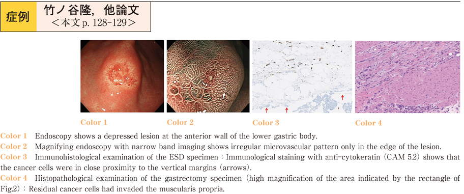
    2015Volume 87Issue 1 Pages 128-129
    Published: December 12, 2015
    Released on J-STAGE: January 06, 2016
    JOURNAL FREE ACCESS
    A 59-years-old man was referred to our hospital for treatment of gastric cancer. Esophagogastroduodenoscopy revealed that the lesion was depressed, 10 mm in diameter, and was located at the anterior wall of the lower gastric body. Signet-ring cell carcinoma was diagnosed using endoscopic biopsy. The lesion was diagnosed as the expanded indication lesion of endoscopic submucosal dissection (ESD) , and ESD was performed. The cancer invaded the submucosa to a depth of 1800 µm and vertical margins were negative ; however, the cancer cells were in close proximity to the vertical margins. The lesion was diagnosed as a non-curative resection. Additional gastrectomy was performed. The gastrectomy specimen revealed that residual cancer cells had invaded the muscularis propria. Since it was possible that the invasive cancer persisted in the deeper tissues of the submucosa, additional gastrectomy was suggested.
    Download PDF (776K)
  • Kotaro Matsumoto, Masatoshi Mabuchi, Hanae Yamada, Hiromichi Tsunashim ...
    2015Volume 87Issue 1 Pages 130-131
    Published: December 12, 2015
    Released on J-STAGE: January 06, 2016
    JOURNAL FREE ACCESS
    The patient was a 43-year-old woman who was admitted to our hospital for an abdominal ultrasonography, that revealed a liver tumor. Computed tomography and magnetic resonance imaging showed a solid hepatic tumor, with adipose components, measuring 39 mm in her caudate lobe. We could not rule out the possibility of hepatocellular carcinoma.
    Histopathological examination of the tumor specimen obtained by using endoscopic ultrasound-guided fine-needle aspiration showed enlarged alveolars cells with oval nuclei and eosinophilic cytoplasm, and enlarged clear cells with clear cytoplasm. The tumor cells stained positive for α-SMA and HMB-45, and thereby a diagnosis of PEComa was made.
    Download PDF (811K)
  • Junichi Akao, Yukiko Takayama, Takahiro Ajihara, Kenta Nagao, Kyoko Sh ...
    2015Volume 87Issue 1 Pages 132-133
    Published: December 12, 2015
    Released on J-STAGE: January 06, 2016
    JOURNAL FREE ACCESS
    A 40-year-old man came to our hospital because of jaundice. CT showed a contrast-enhanced tumor in the lower bile duct, and peripheral dilatation. Cholangiography revealed a filling defect in the lower bile duct. A transpapillary biopsy revealed no evidence of neoplastic cells. Endoscopic ultrasonography showed a hypoechoic tumor in the lower bile duct. We made a diagnosis of small cell carcinoma based on the EUS-FNA findings and treated the patient with chemotherapy.
    Small cell carcinoma of the bile duct system is rare and difficult to diagnose. Based on our experience, EUS-FNA can be useful for making a histological diagnosis of bile duct tumors that are impossible to diagnose by transpapillary biopsy or cytology.
    Download PDF (690K)
  • Satoshi Nakao, Jun Hamanaka, Hiroyuki Oka, Hiroshi Okazaki
    2015Volume 87Issue 1 Pages 134-135
    Published: December 12, 2015
    Released on J-STAGE: January 06, 2016
    JOURNAL FREE ACCESS
    A 70-year-old man visited our hospital with vomiting and hematemesis. Abdominal CT scan showed common bile duct stone and enlarged gallbladder contained high density fluid and air. After admission, contrast enhanced CT scan showed cholecystoduodenal and cholecystocolonic fistula. Esophagogastroduodenoscopy and colonoscopy revealed fistulas in the duodenum and transverse colon near the hepatic flexure. Fistulography showed gallbladder. We performed endoscopic closure of the cholecystocolonic fistula using an Over the Scope Clip (OTSC) system. After closure the patient developed cholangitis, endoscopic retrograde cholangiopancreatography and removal of stones was performed. Cholecystoduodenal fistula reduced the size gradually, and cholecystocolonic fistula was closed completely.
    Download PDF (1113K)
  • Masashi Tanaka, Taku Yabuki, Sayaka Chishima, Iwao Chishima, Yoshitami ...
    2015Volume 87Issue 1 Pages 136-137
    Published: December 12, 2015
    Released on J-STAGE: January 06, 2016
    JOURNAL FREE ACCESS
    An 83-year-old women was admitted to our hospital because of a renal abscess, and she was given ceftriaxone for four weeks. After 36 days of admission, abdominal CT revealed high-density structures in the gallbladder and common bile duct (CBD) , which were not detected earlier. We diagnosed the abnormality as biliary pseudolithiasis caused by ceftriaxone and stopped ceftriaxone. A total of 144 days after diagnosis of pseudolithiasis, the patient complained of epigastralgia and high fever with elevated biliary enzyme levels. Abdominal CT once again revealed high-density structures in the gallbladder and CBD, and a diagnosis of CBD stones and cholangitis was made. Endoscopic sphincterotomy was performed for removal of the CBD stones, and laparoscopic cholecystectomy was subsequently performed. Typically, biliary pseudolithiasis has been reported to disappear a short time after discontinuing ceftriaxone. However, our case remained long-term, and caused biliary obstruction and colic pain.
    Download PDF (668K)
  • Minoru Tomizawa, Fuminobu Shinozaki, Yasufumi Motoyoshi, Takao Sugiyam ...
    2015Volume 87Issue 1 Pages 138-139
    Published: December 12, 2015
    Released on J-STAGE: January 06, 2016
    JOURNAL FREE ACCESS
    A 72-year-old man visited our hospital for follow-up after treatment with interferon for hepatitis C virus infection. Ultrasonography screening demonstrated wall thickening in the fundus of the gallbladder. Magnetic resonance cholangiopancreatography showed the pearl-necklace sign, typical of adenomyomatosis of the gallbladder. However, it was possible that cancer was associated with the thickened wall. The thickened wall was investigated further by endoscopic ultrasonography (EUS) , using perfluorobutane as a contrast agent. EUS clearly demonstrated cystic lesions, suggesting Rokitansky-Aschoff sinuses in the thickened wall, consistent with the pearl necklace sign. The thickened wall was homogenously enhanced. These observations confirmed adenomyomatosis of the gallbladder, and suggested a low possibility of associated cancer. The patient was followed up with abdominal ultrasonography to confirm the diagnosis.
    Download PDF (935K)
  • Katsunori Sekine, Toshiyuki Sakurai, Naoyoshi Nagata, Kazuhiro Watanab ...
    2015Volume 87Issue 1 Pages 140-141
    Published: December 12, 2015
    Released on J-STAGE: January 06, 2016
    JOURNAL FREE ACCESS
    Endoscopic retrograde cholangiopancreatography (ERCP) has some complications. Especially, retroperitoneum perforation as a result of ERCP or endoscopic sphincterotomy (EST) is one of rare complications. On the other side, it has a higher risk of mortality. The treatment of retroperitoneum perforation is basically biliary drainage by using endoscopic nasobiliary drainage or ERBD. Recently, endoscopic techniques have been described to close a variety of perforations. In this case report, we describe the closure of a persistent retroperitoneum perforation by using a Fully covered metal stent.
    Download PDF (848K)
  • Yoshihito Kaneko, Kouhei Tsuchida, Takahito Minaguchi, Mari Iwasaki, T ...
    2015Volume 87Issue 1 Pages 142-143
    Published: December 12, 2015
    Released on J-STAGE: January 06, 2016
    JOURNAL FREE ACCESS
    A 64-year-old woman underwent Roux-en-Y gastric bypass surgery for the treatment of antral obstruction due to type 3 gastric cancer. Two months later, liver dysfunction due to malignant biliary tract stenosis was noted. The endoscopic retrograde cholangiography with a double balloon endoscope (DBE) and the percutaneous transhepatic biliary drainage for biliary obstruction were unsuccsessful. Percutaneous transhepatic gallbladder biliary drainage (PTGBD) was performed,and thereafter self expanded metalic stent using DBE was successfully placed into the common bile duct with the Rendezvous technique from the PTGBD route.this Rendezvous procedure using DBE may be one of the therapeutic options for biliary obstructidon after gastric surgery.
    Download PDF (749K)
  • Isao Arima, Hisafumi Yamagata, Masateru Hataji, Sae Nakayama, Atsuki M ...
    2015Volume 87Issue 1 Pages 144-145
    Published: December 12, 2015
    Released on J-STAGE: January 06, 2016
    JOURNAL FREE ACCESS
    A 66-year-old man with fever and jaundice came our hospital. Blood tests showed increased inflammatory response and elevated enzymes of the hepatobiliary tract Pancreatic head tumors and distention of the common bile duct were observed on computed tomography scan. The patient was diagnosed as obstructive cholangitis triggered by pancreatic cancer and was hospitalized. Because endoscopic retrograde cholangiopancreatography failed, a plastic stent was placed into a stricture by the percutaneous transhepatic biliary drainage (PTBD) -guided rendezvous procedure. Because arterial hemorrhage occurred at the time of PTBD tube removal, coil embolization was performed after emergency hepatic arteriography. Fasting, replacement fluid, and antimicrobial administration improved cholangitis and jaundice. We report a case of internal fistulization performed by PTBD-guided rendezvous procedure to obstructive jaundice induced by malignant biliary stricture
    Download PDF (941K)
  • Katsumasa Kobayashi, Toru Asano, Ai Minamidate, Takahito Nozaka, Mana ...
    2015Volume 87Issue 1 Pages 146-147
    Published: December 12, 2015
    Released on J-STAGE: January 06, 2016
    JOURNAL FREE ACCESS
    A 52-year-old male, who was kicked by his son 2 days previously, was taken to our hospital with the chief complaint of abdominal pain. Abdominal computed tomography (CT) showed pancreatic head injury and fluid collection extend to the inferior pole of right kidney. Endoscopic retrograde pancreatography (ERP) showed main pancreatic duct injury with leakage of contrast medium at the pancreatic head. We placed a 5-Fr endoscopic naso-pancreatic drainage (ENPD) tube. On the same day, operation was performed for intraperitoneal drainage. Fever and abdominal pain got better day by day. On the 20 th day, the ENPD tube was exchanged to pancreatic duct stent. There were no serious complications, and he was discharged on the 36 th hospital day.
    Download PDF (967K)
  • Koichi Ito, Tomoyuki Yada, Naomi Uemura
    2015Volume 87Issue 1 Pages 148-149
    Published: December 12, 2015
    Released on J-STAGE: January 06, 2016
    JOURNAL FREE ACCESS
    A 76-year-old man was admitted to our hospital with melena and loss of consciousness. A contrast-enhanced computed tomography revealed a duodenal diverticulum with extravasation. We then attempted hemostasis by esophagogastroduodenoscopy (EGD) , which revealed blood in the duodenum and a clot in the duodenal diverticulum. Despite using direct and side-viewing endoscopy, the bleeding point was not detected. An emergency angiography of the inferior pancreaticoduodenal artery revealed extravasation near the pancreas head and embolization was performed using a gelatin sponge. Following embolization, there was no extravasation from that area. EGD on the second day of hospitalization revealed an ulcer in the duodenal diverticulum, but there was no bleeding. Oral intake was resumed and the patient was discharged on the 16 th day of hospitalization.
    This case demonstrates the effectiveness of transcatheter arterial embolization for duodenal diverticulum hemorrhage, which is difficult to manage using EGD.
    Download PDF (784K)
  • Masakazu Abe, Chika Kusano, Sho Suzuki, Mikinori Koga, Shin Kono, Take ...
    2015Volume 87Issue 1 Pages 150-151
    Published: December 12, 2015
    Released on J-STAGE: January 06, 2016
    JOURNAL FREE ACCESS
    A lot of adverse event were happened after endoscopic resection for nonampullary duodenal tumors. We suggest that useful of PGA sheets and fibrin glue these two studies.
    [Case 1] The patient was a 77-year-old man. Endoscopic mucosal resection was performed for a type 0-IIa lesion located in 2nd portion of duodenum. A PGA sheet was applied to the opening of the ulcer base after resection via biopsy forceps, after which fibrin glue was applied. [Case 2] The patient was a 53-year-old man. Endoscopic submucosal dissection was performed for a type 0-IIc lesion located in 2nd portion of duodenum. The ulcer was covered the same way as described for Case 1.
    These cases indicate that shielding method with PGA sheets and fibrin glue can be effective and safe to prevent adverse events such as delayed bleeding and perforation after endoscopic resection for nonampullary duodenal tumors.
    Download PDF (902K)
  • Taikan Yamamoto, Takashi Sekikawa, Ken Shimada, Akio miyokawa, Yasutak ...
    2015Volume 87Issue 1 Pages 152-153
    Published: December 12, 2015
    Released on J-STAGE: January 06, 2016
    JOURNAL FREE ACCESS
    Gastric outlet obstruction (GOO) is a late complication of advanced and metastatic malignant tumors. Self-expanding metal stent (SEMS) placement is a safe and effective palliative treatment for patients with GOO. In general, the median survival time after SEMS placement for GOO is approximately 100 days. Eight patients with symptomatic malignant GOO were treated by SEMS placement in our hospital. Surgical bypass could not be performed due to severe advanced malignant tumors. We estimated the gastric outlet obstruction scoring system (GOOSS) as the main outcomes. We performed SEMS placement using endoscopy and got success in all cases. No complications occurred after the procedure. Our study findings indicate that SEMS placement is a safe and effective method for managing malignant GOO.
    Download PDF (840K)
  • Chika Nigauri, Kazuya Sugimori, Makoto Sugimori, Ayako Takeda, Yuniba ...
    2015Volume 87Issue 1 Pages 154-155
    Published: December 12, 2015
    Released on J-STAGE: January 06, 2016
    JOURNAL FREE ACCESS
    An 81-year-old male patient was diagnosed with carcinoma of the ampulla of Vater. At a previous hospital, he had undergone placement of a metallic stent in both the bile duct and duodenum in a side-to-end positional relationship for stenosis of the lower bile duct and duodenum. However, he visited our hospital because he repeatedly developed cholangitis caused by multiple bile duct stones. We planned endoscopic ultrasound-guided biliary drainage (EUS-BD) with an approach from the first part of the duodenum. First, we used argon plasma coagulation to trim the duodenal metallic stent. Next, using forceps to grip the end of the biliary stent exposed by the trimming, we removed the biliary metallic stent, which was impeding punctures. This ingenuity enabled us to succeed in EUS-BD.
    Download PDF (934K)
  • Kana Nakayama, Shigeaki Nagao, Kazuki Horiuchi, Chihiro Yasue, Akikiko ...
    2015Volume 87Issue 1 Pages 156-157
    Published: December 12, 2015
    Released on J-STAGE: January 06, 2016
    JOURNAL FREE ACCESS
    An 80-year-old man, who had been suffering from anorexia and upper abdominal pain since 6 months ago, was sent to our hospital suspected of protein loosing enteritis (PLE) because of the marked hypoalbuminemia and the edematous states in the jejunum found by computed tomography.
    α1-antitrypsin clearance test was positively high and 99mTc-HAS-D scintigraphy showed leakages mainly in the jejunum and slightly in the ascending colon. Multiple ulcers were found in the jejunum by double balloon endoscopy. The histopathological findings showed owl’s eye including bodies in the epithelium cells of the jejunum and these cells were positive for anti-CMV antibodies. Therefore we supposed CMV had induced the membrane inflammation in the jejunum and it had caused PLE.
    Download PDF (745K)
  • Kazuma Sekiba, Makoto Okamoto, Nariaki Odawara, Makoto Moriyama, Sachi ...
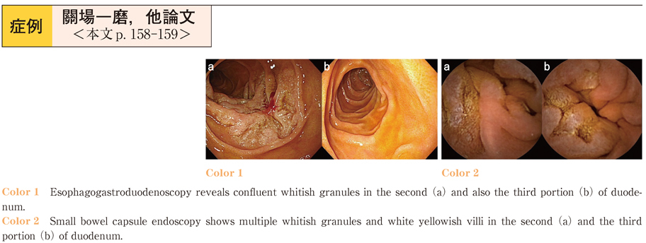
    2015Volume 87Issue 1 Pages 158-159
    Published: December 12, 2015
    Released on J-STAGE: January 06, 2016
    JOURNAL FREE ACCESS
    Duodenal follicular lymphoma (dFL) is a rare disease, accounting for only 1% to 3% of gastrointestinal non-Hodgkin’s lymphoma. Neither standard surveillance nor treatment for dFL has been established. In this article, we report a case of dFL successfully detected by capsule endoscopy, and followed with a watch-and-wait strategy.
    A 59-year-old asymptomatic male was admitted to our hospital for evaluation of confluent whitish granules in the second portion of duodenum detected on screening esophagogastroduodenoscopy. Biopsy specimens showed a nodular lymphocyte expansion, constituted by small cleaved lymphoid cells that were positive for CD20, CD79a, CD10 and BCL2, but negative for CD5 and Cyclin D1. Capsule endoscopy detected the tumors in the second and third portion of duodenum. The final diagnosis was stage I follicular lymphoma, grade 1, of the duodenum. As the lymphoma caused no symptoms, no treatment was performed in accordance of the watch-and-wait policy.
    Download PDF (588K)
  • Atushi Ohsaki, Keigo Ashitani, Hideki Ohgo, Minoru Yamaoka, Takaya Ich ...
    2015Volume 87Issue 1 Pages 160-161
    Published: December 12, 2015
    Released on J-STAGE: January 06, 2016
    JOURNAL FREE ACCESS
    A 60-oyear-oold male visited a clinic because of gastric discomfort. This symptom was temporarily improved by a proton pump inhibitor, but it was worsened by discontinuation of the drug. He was referred to our hospital. Esophagogastroduodenoscopy showed an elevated lesion with multiple whitish small granular protrusions in the duodenal second portion, occupying two thirds of the circumference. The lesion was diagnosed to be a follicular lymphoma by histopathological examination including immunostaining of the biopsy specimens. He was admitted to our hospital. Abdominal CT scan showed no lymph node metastasis. Capsule endoscopy of the small intestine showed lymphoid follicles in the distal ileum in addition to the duodenal lesion. Bone marrow aspiration showed no invasion of lymphoma cells. This case was diagnosed as stage I according to the Lugano international conference classification. He underwent monotherapy using rituximab four times. However, the lesion did not respond. Therefore, radiotherapy was added which induced regression of the duodenal lesion. Follow-oup capsule endoscopy did not show any lesion in the distal ileum. As long-term outcome after treatment for duodenal follicular lymphoma is not known, strict observation is considered to be necessary.
    Download PDF (692K)
feedback
Top