Progress of Digestive Endoscopy
Online ISSN : 2187-4999
Print ISSN : 1348-9844
ISSN-L : 1348-9844
Volume 91, Issue 1
Displaying 1-50 of 60 articles from this issue
Clinical study
  • Izumi Inoue, Masashi Oka, Masao Ichinose
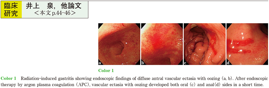
    2017Volume 91Issue 1 Pages 44-46
    Published: December 08, 2017
    Released on J-STAGE: December 21, 2017
    JOURNAL FREE ACCESS

    Radiation-induced gastritis is a rare cause of gastrointestinal bleeding. The aim of this study was to evaluate the clinical features associated with radiation-induced gastritis in cirrhosis treated with radiation therapy for hepatocellular carcinoma. Radiation-induced gastritis was observed in 6 of 12 patients showing endoscopic findings of diffuse antral vascular ectasia and correlated significantly with radiation therapy for recurrence of hepatocellular carcinoma in abdominal lymph nodes. Argon plasma coagulation was performed in 4 patients with radiation-induced hemorrhagic gastritis and 3 patients with radiation therapy for abdominal lymph nodes showed refractory radiation-induced hemorrhagic gastritis. We suggest that radiation therapy for abdominal lymph nodes in cirrhosis could be associated with refractory radiation-induced hemorrhagic gastritis.

    Download PDF (708K)
  • Atsuko Takahashi, Kumiko Momma, Junko Fujiwara, Akinori Miura
    2017Volume 91Issue 1 Pages 47-51
    Published: December 08, 2017
    Released on J-STAGE: December 21, 2017
    JOURNAL FREE ACCESS

    To clarify treatment strategy of synchronous double cancers at esophagus and pharynx, 50 cases were studied. Early cancers had no symptom and 70〜75% of them were detected in yearly endoscopic checkup. On the other hand, 80〜90% of all advanced cancers had complaint and detected through examination for them. 1) In cases with early cancer both in pharynx and esophagus, Endoscopic Resection (ER) was applied for 75% of all pharyngeal cancer and 85% of esophageal cancer. Only one case was lost by cancer recurrence. 2) In 11 cases with early pharyngeal cancer and advanced esophageal cancer, 64% of them were lost by cancer death. 55% of 11 patients with advanced pharyngeal cancers and early esophageal cancers were lost by cancer death. 3) Only one patient among 8 with advanced cancer both in pharynx and esophagus survived after treatments.
    In cases with synchronous pharyngeal and esophageal cancer, excellent results and function preservation were expected for patients with early cancer. Advance cancer strongly suggested poor prognosis and that good quality of life after treatment was most important.

    Download PDF (1305K)
  • Kazutake Oguma, Sadanori Kubo, Fumihiko Nozu, Hiroaki Shiraishi, Takes ...
    2017Volume 91Issue 1 Pages 52-56
    Published: December 08, 2017
    Released on J-STAGE: December 21, 2017
    JOURNAL FREE ACCESS

    Purpose : Upper endoscopy has been performed at our hospital since February 2013. In cases in which Helicobacter pylori (H. pylori) -associated gastritis was suspected, H. pylori testing and eradication therapy were covered by national health insurance. We aimed to examine changes in digestive symptoms before and after eradication therapy for H. pylori-associated gastritis. Participants and Methods : Of 61 patients who were diagnosed with atrophic gastritis by esophagogastroduodenoscopy according to the Kimura-Takemoto classification and who were subsequently diagnosed with H. pylori infection using the urea breath test from March 2013 to March 2017, 60 who achieved successful primary and secondary eradication covered by the Japanese national health scheme were selected for the study. The patients were interviewed both before and after eradication therapy, and the Global Overall Symptom (GOS) scale was used to evaluate their digestive symptoms. H. pylori eradication regimens were composed of the followings : 7-day first-line therapy [esomeprazole (EPZ) 20 mg b.i.d. , amoxicillin (AMPC) 750 mg b.i.d. , and clarithromycin (CAM) 400 mg b.i.d.] and 7-day second-line therapy [EPZ 20 mg b.i.d. , AMPC 750 mg b.i.d. , and metronidazole (MNZ) 250 mg b.i.d.]. Results : The success rates of the eradication regimens were 67.2% (41/61) in the first-line therapy, 94.4% (17/18) in the second-line therapy. The GOS scores after eradication therapy significantly decreased in stomach oppression, stomach ache, heartburn, feeling of gastric distension, loss of appetite, belching, acid reflux, and nausea (p<0.05 each) . Conclusion : H. pylori eradication therapy conferred a benefit of quality of life (QOL) improvement because stomach ache, stomach oppression, heartburn, feeling of gastric distension, loss of appetite, belching, and gastroesophageal reflux, nausea were significantly relieved.

    Download PDF (1062K)
  • Mikito Mori, Kiyohiko Shuto, Tomoki Michida, Hisahiro Matsubara, Keiji ...
    2017Volume 91Issue 1 Pages 57-61
    Published: December 08, 2017
    Released on J-STAGE: December 21, 2017
    JOURNAL FREE ACCESS

    Laparoscopy and endoscopy cooperative surgery (LECS) allows for easy access any tumor and is effective for treating of gastric submucosal tumors (SMTs) . We experienced 36 patients (male, n=17 ; female, n=19) who underwent LECS for gastric SMT. The median operation time was 189 min and the median blood loss was 5 ml. Three patients had postoperative complications and no patients showed any signs of recurrence. Inverted LECS with sentinel node basin dissection was applied to 3 patients (male, n=2 ; female, n=1) with early gastric cancer. The median operation time was 280 min and the median blood loss was 70 ml. No patients had any postoperative complications or signs of recurrence. Although further work is still required, LECS is considered to be safe and useful for the treatment of early gastric cancer and other epithelial malignancies.

    Download PDF (836K)
  • Yuko Fujii, Makoto Nishimura, Takuya Takemura, Miho Matsukawa, Junko M ...
    2017Volume 91Issue 1 Pages 62-66
    Published: December 08, 2017
    Released on J-STAGE: December 21, 2017
    JOURNAL FREE ACCESS

    Endoscopic submucosal dissection (ESD) is the widely prevailing treatment for colorectal neoplasm. However, it is still technically difficult especially for lesions with fibrosis. We report the safety and feasibility of a pocket creation method (PCM) using DualJKnife, which enables additional submucosal injection between ESD, for colorectal lesions. Between June 2013 and March 2017, 13 of 184 cases of colorectal lesions were treated using ESD with PCM. As a result, the mean procedure time was 47.8 minutes, and the mean resected specimen size was 34.1 mm. The en bloc and complete resection rates were both 100%. As complication, no perforation (0%) occurred, but postoperative bleeding occurred in 1 case (7.7%) . Two lesions had previous biopsies, and 1 lesion had a fibrosis in the submucosal layer. However, a submucosal pocket was created in all the cases without any difficulty. In conclusion, ESD using PCM is reliable and safe for colorectal lesions and has good affinity with DualJKnife.

    Download PDF (874K)
  • Yuichi Takano, Masatsugu Nagahama, Fumitaka Niiya, Takahiro Kobayashi, ...
    2017Volume 91Issue 1 Pages 67-71
    Published: December 08, 2017
    Released on J-STAGE: December 21, 2017
    JOURNAL FREE ACCESS

    Ultrasound-guided percutaneous biopsies are regularly performed for hepatic tumors requiring diagnostic histological examinations. However, in some patients, percutaneous biopsies are difficult to perform. We retrospectively studied 16 patients who underwent hepatic tumor biopsy using endoscopic ultrasound-guided fine needle aspiration (EUS-FNA) between 2013 and 2016 because percutaneous biopsy was difficult to perform. The patients included nine men and seven women ; their mean age was 67.5 years (range, 43-82 years) . Reasons for avoiding percutaneous liver biopsy were ascites in 12 patients, Chilaiditi syndrome in 2, caudate lobe tumors in 2, and small tumors (<15 mm) in 2 (with duplications) . The mean diameter of target lesions was 30.3 mm. The lesions were more commonly located in the left lobe in 12 patients, right lobe in 2, and caudate lobe in 2. Making a histological diagnosis was possible in all patients, and immunostaining was performed in 13 patients (81%) . No complications (bleeding, infection, or perforation) were observed. EUS-FNA for hepatic tumors is a safe and useful method. When performing percutaneous biopsy is difficult, EUS-FNA may be an effective alternative and should be considered.

    Download PDF (1161K)
  • Daiichiro Kikuta, Hiroshi Nakagawara, Shuzo Nomura, Kunio Iwatsuka, To ...
    2017Volume 91Issue 1 Pages 72-75
    Published: December 08, 2017
    Released on J-STAGE: December 21, 2017
    JOURNAL FREE ACCESS

    Histopathological diagnosis of malignant biliary strictures often have difficulty, because sampling is not easy. Thus we have sought to improve the accuracy by combining transpapillary biliary cytology, brushing cytology, and bile duct biopsy. We conducted a retrospective study to assess the diagnosability in 41 patients with malignant biliary strictures. Diagnostic accuracy was compared among the specimens from transpapillary aspiration cytology, brushing cytology, bile duct biopsy, biliary cytology immediately after brushing, and cytology of bile previously collected via endoscopic nasobiliary drainage (ENBD) . Bile duct biopsy had the highest rate of diagnostic accuracy at 85.7%, and cytology of bile previously collected via ENBD had the lowest rate at 25.0%. Bile duct biopsy was most useful for diagnosis of malignant biliary strictures. However, use of Endoscopic ultrasound-guided fine needle aspiration biopsy and biopsy under peroral cholangioscopy was to be considered for the difficult cases in collectingspecimens.

    Download PDF (724K)
  • Eisuke Iwasaki, Seiichiro Fukuhara, Takahiro Matsunaga, Yujiro Machida ...
    2017Volume 91Issue 1 Pages 76-80
    Published: December 08, 2017
    Released on J-STAGE: December 21, 2017
    JOURNAL FREE ACCESS

    We sometimes experience an acute cardiorespiratory complication such as hypotension, bradycardia, hypoxia or cardiac arrest during ERCP treatment under deep sedation. The incidence is known to be high especially in patients with comorbid diseases. We first evaluated the relationship during American Society of Anesthesiologists physical status (ASA-PS) and cardiorespiratory complication in our institution. The ASA-PS classification was significantly higher in the patients with a cardiorespiratory vital change requiring intervention during ERCP compared to the control. Therefore, it is necessary to check the underlying disease of the patient at timeout, and to decide the dose of sedative agent according to patient’s condition. Secondary, we evaluated an introduction of a simulation-based crisis resource management training (SBT) for endoscopic unit, under cooperation of many medical professions. We successfully construct a safer ERCP examination based on the experience of SBT, by discussing multiple effective safety managements. However, standardization and evaluation of SBT has not been still sufficient, and further study is required. The carefully planned SBT was useful for promoting safe and reliable endoscopic examination, and is expected to be introduced in many facilities in the future.

    Download PDF (847K)
  • Toru Kaneko, Hiroshi Imaizumi, Mitsuhiro Kida, Eiji Miyata, Rikiya Has ...
    2017Volume 91Issue 1 Pages 81-84
    Published: December 08, 2017
    Released on J-STAGE: December 21, 2017
    JOURNAL FREE ACCESS

    Background : In recent years, pancreatic/peripancreatic fluid collection (PFC) has been performed by endoscopic ultrasound-guided cyst-drainage (EUS-CD) , sometimes followed by endoscopic necrosectomy (EN) , and good results have been obtained. Minimally invasive treatment by only endoscopic procedures has become possible even in patients who previously required open surgery.
    Objective : To report the treatment outcomes of EUS-CD and EN performed in our hospital.
    Results : EUS-CD was performed in 67 patients in our hospital from February 2000 through December 2016. EN was performed in 22 patients who initially showed no clinical improvement. Percutaneous drainage was additionally performed in 4 patients. Clinical improvement was obtained in all patients after EN. Short-term procedural complications of EUS-CD occurred in 7 patients (1.9%) and included bleeding in 4 patients, leakage in 1 patient, stent migration in 1 patient, and guidewire migration in 1 patient. EN was associated with bleeding in 3 patients. Air embolism and aspiration pneumonia did not occur as a complication of EN in any patient.
    Conclusions : EUS-CD or EN is often performed in many hospitals in patients who undergo PFC, and a consensus is being obtained regarding the usefulness of these procedures. Although minimally invasive, these procedures can sometimes be life-threatening. Further studies of larger numbers of patients are needed to ensure that treatment can be performed safely.

    Download PDF (781K)
  • Akitoshi Kobayashi, Toshiyuki Natsume, Shinichiro Shimizu, Takashi Mar ...
    2017Volume 91Issue 1 Pages 85-89
    Published: December 08, 2017
    Released on J-STAGE: December 21, 2017
    JOURNAL FREE ACCESS

    We report the differential diagnosis between benign and malignant for branch duct intraductal papillary mucinous neoplasm (BD-IPMN) by endoscopic ultrasonography (EUS) . The subjects were 44 patients consist mucin plug 9 cases, granule 11 cases, and mural nodule 24 cases. In the mucin plug group, the operation was one case, and that was benign. In the granule group, the operation was 4 cases, and all were benign. In mural nodule group, the operations were 15 cases, and malignant were 6 cases. The nodule was higher with a malignancy than benign. Accuracy was 96.9% when we assumed cut off level of nodule 7mm. The nodule was seen only in cyst diameter 20mm size or more. In mucin plug and granular group, follow-up was proper. In mural nodule group, an operation is recommended in case with more than 7mm of nodule height. Cyst diameter might have a nodule in case more than 20mm, and EUS was recommended.

    Download PDF (1036K)
  • Teppei Akimoto, Osamu Goto, Hirofumi Kawakubo, Yuko Kitagawa, Naohisa ...
    2017Volume 91Issue 1 Pages 90-93
    Published: December 08, 2017
    Released on J-STAGE: December 21, 2017
    JOURNAL FREE ACCESS

    In our institution, treatment plans for gastrointestinal tumors are decided on a weekly conference with staffs concerned in departments of gastroenterology, endoscopy, surgery and radiology. Furthermore, collaborative surgery by endoscopists and surgeons is also clinically performed as laparoscopic and endoscopic cooperative surgery (LECS) . Sentinel node navigation surgery (SNNS) for cT1N0M0 early gastric cancer is a typical case of LECS. In SNNS, minimally invasive surgery can be applied to SN-negative cases. Particularly, we conduct non-exposed endoscopic wall-inversion surgery in cases which have localized SNs and lymphatic flows. The collaboration is effective not only for diagnosis and treatment but also for development of new technique. Endoscopic hand suturing, which enables to suture the mucosal defect using curved needle and thread in the stomach, has been developed by the inspiration of laparoscopic hand suturing performed. Collaboration with different specialties is important for a clinical practice as well as development of novel treatments.

    Download PDF (981K)
Case report
  • Sho Sato, Shunsuke Sato, Nozomi Amano, Ayato Murata, Hironori Tsuzura, ...
    2017Volume 91Issue 1 Pages 94-97
    Published: December 08, 2017
    Released on J-STAGE: December 21, 2017
    JOURNAL FREE ACCESS

    A 75-year-old male presented with black stool and severe anemia. Upper gastrointestinal endoscopic examination revealed a 10-cm large exophytic tumor on the upper posterior wall of the stomach, which was not found in an examination 2 years before. Biopsy specimens showed an undifferentiated sarcoma or carcinosarcoma. A total gastrectomy was performed for bleeding management. Histological findings revealed that most of the tumor was composed of various atypical cells such as polygonal, elliptical, and spindle-shaped cells, and differentiation into specific mesenchymal tissue was not observed. In the immunohistological study, the mesenchymal marker was negative, whereas the epithelial marker such as cytokeratin (AE1/AE3) was positive in some cells. Based on these results, the tumor was diagnosed as gastric sarcomatoid carcinoma. Gastric sarcomatoid carcinoma is a rare disease, and much is unknown about its development process. In this case, as a part of sarcoma-like components was positive in the epithelial marker, we speculated that an epithelial malignant tumor had changed to a sarcoma-like lesion. Because endoscopic follow-up of rapid progression of gastric carcinosarcoma has not been reported yet, the present case is considered valuable.

    Download PDF (818K)
  • Kazutake Oguma, Masashi Sato, Sadanori Kubo, Fumihiko Nozu, Hiroaki Sh ...
    2017Volume 91Issue 1 Pages 98-101
    Published: December 08, 2017
    Released on J-STAGE: December 21, 2017
    JOURNAL FREE ACCESS

    We report of a case report of four elderly patients who underwent duodenal stent placement for advanced gastric cancer with pyloric stenosis. The average values were as follows : age, : 85.8 years, stent insertion time : 19 minutes, days required for starting safe postoperative oral feeding : 2 ; days between stent insertion and hospital discharge : 50 (range : 9-92) , and survival time after stent insertion (as of October 6, 2017) : 372 days (range : 98-927) . Particularly a 96-year-old woman who had type 2 advanced gastric cancer in the pyloric antrum can still eat orally and is alive. She is an outpatient, and 927 days have passed since the stent was placed.

    Download PDF (1051K)
  • Junko Nagata, Takayuki Shirai, Yoko Tukune, Hiroyuki Ito, Hitoshi Ichi ...
    2017Volume 91Issue 1 Pages 102-105
    Published: December 08, 2017
    Released on J-STAGE: December 21, 2017
    JOURNAL FREE ACCESS

    A 69-year-old man was referred to another hospital for consultation of atrophic gastritisdetected by upper gastrointestinal series performed at a medical checkup. Malignant lymphoma was suspected on esophagogastroduodenoscopy, and he was referred to our hospital. Twenty-nine years previously, he had been diagnosed with pulmonary sarcoidosis. On esophagogastroduodenoscopy at our hospital, two lesions were detected in the anterior wall of the lower gastric body and posterior wall of the middle gastric body. The mucosa over each lesion had a reddish scar and a slightly elevated edge. Histopathology of biopsy specimens showed noncaseating epithelioid cell granuloma. Gastric sarcoidosis was diagnosed. On blood examination, serum calcium, angiotensin-converting enzyme (ACE) , lysozyme and γ-globulin levels were normal. Interferon-γ for pulmonary tuberculosis was negative. Soluble interleukin-2 receptor level was 1020 U/ml and was higher than four months previously. On follow-up esophagogastroduodenoscopy, the number of lesions had increased in the posterior wall of the fornix. He was asymptomatic. Helicobacter pylori infection was eradicated with triple therapy. He was also treated with a proton pump inhibitor, and the lesions did not increase in size and number.

    Download PDF (783K)
  • Masao Takatori, Yusaku Takatori, Katsuhiro Miura, Hideyuki Suzuki
    2017Volume 91Issue 1 Pages 106-108
    Published: December 08, 2017
    Released on J-STAGE: December 21, 2017
    JOURNAL FREE ACCESS

    A 68-year-old woman was diagnosed as having rare primary AL amyloidosis localized in a small IIc-like gastric ulcer at the posterior wall of the antrum. The remarkable infiltration of plasma cells was found ; the lambda positive cells were dominant than the kappa positive cells. Six months after the diagnosis, the IIc-like legion changed to the shallow and broad ulcer, resulting in no definite changes during the several-year observation. Narrow Band Imaging (NBI) identified the margin of these amyloidosis related ulcer more distinctively than normal lights with diffuse whitish appearance. In the similar case with the shallow ulcer induced by microcirculation disturbance with amyloid deposit, NBI was also useful to distinguish the margin of amyloid deposit from non-deposit legion. To establish the clinical significance, the accumulation of data based on pathological analysis is thus called for.

    Download PDF (849K)
  • Mitsuru Nagata
    2017Volume 91Issue 1 Pages 109-113
    Published: December 08, 2017
    Released on J-STAGE: December 21, 2017
    JOURNAL FREE ACCESS

    The patient was a 78-year-old woman who had bloody stool and visited our hospital the same day she noticed this. She was suspected of having small intestinal bleeding because of extravasation in the small intestine on computed tomography, and emergency double balloon endoscopy was performed. Small intestinal Dieulafoy’s ulcer with active bleeding was detected, which was stopped by the clip method. While diagnosing small intestinal bleeding can be difficult, we were able to obtain an early diagnosis and start therapy thanks to double balloon endoscopy with good timing. Double balloon endoscopy is considered to be the first choice of treatment for small intestinal bleeding because the diagnosis and treatment can be performed at the same time. Given that few reports have described small intestinal Dieulafoy’s ulcer, we feel that the findings of this case are important.

    Download PDF (943K)
  • Nao Kume, Ryuichi Yamamoto
    2017Volume 91Issue 1 Pages 114-117
    Published: December 08, 2017
    Released on J-STAGE: December 21, 2017
    JOURNAL FREE ACCESS

    Two cases of endoscopic treatment in very elderly patients aged 100 years old are reported. The first patient, a 100-year-old-man, cholangitis with common bile duct stone, was admitted. Abdominal computed tomography at admission showing the dilatation of intrahepatic bile duct.
    Endoscopic theraphy for common bile duct stone (10 mm) was performed with endoscopic sphincterotomy. The second patient, a 100-year-old-woman, ileus with a colon cancer, was admitted. Abdominal computed tomography showing asending colon cancer and a colon cancer ileus. Endoscopic colon stenting was performed.With regard to the oldest old, endoscopic theraphy can be considered safe and effective.

    Download PDF (1115K)
  • Yuichi Shinya
    2017Volume 91Issue 1 Pages 118-119
    Published: December 08, 2017
    Released on J-STAGE: December 21, 2017
    JOURNAL FREE ACCESS

    Since Helicobacter pylori (H. pylori) eradication therapy in gastritis patients was covered by health insurance in February 2013, endoscopic findings of black spots in the gastric mucosa have increased. This study was conducted to clarify the relationship between H. pylori eradication and black spots. Subjects were 587 consecutive cases who underwent esophagogastroduodenoscopy. 82.5% (226/274) of those with black spots were cases of successful eradication, and 71.5% (226/316) of successful H. pylori eradication patients showed black spots ; the differences were significant. Therefore, black spots were likely to be observed in patients with successful H. pylori eradication. Among the 587 cases who underwent esophagogastroduodenoscopy, only four H. pylori-infected patients before eradication therapy showed black spots. The other cases with black spots were H. pylori-eradicated or H. pylori-uninfected. The mechanism of the generation of black spots is unclear. Although black spots are not mentioned in the Kyoto Classification of Gastritis as a finding in patients after successful eradication, black spots are suggested to be a useful indicator of successful H. pylori eradication.

    Download PDF (724K)
  • Mifuji Tomioku, Hideo Shimada, Takayuki Nishi, Takashi Ohgimi, Takayuk ...
    2017Volume 91Issue 1 Pages 120-121
    Published: December 08, 2017
    Released on J-STAGE: December 21, 2017
    JOURNAL FREE ACCESS

    We here report two patients with esophageal intramural pseudodiverticulosis (EIPD) that showed typical findings by esophagography and endoscopy. Case 1 was a 70-year-old man, and Case 2 was an 83-year-old man. The initial symptom of both patients was dysphagia. Esophagography revealed pseudodiverticula which were observed as multiple small flask-shaped outpouchings. The opening of the pseudodiverticulum was observed as small, pinhole-like depressions by endoscopic examination. Histologically, a pseudodiverticulum does not extend beyond the proper muscle layer, and is different from the usual esophageal diverticulum. EIPD is important as one of the diseases causing esophageal stenosis, but it is not well known in Japan.

    Download PDF (695K)
  • Aya Terane, Akiko Sasaki, Hideto Egashira, Toshiki Tsukiyama, Haruka I ...
    2017Volume 91Issue 1 Pages 122-123
    Published: December 08, 2017
    Released on J-STAGE: December 21, 2017
    JOURNAL FREE ACCESS

    A 69-year-old man was admitted to our hospital due to worsening dysphagia. He had esophageal achalasia 12 years prior and underwent endoscopic balloon dilatation (EBD) twice. Non-steroidal anti-inflammatory drugs (NSAIDs) were also administered because of lower foot fracture 10 days before hospitalization. On the second day, he had hematemesis, and emergency endoscopy revealed an actively oozing Dieulafoy’s ulcer on the left wall of the lower thoracic esophagus. Although the bleeding temporarily stopped by clipping, four hemostatic procedures were required four days thereafter. Because endoscopic hemostasis was considered difficult, transcatheter arterial embolization (TAE) was performed on the esophageal artery branch and left bronchial artery from the left gastric artery. Subsequently, rebleeding did not occur, and EBD was applied to the achalasia. Dysphagia improved, and he was discharged. Dieulafoy’s ulcers associated with esophageal achalasia are rare, and chronic food residue and stagnation of NSAIDs associated with transit disorder may have possibly contributed to ulceration in this case. TAE was effective for this case, wherein the feeder was considered to be a relatively dilated artery and endoscopically difficult to stop the bleeding.

    Download PDF (846K)
  • Tomoaki Horiuchi, Akinori Mizoguchi, Reiichi Iida, Nanoka Chiya, Yuta ...
    2017Volume 91Issue 1 Pages 124-125
    Published: December 08, 2017
    Released on J-STAGE: December 21, 2017
    JOURNAL FREE ACCESS

    A 66-year-old man experienced epigastric pain after massive vomiting. He was diagnosed with spontaneous perforation of the esophagus. Laparoscopic transhiatal repair was immediately performed. After surgery, esophageal-mediastinal fistula resulting from the anastomotic insufficiency remained and didn’t improve with conservative treatment. We attempted to close the fistula with a polyglycolic acid sheet and fibrin glue via endoscopy. First, a polyglycolic acid sheet was packed in the fistula. Then, we sprayed fibrin glue on the sheet. Finally, we used endoscopic clips to prevent from falling off the sheet. As a result, leakage stopped completely and the endoscopy confirmed closure of the fistula. We report endoscopic closure of the remaining esophageal fistula after surgical repair with literature reviews.

    Download PDF (629K)
  • Kouji Shimada, Hitoshi Kino, Fumi Sakuma, Takanao Tanaka, Fuminari Tak ...
    2017Volume 91Issue 1 Pages 126-127
    Published: December 08, 2017
    Released on J-STAGE: December 21, 2017
    JOURNAL FREE ACCESS

    The patient was a 63-year-old man. He vomited black-colored hematemesis the day after his heavy alcohol consumption. Due to falling into shock afterward, he was refered to Dokkyo Medical University Hospital. After administration of intravenous fluid to manage dehydration, his condition was improved. Since upper gastrointestinal bleeding was suspected, endoscopy was performed, which revealed extensive black necrosis in the mucosa of the lower esophagus. He was thus diagnosed as acute esophageal necrosis. He was kept in NPO and treated with intravenous administration of proton pump inhibitor. Examination by upper gastrointestinal endoscopy performed 12- and 22 days after the treatment demonstrated normal esophageal mucosa with regeneration. He began to intake foods without problems and discharged 23 days after admission. We herein report a rare case of acute necrotizing esophagitis presenting as a black esophagus on endoscopy.

    Download PDF (694K)
  • Shuji Uda, Soichiro Yamamoto, Daiki Yokoyama, Sayuri Hasegawa, Masaya ...
    2017Volume 91Issue 1 Pages 128-129
    Published: December 08, 2017
    Released on J-STAGE: December 21, 2017
    JOURNAL FREE ACCESS

    A 60-year-old male with esophageal cancer underwent endoscopic mucosal resection and thereafter developed esophageal stricture. He was treated with oral steroids and underwent endoscopic balloon dilatation (EBD) several times. The esophageal stricture did not improve after two EBD treatments. Esophageal perforation occurred during the third EBD treatment, and the esophageal muscular layer was rupturv ed. The perforation became larger and a mediastinal abscess that reached the abdomen was observed by a naso-esophageal extraluminal drainage catheter. Therefore, esophageal and mediastinal drainage was performed. The patient was treated with antibiotics and drainage. The perforation closed, but the patient again developed esophageal stricture. A covered removable esophageal stent was placed considering the risk of EBD-induced perforation. The stent was retrieved two weeks after placement without complications. Fifteen months have passed since stent retrieval, and the patient has had no recurrence of esophageal stricture.

    Download PDF (753K)
  • Masashi Takeuchi, Rieko Nakamura, Shuhei Mayanagi, Koichi Suda, Kazuma ...
    2017Volume 91Issue 1 Pages 130-131
    Published: December 08, 2017
    Released on J-STAGE: December 21, 2017
    JOURNAL FREE ACCESS

    A 73year-old man underwent chemoradiotherapy for cervical esophageal cancer 10 years ago. The effect of treatment was clinical complete response, and he was followed up by endoscopy and CT for 9 years. However, he came to hospital 9years after CRT because of dysphagia. We found local recurrence in cervical esophagus. At first, we could not detect cancer cell in specimen with pathologic findings, finally we diagnosed it suspicion of squamous cell carcinoma. We carried out laryngopharyngoesophagectomy. Pathologic findings revealed pT3, INFb, pIM0, ly0, v0, pPM0, pDM0, pRM0, pN0, pStage II. We report here our clinical experience, local recurrences 10 years after chemoradiotherapy for cervical esophageal cancer, together with some literature review.

    Download PDF (868K)
  • Atsushi Inukai, Takeshi Matsutani, Nobutoshi Hagiwara, Tsutomu Nomura, ...
    2017Volume 91Issue 1 Pages 132-133
    Published: December 08, 2017
    Released on J-STAGE: December 21, 2017
    JOURNAL FREE ACCESS

    A 68-year-old man had undergone radical resection for thoracic esophageal squamous cell carcinoma (fT1bN0M0, fStage I) , and received chemoradiation therapy (CRT) for cervical spine metastasis on 32 months after operation. Gastrointestinal endoscopy 22 months after CRT revealed an ulcerative and localized type tumor in the gastric conduit. A biopsy yielded a diagnosis of squamous cell carcinoma, consistent with metastasis from the esophageal carcinoma. We conducted photodynamic therapy (PDT) for the recurrent tumor as salvage treatment. PDT was fairly efficacious, prolonging survival concomitant with tumor stasis. During 6 times PDT, the patient developed no adverse effect for this treatment with favorable activities of daily life. Salvage PDT, minimally invasive tool as multimodality therapy, may improve prognosis of the patients with a difficult-to-treat recurrent esophageal cancer after CRT

    Download PDF (672K)
  • Yasuhide Muto, Masaya Uesato, Akiko Kagaya, Tetsurou Maruyama, Yukiko ...
    2017Volume 91Issue 1 Pages 134-135
    Published: December 08, 2017
    Released on J-STAGE: December 21, 2017
    JOURNAL FREE ACCESS
  • Terunao Iwanaga, Katsushi Seza, Mayu Oouchi, Ariki Nagashima, Susumu M ...
    2017Volume 91Issue 1 Pages 136-137
    Published: December 08, 2017
    Released on J-STAGE: December 21, 2017
    JOURNAL FREE ACCESS

    Esophagogastroduodenoscopy on a 44-year-old man revealed numerous white flattened polypoid lesions in the esophagus. Those elevations stained by lugol was classified as glycogenic acanthosis based from histopathological findings. Cowden’s disease was strongly suspected from the typical endoscopic findings. Since Cowden’s disease is one of multiple hamartoma syndrome, which manifest high incidence of benign/malignant tumors in skin mucosal abnormalities, breast, thyroid, endometrium, in addition to tumors in the gastrointestinal tract, thus other signs of tumor for other body parts of the patient was investigated. As a result, goiter, skin lesion, macrocephaly, mental development retardation and family history were confirmed from the investigation. Thus confirming the diagnostic criteria of a Cowden’s disease, and it is currently undergoing a follow-up observation. Since an example of Cowden’s disease, which is a relatively rare disease was experienced, thus a report including some literature considerations will be presented in this paper.

    Download PDF (705K)
  • Taira Sato, Masanori Takahashi, Kei Funada, Iichiro Ohtsu, Hirosato Do ...
    2017Volume 91Issue 1 Pages 138-139
    Published: December 08, 2017
    Released on J-STAGE: December 21, 2017
    JOURNAL FREE ACCESS

    A 50-year-old male was referred to our hospital for further examination of non-cancerous, irregular-shaped gastric ulcers. Endoscopic examination showed friable and confluent ulcers covered with dirty white mucous in the gastric body and antrum, and pyloric stenosis was observed with normal pliability. Several verrucous lesions resembling syphilitic eruptions were observed in the middle body. Although was not identified in gastric biopsy specimens, the patient was diagnosed with secondary syphilis based on positive serologic tests for syphilis, presence of generalized eruptions in the past month and typical endoscopic findings of gastric syphilis. After administration of ampicillin (1500 mg/day for one week) , gastric edema, erythema, ulceration and pyloric stenosis significantly improved upon examination four months later. Recently, the incidence of syphilis has been gradually increasing, and it is important to be able to recognize the typical endoscopic findings of gastric syphilis.

    Download PDF (668K)
  • Ayako Kobayashi, Maiko Kishino, Rie Kure, Yukiko Himukai, Hirotaka Yam ...
    2017Volume 91Issue 1 Pages 140-141
    Published: December 08, 2017
    Released on J-STAGE: December 21, 2017
    JOURNAL FREE ACCESS

    A 52-year-old woman was diagnosed with esophageal perforation, sepsis and infective endocarditis (IE) , and she was hospitalized for more than half a year. As oral ingestion proved difficult, intestinal nutrition via naso-gastric tube was initiated. Two days later, she developed hematemesis. Esophagogastroendoscopy revealed mucosal congestion, edema and multiple erosive lesions in the posterior gastric wall. Gas in the gastric wall and wall thickening, accompanied by hepatic portal venous gas, were demonstrated by enhanced CT scan. Based on these findings, the patient was diagnosed with emphysematous gastritis with hepatic portal venous gas. E.coli, gas-producing bacteria, developed in the patient’s gastric juice. It was thought that mucosal damage was caused by the tube. Her clinical symptoms improved with the administration of antibiotics.
    Gastric tube placement is one of the risks of emphysematous gastritis, and early diagnosis and treatment appear to be crucial when this disease is suspected.

    Download PDF (936K)
  • Mitsunori Maeda, Hideyuki Hiraishi
    2017Volume 91Issue 1 Pages 142-143
    Published: December 08, 2017
    Released on J-STAGE: December 21, 2017
    JOURNAL FREE ACCESS

    A 71-year-old man was admitted to our hospital because of dehydration, rhabdomyolysis, decubitus ulcers, and severe dementia. The patient, who suddenly passed black tarry stools and had a drop in the hemoglobin level on the third day of hospitalization, underwent emergency upper endoscopy that revealed an ulcer without an exposed vessel at the gastric angle. The patient received blood transfusions and proton pump inhibitors (PPI) . The upper endoscopy after 2 weeks showed a pseudoaneurysm measuring approximately 10 mm at the ulcer floor. CT angiography indicated left gastric artery pseudoaneurysm. In consideration of the fact that the patients had dementia and poor performance status, conservative treatment continued. After 1 month, an endoscopy revealed that the aneurysm was tending toward a decrease in size, and after 2 months, it revealed the disappearance of the aneurysm. Here, we report a rare case of pseudoaneurysm.

    Download PDF (907K)
  • Yasunori Yamamoto, Akiyoshi Ishiyama, Sho Shiroma, Kaoru Nakano, Akira ...
    2017Volume 91Issue 1 Pages 144-145
    Published: December 08, 2017
    Released on J-STAGE: December 21, 2017
    JOURNAL FREE ACCESS

    A 56-year-old female and a 62-year-old male were referred to our hospital with gastric elevated lesions. White light imaging revealed a submucosal tumor-like lesion located at the upper gastric body, and endoscopic ultrasound sonography revealed multiple anechoic areas in the second and third layers in both patients. The masses in the female and male patients were resected by ESD and LECS, respectively. Microscopic findings of resected specimens showed cystic proliferation of glands and formation of ectopic duct-like structures, which was diagnosed as a gastric hamartomatous inverted polyp (GHIP) .
    GHIPs are rare and difficult to diagnose based on forceps biopsy. We describe GHIPs with characteristic endoscopic findings in two patients.

    Download PDF (866K)
  • Masami Tanaka, Toshiro Iizuka, Shu Hoteya, Naoko Inoshita, Yorinari Oc ...
    2017Volume 91Issue 1 Pages 146-147
    Published: December 08, 2017
    Released on J-STAGE: December 21, 2017
    JOURNAL FREE ACCESS

    A sixties woman was found to have a about 25mm gastric submucosal tumor (SMT) on the anterior wall of the antrum on esophagogastroduodenoscopy (EGD) , which had not been observed on EGD one year previously. Endoscopic ultrasonography (EUS) showed that the SMT was a heterogenous and echogenic lesion with an anechoic area and it was mainly located in the third layer. Endoscopic submucosal dissection (ESD) was performed in order to diagnose the tumor.
    Pathological examination revealed heterotopic gastric glands (HGG) with hyperplastic mucosa. HGG are sometimes accompanied by gastric cancer. Our case was not complicated with gastric cancer.
    We experienced a case of HGG whose size increased rapidly within one year, in which ESD was performed in order to obtain a diagnosis.

    Download PDF (687K)
  • Masaru Mizutani, Yousuke Iriguchi, Johji Oda, Tetsurou Yamazato, Akihi ...
    2017Volume 91Issue 1 Pages 148-149
    Published: December 08, 2017
    Released on J-STAGE: December 21, 2017
    JOURNAL FREE ACCESS

    A 50-year-old woman was found to have a tumor shadow on the posterior wall of the gastric cardia on X-ray examination. Gastroendoscopy revealed a submucosal tumor-like, elevated lesion with a depression. Magnifying endoscopy showed an abnormal microvascular pattern in the depression. Biopsy examination of the lesion revealed signet ring cell carcinoma. Endoscopic ultrasonography revealed a low-echogenic, solid mass mainly in the third layer. Interpretation of images of the 4th layer and deeper tissues was difficult due to the location. Its invasion depth was diagnosed as T2. Proximal gastrectomy was performed. Pathological examination revealed infiltration of signet ring cell carcinoma in the mucosal layer, and poorly differentiated solid adenocarcinoma with lymphoid stroma in the submucosal layer. The final diagnosis was T3 cancer, 23×20 mm in size, without lymph node metastasis. The lesion was positive for Helicobacter pylori but negative for Epstein-Barr virus.

    Download PDF (833K)
  • Tomohiro Mitsui, Yoshimasa Suzuki, Yusuke Sano, Sho Takagi, Sae Nakama ...
    2017Volume 91Issue 1 Pages 150-151
    Published: December 08, 2017
    Released on J-STAGE: December 21, 2017
    JOURNAL FREE ACCESS

    Patient is an 82 years old male with a chief complaint of chest disturbance and vomiting. Gastroendoscopy was performed and erupting nodule was detected on gastric antral wall, suspicious of submucosal tumor. Biopsy showed Group 1 study. CA19-9 and CEA was both positive, and thickening of antral wall was shown on abdominal CT with contrast. Repeat biopsy showed no malignancy. EUS-FNA was performed, and biopsy specimen showed Group 5 to confirm malignancy.Therefore, EUS-FNA may be considered to be the next tool for diagnosis.

    Download PDF (696K)
  • Shuhei Tazaki, Kazuhiko Hayashi
    2017Volume 91Issue 1 Pages 152-153
    Published: December 08, 2017
    Released on J-STAGE: December 21, 2017
    JOURNAL FREE ACCESS

    A 53-year-old woman with Hashimoto’s disease visited our clinic because of abnormality in the gastric barium examination. In blood biochemistry, IgE RAST of wheat was positive ; H. pylori IgG antibody and pepsinogen were classified into group D of the ABC screening method. Upper gastrointestinal endoscopy revealed chronic gastritis with reverse atrophy. Histopathological findings showed eosinophil infiltration of more than 20 / HPF in the gastric and duodenal mucosa, endocrine cell micronest with chromogranin A staining. Additionally, hyper gastrinemia and anti-parietal cell antibody, anti-intrinsic factor antibody were detected. Therefore, this case was diagnosed as type A gastritis accompanied by eosinophil infiltration in the gastric and duodenal mucosa Eosinophilic gastroenteritis and type A gastritis are still rare, but it is reported that type A gastritis is present in 25 % of group D of the ABC screening method, so type A gastritis is expected to increase by spread of the ABC screening method.

    Download PDF (756K)
  • Nanoka Chiya, Shin Nishii, Suguru Ito, Akinori Mizoguchi, Hisato Terad ...
    2017Volume 91Issue 1 Pages 154-155
    Published: December 08, 2017
    Released on J-STAGE: December 21, 2017
    JOURNAL FREE ACCESS

    A 70-year-old man with past medical history of left renal cell carcinoma status post nephrectomy in 1988 presented to our department, complaining of melena and anemia. He underwent partial gastrectomy for gastric metastasis in 2007, followed by pancreatectomy for pancreatic metastasis, by radiofrequency ablation for right renal metastasis (2009) , and by molecular target therapy for multiple liver metastasis and retroperitoneal metastasis (2012) . He also had an evidence of right adrenal metastasis in 2013. On gastrointestinal endoscopy, a protruding lesion covered with white moss was found on greater curvature of the upper part of gastric body (color 1, 2, 3) . The lesion showed bleeding diathesis during the procedure. The pathological findings of the lesion were consistent with the gastric metastasis of renal cell carcinoma. Although the patient’s initial symptoms, melena and anemia, were disappeared spontaneously after the biopsy, the patient decided to take palliative care including the deep sedation. Here, we report a case of gastric metastasis of renal cell carcinoma, which presented with gastrointestinal hemorrhage.

    Download PDF (592K)
  • Yuta Sugiyama, Keita Fukuda, Taro Shimizu, Minami Endo, Masako Hashigu ...
    2017Volume 91Issue 1 Pages 156-157
    Published: December 08, 2017
    Released on J-STAGE: December 21, 2017
    JOURNAL FREE ACCESS

    A 71-year-old man with a history of distal gastrectomy (Billroth-I reconstruction) presented to our hospital with frequent vomiting and was hospitalized. On the third day after admission, an upper endoscopy showed gastric bezoars in the descending portion of the duodenum, hence we performed endoscopic lithotripsy, but it failed to achieve complete crushing, and cola dissolution therapy was initiated via the gastric tube. Similarly, on the eighth day after admission, we performed an additional endoscopic lithotripsy but failed to achieve complete crushing. An ileus tube was then placed and cola dissolution therapy was continued. On the eleventh day after admission, the patient complained of abdominal pain, and abdominal CT scan revealed that ascites appeared around the liver and the bezoars had migrated to the distal jejunum. Further conservative treatment was considered difficult, so the bezoars were removed by performing an emergency surgery. There are reports suggesting that surgery for intestinal obstruction caused by bezoars could be avoided by dissolution therapy with cola via an ileus tube, while other reports indicate that intestinal necrosis may occur during dissolution therapy. Therefore, it is important to carefully judge the indications for dissolution therapy and not miss the timing of surgery.

    Download PDF (667K)
  • Tetsuo Nakayama, Shiko Kuribayashi, Yasumori Fukai, Koki Hoshi, Emi Ir ...
    2017Volume 91Issue 1 Pages 158-159
    Published: December 08, 2017
    Released on J-STAGE: December 21, 2017
    JOURNAL FREE ACCESS

    A 64-year-old male patient presented with severe anemia and hematochezia following sunitinib administration for the treatment of pulmonary metastasis of renal cell carcinoma. Sunitinib administration was discontinued because of digestive hemorrhage and pancytopenia. Since a bleeding site could not be detected by esophagogastroduodenoscopy and colonoscopy, small bowel capsule endoscopy (CE) was performed. Angioectasia and blackish intestinal contents around the angioectasia in the middle intestine were found with the CE. We performed transanal double-balloon endoscopy (DBE) , but it could not reach the site of angioectasia. Argon plasma coagulation was then performed during the DBE at distal sites of ileum which were not detected by the CE. After discontinuation of sunitinib and DBE, bleeding stopped and anemia was improved.

    Download PDF (668K)
  • Tomofumi Horie, Naoki Hosoe, Keiko Ishikawa, Ryoichi Miyanaga, Kayoko ...
    2017Volume 91Issue 1 Pages 160-161
    Published: December 08, 2017
    Released on J-STAGE: December 21, 2017
    JOURNAL FREE ACCESS

    A 71-year-old woman who had been diagnosed as chronic nonspecific multiple ulcers of the small intestine (CNSU) at the age of 25, and ileocecal resection was performed for the ileal stenosis in total three times. The patient had abdominal pain, vomiting from March X year. Colonoscopy was performed to investigate these symptoms. The shallow ulcer with stenosis was observed in the 3 to 4 cm oral side from the ileocecal anastomosis. The stenosis was successfully treated by endoscopic balloon dilatation, and this case was avoided surgical resection. This case was positive for SLCO2A1 gene mutation which was considered as a causative gene mutation of CNSU.

    Download PDF (784K)
  • Kastushi Seza, Terunao Iwanaga, Mayu Ouchi, Ariki Nagashima, Susumu Ma ...
    2017Volume 91Issue 1 Pages 162-163
    Published: December 08, 2017
    Released on J-STAGE: December 21, 2017
    JOURNAL FREE ACCESS

    An upper gastrointestinal endoscopy showed a submucosal tumor of the papilla of Vater for a 38 year-old woman. Endoscopic ultrasonography (EUS) showed a mixed echoic mass (12mm) in the submucosal layer of papilla of Vater and showed no remarkable findings in the common bile duct and the main pancreatic duct. Endoscopic biopsy was performed, but pathologic findings showed normal duodenal mucosa. Abdominal computed tomography showed a well-enhanced tumor in the papilla of Vater. These findings led us to suspect a neuroendocrine tumor or gangliocytic paraganglioma. EUS-FNA was not performed because of high level of catecholamine and a transduodenal extirpation of the tumor was performed. The yellowish tumor was located in the duodenal submucosal layer. We identified three components in the tumor : epithelioid cells, ganglion-like cells, and spindle cells. The tumor was finally diagnosed as a gangliocytic paraganglioma of the duodenum according to the histological findings. Gangliocytic paraganglioma is a rare disease in the duodenum. Therefore, we report a case of gangliocytic paraganglioma with a review of the literature.

    Download PDF (1022K)
  • Yukiko Himukai, Hirotaka Yamamoto, Rie Kure, Ayako Kobayashi, Maiko Ki ...
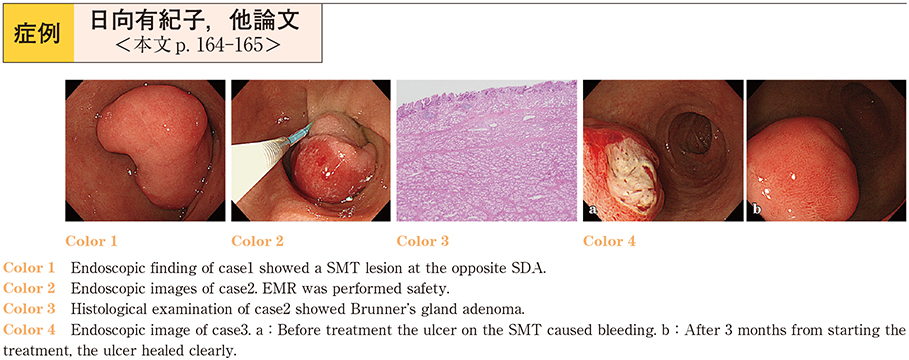
    2017Volume 91Issue 1 Pages 164-165
    Published: December 08, 2017
    Released on J-STAGE: December 21, 2017
    JOURNAL FREE ACCESS

    A 49-year-old man visited our hospital for investigation of duodenal SMT. Compared with endoscopic findings 2 years earlier, the lesion showed enlargement. LECS was performed. Pathological diagnosis was Brunner’s gland adenoma. A 72-year-old woman visited our hospital for investigation of anemia.
    An EGDS revealed SMT with erosion on the duodenum. We diagnosed the lesion was the cause of anemia. EMR was performed. Pathological diagnosis was Brunner’s gland adenoma. A 67-year-old-man visited our hospital for investigation of hematemesis. An EGDS revealed the ulcer on the known duodenal SMT. Because of the patient’s complications, we chose oral PPI administration was begun. The patient has been clear of recurrence since. Brunner’s gland adenoma and hyperplasia are basically benign lesions. When we decide their treatment policy, we must take into consideration the risk of malignancy and the patient’s ability to tolerate an operation.

    Download PDF (913K)
  • Eriko Nakagomi, Yusuke Nishikawa, Kazuhiro Fuchinoue, Tomoko Kobayashi ...
    2017Volume 91Issue 1 Pages 166-167
    Published: December 08, 2017
    Released on J-STAGE: December 21, 2017
    JOURNAL FREE ACCESS

    A 17-year-old female was admitted to our hospital with abdominal pain, vomiting and abdominal swelling. On the same day, we performed computed tomography (CT) and diagnosed it as small intestine ileus, so an ileus tube was inserted. As both small bowel X-Ray examination through the ileus tube and single balloon enteroscopy showed a stenosis of the upper jejunum, we took a biopsy specimen. Histological findings showed that the stenosis was an adenocarcinoma. We diagnosed it as primary small intestinal cancer and performed a surgical operation one month later. We will report on this rare case of juvenile small intestinal cancer, including some review of literature.

    Download PDF (972K)
  • Satoshi Kinoshita, Kaoru Takabayashi, Yuichiro Hirai, Yoko Kubosawa, Y ...
    2017Volume 91Issue 1 Pages 168-169
    Published: December 08, 2017
    Released on J-STAGE: December 21, 2017
    JOURNAL FREE ACCESS

    A 86-year-old woman was admitted to our hospital with hematochezia. Colonoscopy was performed, and showed multiple telangiectasia and congestive bleeding on sigmoid colon. She has a medical history of uteri cancer and treated with radiation therapy. Thus, she was diagnosed radiation sigmoiditis. Argon plasma coagulation (APC) was performed, but because of its wide range of lesion, sufficient therapeutic effect could not obtained. The steroid suppository and enema are commonly used to treat proctitis and sigmoiditis, and which is considered to be effective to improve edema and inflammation of intestinal tract. Because of it, we started the steroid enema. After 10 days of treatment, colonoscopy was performed, and it shows the mucosal healing of sigmoid colon. This case suggests that combination of APC and the steroid enema are useful to the treatment of chronic hemorrhagic radiation sigmoiditis.

    Download PDF (737K)
  • Ryuta Kitagawa, Osamu Kobayashi, Kazuki Ohta, Yuki Suyama, Shutaro Has ...
    2017Volume 91Issue 1 Pages 170-171
    Published: December 08, 2017
    Released on J-STAGE: December 21, 2017
    JOURNAL FREE ACCESS

    A 74-year-old man admitted to our hospital with diarrhea and alopecia. Gastroendoscopy and colonoscopy revealed diffuse reddish, inflammatory polyps and adjacent edematous mucosa in the stomach and colon. Histopathology of biopsy specimens showed cystic dilatation of mucosal glands and invasion by inflammatory cells, compatible with Cronkhite-Canada syndrome. Narrow band imaging identified eleven polyps in the colon showing NBI International Colorectal Endoscopic (NICE) Classification type 2, and histopathology of biopsy specimens from the polyps showed several adenoma cells. He was treated intravenously with prednisolone (PSL) at 50 mg/day, and his symptoms improved. PSL was gradually tapered and was continued orally at 10 mg/day. Six months after initiating steroid therapy, repeat colonoscopy revealed resolution of inflammatory polyposis and could not detect NICE Classification type 2 polyps.

    Download PDF (1387K)
  • Masao Kusano, Daisuke Komazawa, Hiromichi Ito, Masaki Tosa, Kaname Uno ...
    2017Volume 91Issue 1 Pages 172-173
    Published: December 08, 2017
    Released on J-STAGE: December 21, 2017
    JOURNAL FREE ACCESS

    A 56-year-old woman presented with abdominal pain after undergoing a barium study for gastric cancer screening 2 days prior. On day 3 after the examination, she visited an emergency room and a glycerin enema was performed as the abdominal radiography revealed stagnation of barium in the pelvic region. On day 7 after the examination, she was referred to our department because of lower abdominal pain and a sensation of retained feces. Abdominal radiography revealed a small amount of barium pooled in the pelvic region and splenic flexure without free air. Colonoscopy revealed a deep ulcer with barium stagnation. The barium was removed with forceps. After 1 month, the ulcer lesion was covered elevated mucosa with dilated and tortuous surface vessels. Histopathology revealed inflammatory granulation. Five months after the initial colonoscopy, the elevated lesion was improved slightly and was incidentally biopsied. Histopathological findings remained unchanged. Eleven months after the initial colonoscopy, the elevated lesion was replaced by an ulcer scar.

    Download PDF (787K)
  • Masao Kusano, Daisuke Komazawa, Hiromichi Ito, Masaki Tosa, Kaname Uno ...
    2017Volume 91Issue 1 Pages 174-175
    Published: December 08, 2017
    Released on J-STAGE: December 21, 2017
    JOURNAL FREE ACCESS

    A 40-year-old woman was referred to our department because of constipation and hematochezia about 3 days before menstruation. Colonoscopy revealed a slightly elevated lesion in the sigmoid colon. Histopathological examination revealed non-specific colitis. Three months later, a second colonoscopy during menstruation revealed an elevated lesion with an irregular surface and erythematous mucosa. Histopathological examination revealed ectopic glands in the muscle layer.
    Estrogen and progesterone-receptors, and CD10 stained positively on immunostaining. Therefore, we made a diagnosis of endometriosis of the sigmoid colon. Gonadotropin-releasing hormone agonist treatment was performed once a month for 6 months. Endoscopy revealed reduction of the elevated lesion with irregular surface and erythematous mucosa. Histopathological examination showed no evidence of endometriosis. The patient is still being followed-up regularly.

    Download PDF (905K)
  • Rikako Nakajima, Yuri Kumakura, Hiroyuki Ariga, Junya Kashimura, Norio ...
    2017Volume 91Issue 1 Pages 176-177
    Published: December 08, 2017
    Released on J-STAGE: December 21, 2017
    JOURNAL FREE ACCESS

    An 82-year-old woman with disturbance of consciousness was admitted to our hospital. Blood examination results showed renal failure, hyponatremia, and hypokalemia. Endocrine function was normal. Abdominal computed tomography images revealed a 53-mm tumor and intussusception in the recto-sigmoid. Contrast enema results revealed a concave pressure defect. Endoscopic examination revealed a continuous circumferential villous tumor located at 5 cm from the lower rectum, which could have intussuscepted due to peristalsis. We diagnosed EDS due to villous tumor of rectum and performed a Hartmann procedure ; the resected specimen showed a 140 × 110 mm circumferential villous tumor, and pathological investigation revealed adenocarcinoma with an adenoma component. EDS is a rare disorder characterized by a villous tumor leading to chronic secretory diarrhea which in turn leads hypokalemia, hyponatremia, and renal failure. Intussusception in adults is rare in comparison with children.

    Download PDF (837K)
  • Takashi Ogimi, Hideo Shimada, Takayuki Nishi, Takayuki Tajima, Hiroshi ...
    2017Volume 91Issue 1 Pages 178-179
    Published: December 08, 2017
    Released on J-STAGE: December 21, 2017
    JOURNAL FREE ACCESS

    A 58-year-old woman visited our hospital with the complaint of bleeding on defecation. Colorectal endoscopy showed a 10-mm submucosal tumor with a slight central depression. Histopathological examination of biopsy specimens led to the diagnosis of rectal neuroendocrine tumor (NET) . Thoracoabdominal computed tomography did not reveal lymph node or distant metastasis. We performed endoscopic submucosal dissection. Immunohistopathological examination of the resected tumor led to the diagnosis of synaptophysin-positive, chromogranin-negative, CD56-positive grade 2 NET with a positive resection margin. Therefore, we additionally performed laparoscopic-assisted low anterior resection and lymphadenectomy. No remaining tumor was found in the resected ESD scar. Metastasis was identified in one (No. 251) of the 35 dissected lymph nodes. Her postoperative course was good, and she was discharged on the 12th hospital day. We report a case of a small rectal carcinoid tumor with lymph node metastasis with a literature review.

    Download PDF (645K)
  • Hiroshi Takahashi, Masakatsu Fukuzawa, Hirohito Minami, Tadashi Ichimi ...
    2017Volume 91Issue 1 Pages 180-181
    Published: December 08, 2017
    Released on J-STAGE: December 21, 2017
    JOURNAL FREE ACCESS

    Colonic neuroendocrine cell carcinoma (NEC) is a rare colon cancer. Most NEC patients are diagnosed at an advanced stage and subsequently have a poor prognosis. We report our experience with early-stage NEC.
    A lower gastrointestinal endoscopy was performed on a 77-year-old man with a positive fecal occult blood test. A laterally spreading tumor (25 mm diameter) with nodules was observed in the lower rectum. A biopsy of the nodular region led to a NEC diagnosis after positive MNF-116, CD56, and CG-A immunostains ; the Ki-67 labeling index was approximately 70% by immunohistochemistry. Regional lymph node metastases or distant metastases were not found. The patient underwent a low anterior resection following computed tomographic cystography. The final pathological diagnosis was : rectal NEC, T1b (SM) , ly1, v1, pN0, pM0, and pathological stage I. We describe a rare case of early-stage NEC.

    Download PDF (646K)
  • Sho Takahashi, Hirofumi Fukushima, Kentaro Izumi, Yuzuru Tajima, Takas ...
    2017Volume 91Issue 1 Pages 182-183
    Published: December 08, 2017
    Released on J-STAGE: December 21, 2017
    JOURNAL FREE ACCESS

    An 84-year-old man was monitored after resection of descending colon cancer. During follow-up, colonoscopy showed a flat elevated lesion, 8 mm in size, in the lower rectum. The lesion was slightly reddish and had dilated vessels with branch architecture on the surface. Follow-up colonoscopy performed 10 months later showed that the lesion had progressed to the lateral side and increased to 15 mm. Dilated vessels with branch architecture were further emphasized by magnifying endoscopy with NBI. Biopsy specimens showed MALT lymphoma. Image findings and bone marrow biopsy were negative. We performed an ESD. Immunohistochemically, the lesion was confirmed to be MALT lymphoma. We reported a case of MALT lymphoma that could be observed at an early developmental stage by endoscopy.

    Download PDF (865K)
feedback
Top