Progress of Digestive Endoscopy
Online ISSN : 2187-4999
Print ISSN : 1348-9844
ISSN-L : 1348-9844
Volume 77, Issue 2
Displaying 1-50 of 57 articles from this issue
  • Yutaka Fumizono, Junko Fujisaki, Mizuka Imai, Shin-ichi Imada, Akiyosh ...
    2010 Volume 77 Issue 2 Pages 31-34
    Published: December 10, 2010
    Released on J-STAGE: July 25, 2013
    JOURNAL FREE ACCESS
    The incidence of Barrett′s cancer has been increasing recently, and there are case reports of its resection by ESD. Here, we report five further cases of Barrett′s cancer removed by ESD between March 2005 and December 2008.
    All cases were men mean age 60 years (range 50-74) . Endoscopic macroscopic type was 0-IIa in three patients, 0-IIc in two. All cases originated from SSBE and were well-differentiated adenocarcinomas, four mucosal and one submucosal. The latter required surgical treatment after ESD. Average tumor size was 21mm (11-38mm) . The procedure was as follows : marking by APC, submucosal injection with glyceol or hyaluronic acid, and dissection using an IT knife (R) . We recognized demarcation lines using conventional endoscopy in all cases ; one case had a positive horizontal margin because of cancer invasion under the squamous epithelium which was not diagnosed before ESD. There were no complications in these five cases.
    It is estimated that Barrett′s cancer will continue to increase. ESD can be appropriate for diagnosis and treatment of Barrett′s cancer, but difficulty in recognizing the demarcation line has been reported. In our cohort, we did experience a problem with preoperative evaluation missing cancer invasion under the squamous epithelium. [ESD : endoscopic submucosal dissection, SSBE : short segment Barrett epithelium]
    Download PDF (1130K)
  • Satoshi Hagiwara, Yasuhiro Onozato, Haruhisa Iizuka, Naondo Sohara, Hi ...
    2010 Volume 77 Issue 2 Pages 35-39
    Published: December 10, 2010
    Released on J-STAGE: July 25, 2013
    JOURNAL FREE ACCESS
    Sub-circumferental resection over 75% of the circumference by ESD in the body and angularis is not a risk factor for the occurrence of stricture. Prophylactic endoscopic balloon dilation was considered to be useful to avoid a stricture for the high risk group such as sub-circumferental resection over 90% in the prepylorus or cardia.
    Download PDF (1034K)
  • Makoto Nishimura, Kouichi Nonaka, Takashi Shono, Yasutoshi Ochiai, Osa ...
    2010 Volume 77 Issue 2 Pages 40-43
    Published: December 10, 2010
    Released on J-STAGE: July 25, 2013
    JOURNAL FREE ACCESS
    EUS-guided fine needle aspiration (EUS-FNA) has significantly prevailed in the Western countries, and recently, it has been gradually recognized among Japanese endoscopists. EUS-FNA is useful for not only pancreatic disease, but also mediastinal or abdominal lesions, and many authors have evaluated significantly high diagnostic value as well as safety. In this case series, we performed 61 cases of EUS-FNA, and 13 cases of idiopathic mediastinal or abdominal mass, which revealed 2 cases of sarcoidosis, 2 cases of schwannoma, 4 cases of malignant lymphoma, 1 case of bronchogenic cyst, 1 case of lung cancer, 1 case of cancer from unknown origin, 1 case of benign glanulamatous inflammation, and 1 case of adrenal adenoma. Based on our cases and previous article, EUS-FNA is safe, and a minimally invasive procedure with high diagnostic value.
    Download PDF (890K)
  • Shigetaka Yoshinaga, Hajime Takisawa, Satoru Nonaka, Taku Sakamoto, Te ...
    2010 Volume 77 Issue 2 Pages 44-48
    Published: December 10, 2010
    Released on J-STAGE: July 25, 2013
    JOURNAL FREE ACCESS
    Endoscopic ultrasound-guided fine needle aspiration (EUS-FNA) is useful for the diagnosis of gastrointestinal submucosal tumor and pancreatic tumor and the staging of lung cancers. In this study, we evaluated the effectiveness and safety of EUS-FNA in our institute.
    We performed EUS-FNA for 45 lesions from April 2008 to November 2009 and evaluated the clinical impact and the complication of EUS-FNA. We could achieve specimens from all lesions, and diagnose 41 lesions finally. Two complication occurred, but we could manage conservatively.
    EUS-FNA is a useful procedure that can be performed safely.
    Download PDF (738K)
  • Hiroyuki Fujii, Masami Inase, Maiko Iwata, Shinako Tochitani, Junko Sa ...
    2010 Volume 77 Issue 2 Pages 49-52
    Published: December 10, 2010
    Released on J-STAGE: July 25, 2013
    JOURNAL FREE ACCESS
    A 84-year-old man on hemodialysis for end-stage renal disease was admitted to our hospital with the chief complaint of anemia. Anti-parietal cell antibody was positive on blood test. On gastroendoscopy, three variable forms of elevated lesions were noted, and macro findings were compatible with autoimmune gastritis. Biopsy specimens revealed microscopic proliferation of atypical cells with round nuclei and clear cytoplasm in trabecular pattern. Immunohistochemical staining showed these tumor cells to be uniformly positive for chromogranin A and synaptophysin. Together with MIB1 (Ki67) labeling index result which was not low (>2%) , well differentiated neuroendocrine carcinoma was suspected. Computed tomography showed no signs of metastasis in the liver or extra-gastric lymph nodes. Total gastrectomy was performed. Three tumors were observed. 15mm diameter polyp on the great curvature of upper gastric body was reddish and irregular surface, another was a flat elevated 10mm diameter lesion on the anterial wall of upper gastric body with slight depression, the third was a 40mm diameter submucosal tumor like lesion with a reddish depression on the lesser curvature of upper gastric body. They were all positive for chromogranin A and synaptophysin. MIB1 index were all more than 2%.Thus, they were diagnosed to be differentiated neuroendocrine carcinomas as well. From findings compatible with autoimmune gastritis, and endocrine cell micronests observed on pathology, cancerization from well differentiated neuroendocrine tumor derived from antoimmune gastritis was considered. We also suspect that the laxation between the tumor and the muscle layer by peristalsis made the tumor pedunculated, and ischemic change due to the pressure from growing tumor made the mucosa depressive.
    Download PDF (805K)
  • Koichi Tokai, Hiroyuki Miyatani, Yuji Shindo, Takeshi Uehara, Takashi ...
    2010 Volume 77 Issue 2 Pages 54-55
    Published: December 10, 2010
    Released on J-STAGE: July 25, 2013
    JOURNAL FREE ACCESS
    Purpose : We have studied endoscopic findings and clinical courses of 13 cases of rectal carcinoid that were treated in our hospital from June 2007 to December 2009.
    Method : The resected tumor size was less than 5mm in 5 cases, 5~10mm in 7 cases, and 12mm in one case. The small tumors were hemispherical, Is polyp, or submucosal tumor in shape and white or yellow in color. The 12mm one showed a shape of Isp polyp.
    Results : As an initial therapy, EMR or ESD was performed in 1 or 12 cases, respectively.
    In 12 cases with less than 10mm tumors, the vertical margins were unclear in two cases and negative in other 10 cases. No recurrence has been observed so far in these 12 cases.
    In the case of 12mm tumor, the vertical margin was clearly positive after ESD, so additional rectal resection was performed. After two years, lymphadenopathy was revealed by CT examination and diagnosed as metastatic recurrence by FDG-PET.
    Discussion : We suggest that carcinoid tumors with a size of less than 10mm can be curably removed by endoscopic therapy. If the size is more than 10mm, however, we should consider a possibility of metastasis and FDG-PET is useful for the evaluation.
    Download PDF (749K)
  • Shingo Usui, Shoko Nakamura, Yoshimasa Saito, Yoshiyuki Yamagishi, Hir ...
    2010 Volume 77 Issue 2 Pages 56-57
    Published: December 10, 2010
    Released on J-STAGE: July 25, 2013
    JOURNAL FREE ACCESS
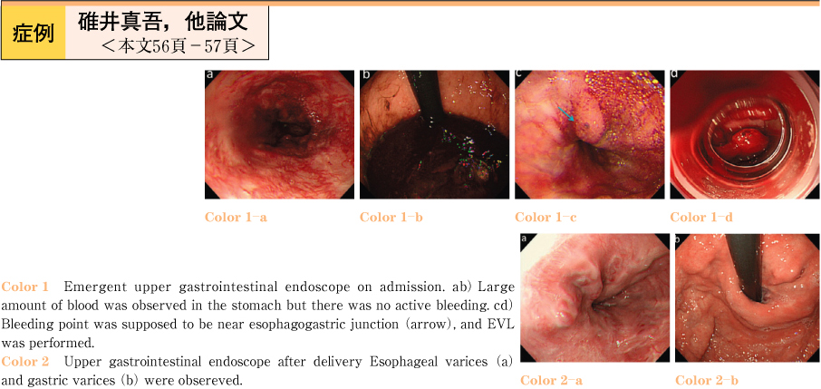
    A 34-years-old woman, who had received Kasai operation due to congenital bile duct atresia at the age of 0, was pointed out esophageal varices and liver cirrhosis at one year and a half before pregnancy. At 25 weeks pregnancy, the esophageal varices was ruptured and she received endoscopic variceal ligation after emergent admission. Although the variceal bleeding was well controlled, the fetus grew up slowly and its growth arrest was recognized by ultrasound examination at 29 weeks pregnancy. Caesarean section was performed and a boy of 842g was delivered. Genital bleeding, however, continued postoperatively because of poor uterine contraction, and a large amount of blood transfusion was necessary. Both of uterine arterial embolization was performed by interventional radiology and she was discharged on the postoperative 15th day. The newborn grew up and was also discharged on the 92nd day of his birth.
    Download PDF (736K)
  • Yasutoshi Ochiai, Koichi Nonaka, Osamu Togawa, Masamitsu Nakao, Makoto ...
    2010 Volume 77 Issue 2 Pages 58-59
    Published: December 10, 2010
    Released on J-STAGE: July 25, 2013
    JOURNAL FREE ACCESS
    In nearly future, the number of patients under antithrombotic therapy could be increasing. In this case report, we experienced a case of Intramural hematoma of the esophagus (IHE) under antithrombotic therapy. This is a 71-year old female with newly diagnosed as Cerebral aneurysm. She is admitted for scheduled coil embolization therapy. The patient had received antithrombotic therapy for one week before admission. After admission, the patient complained of severe epigastralgia,.and emergency enhanced CT scan showed huge IHE located from middle part of the esophagus to the upper area of the stomach. There was also pin-point visible bleeding found inside of the hematoma, by CT scan.
    At the 12 days after admission, endoscopic findings showed esophageal ulcer. The patient has managed conservatively without any symptom. The patient had discharged at 21 days after admission without severe complication. Our case suggests that especially the patient under anticoagulation therapy should be considered IHE and early diagnosed with appropriate modality.
    Download PDF (695K)
  • Nobuhiro Tanno, Kazuo Koyanagi, Satoshi Tabuchi, Hideyuki Tawara, Mako ...
    2010 Volume 77 Issue 2 Pages 60-61
    Published: December 10, 2010
    Released on J-STAGE: July 25, 2013
    JOURNAL FREE ACCESS
    A 73-year-old male patient underwent esophagectomy and reconstruction using a colon via antethoracic route for esophageal cancer in 1994. He had been followed up with an upper gastrointestinal endoscopy for five years after surgery. He presented to his family physician with fever and coughing. His doctor introduced him to the department of the pulmonary disease at our hospital because of the elevation of CEA and CYFRA. He received CT and PET. PET showed SUV uptake in colonic interposition. He received an upper gastrointestinal endoscopy and laterally spreading tumors were identified. These lesions were considered a good candidate for endoscopic resection. The lesions were safely and completely removed by ESD and EMR. Histological examination diagnosed the specimen as high grade tubular adenoma. The patient was hospitalized for 4 days after endoscopic treatment to confirm the absence of complications such as delayed perforation and bleeding, and was then discharged.
    Upper endoscopic screening is recommended after five years follow up of colonic interposition and lesions may be detected early and removed safely by endoscopy.
    Download PDF (674K)
  • Itsuko Hirayama, Nobutake Yamamichi, Chihiro Minatsuki, Kosuke Hirano, ...
    2010 Volume 77 Issue 2 Pages 62-63
    Published: December 10, 2010
    Released on J-STAGE: July 25, 2013
    JOURNAL FREE ACCESS
    A 73-year-old woman was hospitalized due to anemia. Upper gastrointestinal endoscopy was performed and a large gastric phytobezoar which located from gastric fundus to gastric body was found. The phytobezoar firmly adhered to the gastric wall. We chose endoscopic removal witch involved fragmenting the bezoar with Coca-cola water jet, and direct fracture with snares. We found bleeding from the surface of the resection plane of the phytobezoar, then perfomed biopsy. The biopsy sample contained a small number of atypical cells, so we made a diagnosis of the gastric carcinoma which became a core of gastric phytobezoar.
    Download PDF (998K)
  • Tomoko Umakoshi, Kaoru Domon, Yoshinori Igarashi, Naoki Hirano, Takafu ...
    2010 Volume 77 Issue 2 Pages 64-65
    Published: December 10, 2010
    Released on J-STAGE: July 25, 2013
    JOURNAL FREE ACCESS
    A patient is 72 years old woman who complained of epigastric pain. Upper GI endoscopy performed, it found an ulcer in lesser cuveture and a lage gastric calculus which was about 5cm in the diameter. The patient was admitted in our hospital because of removal of gastric calculus.
    Abdominal CT findings showed the stone was 40×26×45mm within air density at the upper body of stomach. After Upper GI X-ray examination, the large stone passed spontaneously because it was solved by sodium hydrogen carbonate. Upper GI endoscopy was performed and it found a healing ulcer and no stone in the stomach.
    This case is very rare because we observed the passing spontaneously of the large gastric stone by endoscopy and CT scan.
    Download PDF (725K)
  • Akiko Narita, Kazuhiro Watanabe, Kaoru Takagi, Satowa Seki, Yasuko And ...
    2010 Volume 77 Issue 2 Pages 66-67
    Published: December 10, 2010
    Released on J-STAGE: July 25, 2013
    JOURNAL FREE ACCESS
    An 85-year-old man presented with acute gastric dilation of upper abdominal pain associated with retching after took too much dinner. Abdominal CT scan showed an“ up side down stomach” as a result of mesenteroaxial volvulus of the stomach. It was treated unsuccessfully with decompression of a nasogastric tube and then, we performed endoscopic reduction of volvulus using esophagogastroduodenoscopy (EGD) and also treated the oozing bleeding ulcer by hemoclips. After 11days on admission, he needed second clipping treatment because of bleeding. But, informed consent couldn't be obtained for operation. Then we transfused the 14units of blood transfusion, and administered intravenous hyperalimentation for 14days without diet. After 26days, EGD showed improvement in ischemic mucosal change at the level of healing stage. In this case, it was possible to see the patient of gastric volvulus with ischemic mucosal change without necrosis after both endoscopic reduction of volvulus and clipping for hemorrhagic ulcer.
    Download PDF (844K)
  • Takaaki Kaneko, Hiroyuki Fukuda, Hidehito Sibasaki, Koutaro Yoshimura, ...
    2010 Volume 77 Issue 2 Pages 68-69
    Published: December 10, 2010
    Released on J-STAGE: July 25, 2013
    JOURNAL FREE ACCESS
    A 63-year-old man, undergoing a distal gastric resection with Billroth II anastomosis for duodenal ulcer 20 years ago, was admitted to our hospital with the complaint of tarry stool. Initial treatments included intravenous fluid resuscitation, proton pump inhibitor, and nothing by mouth. Hemoglobin was 6.8g/dl, so patient received a transfusion of packed red blood cells. Emergent upper gastrointestinal endoscopy showed two marginal ulcers at the saddle area of the jejuna site, one was bleeding with exposed vessel, which was treated successfully by endoscopic hemostasis with hemoclips. The next day, the second endoscopy showed the same ulcer rebleeding, so hemoclips were applied again. On the third day after the hospital admission, the third endoscopy showed the other marginal ulcer bleeding with exposed vessel. So the third endoscopic hemostasis procedure with hemoclips was performed on the visible vessels at the base of the other ulcer to stop the bleeding. On the 10th day after the hospital admission he started feeding himself, discharged on the 17th day and received regular follow-up at the outpatient clinical department. Each value of blood gastorin, anti-Helicobacter pylori IgG antibody was in normal-range. CT angiography showed there was no large blood vessel near the marginal ulcer applied hemoclips. In this paper, we reported this difficult to be treated case and review the literature.
    Download PDF (712K)
  • Masaru Kimata, Hideki Ushiku, Takahisa Yoshikawa, Hisaaki Terauchi, Ka ...
    2010 Volume 77 Issue 2 Pages 70-71
    Published: December 10, 2010
    Released on J-STAGE: July 25, 2013
    JOURNAL FREE ACCESS
    Percutaneous endoscopic gastrostomy (PEG) is popular technique to adult. But it is not so popular for babies and little children because of some difficulties and hazards with PEG. We took CT images after some air filled in a stomach. OsiriX Imaging Software built 3D-CT images. The images were very useful in puncture location and depth. A thin fiberscope did not need general anesthesia. PEG was safely succeeded to one-year girl with hydrocephalus due to bacterial meningitis using thin fiberscope and 3D-CT.
    Download PDF (754K)
  • Yoshiko Kishimoto, Takashi Kawai, Mari Fukuzawa, Sakiko Tsukamoto, Asu ...
    2010 Volume 77 Issue 2 Pages 72-73
    Published: December 10, 2010
    Released on J-STAGE: July 25, 2013
    JOURNAL FREE ACCESS
    The phlegmonous gastritis is a non-peculiarity suppuration-related Inflammation disease which spread led by the mucous membrane lower layer and one of the acute stomach symptoms. It is recognized that I can expect healing by medical treatment with appropriate antimicrobial recently.
    However, as for the case having the underlying disease, early check up, early treatment are important so that there is the death report. I merge phlegmonous gastritis during treatment of MDS this time and report it because I experienced one example that retrograded by conservative medical treatment.
    Download PDF (850K)
  • Yukako Kato, Masaru Nakano, Yasuhisa Ishii, Karin Tanaka, Hiroshi Seri ...
    2010 Volume 77 Issue 2 Pages 74-75
    Published: December 10, 2010
    Released on J-STAGE: July 25, 2013
    JOURNAL FREE ACCESS
    A 62-year-old woman came to our hospital with complaint of epigastralgia for one month. Upper gastrointestinal endoscopy showed scattered nodular lesions 5mm in size in the fornix. Biopsy specimens obtained from the nodular lesions revealed non-caseating granuloma with infiltration of inflammatory cells. One month later, endoscopic examination showed multiple yellowish discolored lesions in the region of greater curvature of the body, and biopsy specimens obtained from the discolored lesion also revealed non-caseating granuloma, H. pylori urea breath test was negative, but serum anti-HP antibody was positive. No definite etiologic factors such as systemic sarcoidosis, Crohn's disease, tuberculosis could be detected. Several reports suggested close relationship between idiopathic granulomatous gastritis and H. pylori, whereas it is not clear in our case. Idiopathic granulomatous gastritis has been rarely reported in Japan. Further clinical investigation of the cases of idiopathic granulomatous gastritis is necessary for clarifying the pathogenesis of the disease.
    Download PDF (869K)
  • Kenichi Takeda, Daisuke Kikuchi, Toshiro Iizuka, Shu Hoteya, Toshifumi ...
    2010 Volume 77 Issue 2 Pages 76-77
    Published: December 10, 2010
    Released on J-STAGE: July 25, 2013
    JOURNAL FREE ACCESS
    In the case of gastric cancers, it is essential to determine the extent and depth of the tumor accurately for deciding on courses of treatment. We have sometimes experienced the cases that pre-therapeutic diagnosis of those factors were difficult. In this report, we describe a case of gastric linitis plastica, which was diagnosed based on the ESD and additional surgical specimens.
    The patient was 60 years old man. A depressed lesion was pointed out at the greater curvature of middle gastric body. Biopsy revealed pooly differentiated and signet ring cell adenocarcinoma. Using magnification and EUS, this lesion was diagnosed as intramucosal cancer and 20mm in size. After informed consent, ESD was performed and the lesion was resected in en bloc fashion. However pathological examination showed incomplete resection of margin positive in endoscopic specimen and isolated cancer cell remained in the muscle layer in surgical resected specimen.
    Finally, this case was diagnosed of gastric linitis plastica based on the pathological examination.
    Download PDF (815K)
  • Maho Iwamoto, Akitake Uno, Seiichi Ishikawa, Kouju Yamada, Yasuaki Kos ...
    2010 Volume 77 Issue 2 Pages 78-79
    Published: December 10, 2010
    Released on J-STAGE: July 25, 2013
    JOURNAL FREE ACCESS
    A 65-year-old woman was going to hospital regularly by rheumatoid arthritis.
    She was reffered for upper gastrointestinal endoscopy (GIE) because of the anemia. First GIE showed a deformity and erosion on the pyloric ring, and pathlogical examination was suspicion of adenocarcinoma as a result of the biopsy.
    So after one month, she underwent 2nd GIE. 2nd GIE showed 2type tumor by the retroflexion method into the duodenal bulb and the biopsy indicated adenocarcinoma.
    At first we diagnosed duodenal cancer, but the lesion was finally shown to be gastric cancer that had prolapsed into the duodenal bulb by distal gastrectomy.
    Result of pathological examination were as follows : moderately differentiated tubular adenocarcinoma, T2 (MP) , 1y1,v0,n (+) .
    We reported a case of advanced gastric cancer prolapsed into the duodenal bulb. So we reviewed about our experience from the literature.
    Download PDF (687K)
  • Naoko Yagi, Yuri Nakamura, Tsutomu Iida, Masataka Nishi, Kenji Nukaga, ...
    2010 Volume 77 Issue 2 Pages 80-81
    Published: December 10, 2010
    Released on J-STAGE: July 25, 2013
    JOURNAL FREE ACCESS
    We report the case of gastric small cell cancer with liver metastasis and lymph node metastasis. The gastric small cell cancer is considered to be poor convalescence and short MST with the special organization type of the gastric cancer. The patient is 81-year-old male and visited with chief complain tarry stool. We diagnosed gastric small cell cancer by the endoscopic findings and histopathological findings of gastric biopsy specimens. Because this case did not have operation adaptation, we performed chemotherapy independent treatment of TS1/CDDP. The improvement and the reduction of the liver metastasis tumors and gastric lesion were recognizes after 3 cycle chemotherapy, but after 8 cycle chemotherapy we recognized the increase of the liver metastasis tumors, However, it passes in 11 months, this patient lives, so we are going to treat by different chemotherapy.
    Download PDF (1024K)
  • Shigeru Matsui, Shigeru Harasawa, Hideaki Sato
    2010 Volume 77 Issue 2 Pages 82-83
    Published: December 10, 2010
    Released on J-STAGE: July 25, 2013
    JOURNAL FREE ACCESS
    A 66-year-old male was diagnosed with a gastric gastrointestinal stromal tumor (GIST) accompanied by peritoneal dissemination and treated with 400mg/day of imatinib. On month after the initiation of administration, both the primary gastric lesion and intraperitoneal mass decreased in size, showing partial remission. Seven weeks after the initiation of administration, drug-induced interstitial pneumonia developed, but improved after steroid pulse therapy. Extensive mediastinal emphysema developed 6 days after the initiation of steroid therapy, but improved after conservative treatment. Subsequently, he died of bacterial pneumonia. During imatinib administration, caution is necessary regarding the possible development of interstitial pneumonia.
    Download PDF (775K)
  • Hirotaka Ishikawa, Yukishige Okamura, Seiichiro Fukuhara, Ken Teramoto ...
    2010 Volume 77 Issue 2 Pages 84-85
    Published: December 10, 2010
    Released on J-STAGE: July 25, 2013
    JOURNAL FREE ACCESS
    We report a case of spontaneous regression of gastric carcinoid tumors associated with gastritis type A.
    The patient was 48-year old man. Endoscopic examination showed many small protruding lesions on the gastric body. Biopsy diagnosed it as a carcinoid tumors.
    His serum gastrin level was remarkably high (≧3000pg/ml) and gastric parietal cell autoantibody was positive.
    These findings suggested that the multiple gastric carcinoids in this patient were associated with gastritis type A.
    We classified the tumors into Type I according to Rindi's report and considered that these tumors were harmless and that there was no indication for major surgery such as total gastrectomy.
    Endoscopically,the carcinoid tumors were shown to be regressed and the level of serum gastrin was decreased during follow-up treatment.
    These findings suggest that gastric carcinoid tumors associated with gastritis type A do not need to be removed immediately.
    Download PDF (656K)
  • Tomonari Kinoshita, Koji Fujita, Tadayuki Sakuragawa, Keiichi Ishikawa ...
    2010 Volume 77 Issue 2 Pages 86-87
    Published: December 10, 2010
    Released on J-STAGE: July 25, 2013
    JOURNAL FREE ACCESS
    A 65-year-old-female was admitted due to chest and abdominal CT finding multiple lung and liver metastases. Upper gastrointestinal scopy′s findings showed multiple maculate lesions and the bull'-eye appearance with blackish ulcer base in the stomach and pathologically diagnosed as metastases of malignant melanoma. Their primary lesion was unidentified, but their findings are typical as malignant melanoma with gastric metastases.
    Download PDF (889K)
  • Kosei Hamano, Taro Watabe, Hidenari Inoue, Keiichiro Nakajyo, Yudai Yo ...
    2010 Volume 77 Issue 2 Pages 88-89
    Published: December 10, 2010
    Released on J-STAGE: July 25, 2013
    JOURNAL FREE ACCESS
    A 66-year-old man with alcoholic cirrhosis was admitted to our hospital with fatigue and weakness for the last few days. He had recent on set of melena with severe anemia. Emergent upper gastrointestinal endoscopy revealed dark-red colored tense varices in the descending portion of duodenum. Multidimensional computed tomography (MDCT) demonstrated the duodenal varices (DV) supplied by the posterior superior pancreaticoduodenal vein (PSPDV) and drained into right testicular vein. We performed balloon-occluded retrograde transvenous obliteration (BRTO) because the ruptured DV were the cause of melena. A balloon catheter was inserted via the right femoral vein and wedged into the rt. testicular vein. Digital subtraction angiography showed DV supplied by the PSPDV and DV collaterals. The collateral veins were embolized with microcoils. The sclerosing agent, 5% solution of ethanolamine oleate with iopamidol (EOI), was continuously infused into DV under the ballon infration until DV were filled with screlosant completely. No major procedure-related complications occurred. Post-BRTO endoscopy and abdominal CT showed successful obliteration of DV. In conclusion, we report a case of DV successfully treated with BRTO. There is no established treatment of DV. Further studies are needed to clarify appropriate treatment for the eradication of DV.
    Download PDF (792K)
  • Hiroto Ishii, Shintaro Tsukinaga, Tetsuyoshi Iwasaki, Shunichi Odawara ...
    2010 Volume 77 Issue 2 Pages 90-91
    Published: December 10, 2010
    Released on J-STAGE: July 25, 2013
    JOURNAL FREE ACCESS
    A 71-year-old man was admitted to our hospital for repetitive vomiting. Only a small intraductal papillary-mucinous neoplasm (IPMN, 28mm in diameter) in the body of pancreas was seen by several practical examinations, however no cause of intestinal obstruction was found. Since he suffered from vomit and nausea for a long time, laparotomy was done. During the laparotomy, several white nodules were found around pancreas, and the nodules were proved to be adenocarcinoma. Therefore, we diagnosed the duodenal obstruction due to peritoneal dissemination of intraductal papillary-mucinous carcinoma (IPMC). Then, gastrojejunostomy was performed. This is a first case of peritoneal dissemination of IPMC without apparent findings of pancreatic invasion.
    Download PDF (773K)
  • Midori Nishio, Ryu Nishiyama, Yuichi Akai, Takeshi Otani, Naoto Kunoki ...
    2010 Volume 77 Issue 2 Pages 92-93
    Published: December 10, 2010
    Released on J-STAGE: July 25, 2013
    JOURNAL FREE ACCESS
    We experienced a rare case that foreign body discovered due to duodenal bleeding with duodenal ulcer, and we successed in picking it up by using gastrointestinal endoscopy. A healthy 50-year-old man had symptoms of tarry stool and feeling dizzy. He was suspected gastrointestinal tract bleeding. So he was introduced to our hospital immediately. On arrival, he was anemic (Hb 9.8g/dl) . We found a duodenal ulcer on second potion by gastrofiberscopy. At the same time, we found a foreign body with penetration through the duodenum at of the opposite wall of the ulcer. We picked up the foreign body by using a forceps. The foreign body was a 25mm long-shaped metallic needle. We treated by proton pomp inhibiter. There were no complications through his duration of hospitalized stays. He traced good passage and discharged after 6 days. By the way, we asked him detailed questions about his clinical history, especially about swallowing foreign bodies, he didn't remember those episodes. So the cause of foreign body ingestion was unidentified.
    Download PDF (683K)
  • Mari Arai, Toshiyuki Tahara, Yasushi Tamabuchi, Kouji Akutsu, Kouhei T ...
    2010 Volume 77 Issue 2 Pages 94-95
    Published: December 10, 2010
    Released on J-STAGE: July 25, 2013
    JOURNAL FREE ACCESS
    We report 2 patients with a diagnosis of duodenal follicular lymphoma. Both patients were underwent esophagogastroduodenoscopy with no presenting symptom, and detected multiple whitish granular lesions in the second portion of the duodenum. They are diagnosed histologically by the biopsy specimen from the lesions. Patients were staged according to the Lugano staging system for gastrointestinal lymphomas. One patient had stage II2 disease and she underwent 3 course of chemotherapy including the administration of rituximab (RCHOP). She achieved complete response and has been found to have no evidence of disease. The other patient had stageIdisease and has been clinically followed. She are alive without disease progression.
    Download PDF (909K)
  • Yutaro Koyama, Takeshi Sugaya, Keiichi Tominaga, Mitsuru Nagata, Naoya ...
    2010 Volume 77 Issue 2 Pages 96-97
    Published: December 10, 2010
    Released on J-STAGE: July 25, 2013
    JOURNAL FREE ACCESS
    A 45-year-old man was admitted to our hospital with the symptom of epigastric pain. Laboratory data on admission was unremarkable.
    Esophagogastroduodenoscopy showed ulcer, which had exposing vessel at the anal side of papilla Vater. In spite of the medical therapy by fasting and prescription of proton pump inhibitor (PPI) for 6 days, epigastric pain persisted and duodenal ulcer did not heal. Abdominal helical dynamic CT showed nodular hypervascular lesion in the pancreatic head, and early filling of portal vein in early phase. Abdominal arterial angiography showed the similar finding to that of dynamic CT : a racemose intrapancreatic vascular network, and early filling of portal vein. Based on the result of CT and angiography, we made a diagnosis of pancreatic arteriovenous malformation (AVM), and subsequently pylorus-preserving pancreatoduodenectomy (PpPD) was done after arterial embolization. Macroscopic findings of resected specimen showed ulcer at the anal and pancreatic side of papilla Vater, and erosion around the ulcer. Pathology of specimen showed venous vasodilation and rupture of the elastic fibers, both of which findings were consistent with pancreatic AVM. Pancreatic AVM is a rare entity and the duodenal ulcer at the descending part accompanied by pancreatic AVM is much less ; there has been only 25 reports in Japan. Duodenal involvement in pancreatic AVM often develops ulcer and bleeding. Therefore, these patients usually complain of epigastric pain and melena. This case report indicated that pancreatic AVM is one of the differential diagnosis in patients with intractable duodenal ulcer.
    Download PDF (802K)
  • Aya Nishida, Tsunao Imamura, Tetsuo Tamura, Yuko Koizumi, Rikako Koyam ...
    2010 Volume 77 Issue 2 Pages 98-99
    Published: December 10, 2010
    Released on J-STAGE: July 25, 2013
    JOURNAL FREE ACCESS
    Follicular lymphomas occasionally arise in the extra-nodal organs and frequently found in the duodenum. We report a case of follicular lymphoma in the duodenal papilla which is followed up with no treatment about 72 months. In February 2004, a 52-year old female with anemia was diagnosed with limphoproliferative disease by upper endoscopy and biopsy. Endoscopy revealed multiple small polyps in the duodenal papilla. After eradication therapy of Helicobacter pylori, she was followed up, and she showed no remarkable endoscopic change. In July 2006, she re-diagnosed with follicular lymphoma by under endoscopic biopsy. Immunohistochemically, the tumor cells were positive for CD20, CD79, CD10, Bcl-2 and Bcl-6, and were negative for CD3 and CD5. MIB-1 positive cells were 10-20%. After the diagnosis, she followed up with no treatment about 72 months because she did not want treatment such as chemotherapy, surgical resection and radiation, and because she showed no remarkable progressive endoscopic change. It is suggested that following up with no treatment is might able to the choice of treatment of follicular lymphoma in the duodenal papilla.
    Download PDF (654K)
  • Yasuhiro Onozato, Haruhisa Iizuka, Naondo Sohara, Satoshi Hagiwara, Ri ...
    2010 Volume 77 Issue 2 Pages 100-101
    Published: December 10, 2010
    Released on J-STAGE: July 25, 2013
    JOURNAL FREE ACCESS
    A 70-year-old female underwent esophagogastroduodeno scopy as part of a routine medical evaluation due to symptoms of epigastric discomfort, which thus revealed a small elevated lesion in the duodenum opposite the duodenal papilla. The diagnosis based on biopsy specimens was group 3, tubular adenoma. Conventional endoscopy using white-light revealed a whitish small elevated lesion. Magnifying endoscopy revealed an irregular villous structure with a white rim. Magnifying endoscopy with narrow-band imaging showed meandering irregular vessels demonstrating an unequal caliber in their villous structures. Because early duodenal cancer was suspected based on the endoscopic findings, an endoscopic mucosal dissection (ESD) was selected for both diagnostic and treatment purposes. The lesion was completely resected by ESD and was sutured by means of endoscopic clipping. The resected specimen measured 7×5×2 mm/11×8mm in size. A histopathological examination revealed well differentiated intra-mucosal adenocarcinoma without vascular invasion. Irregular ductal structures with vascular-rich, atypical nuclei were observed. The atypical nuclei showed positive staining for p-53 and MIB-1 immunohistochemistry. The lesion was complete resected and there were no complications.
    Download PDF (761K)
  • Masao Kusano, Masaki Tosa, Norihiro Shimada, Yamagiwa Tetsuya, Naohiro ...
    2010 Volume 77 Issue 2 Pages 102-103
    Published: December 10, 2010
    Released on J-STAGE: July 25, 2013
    JOURNAL FREE ACCESS
    A 76-year-old female was diagnosed as having adenomas of main papilla, minor papilla and third portion of the duodenum. She was received subtotal colectomy for familial adenomatous polyposis at 41-year-old. Endoscopic retrograde cholangiopancreatogrphy showed stenosis of lower common bile duct and pancreas divism. At first, we resected adenoma of major papilla and inserted plastic tube stent in the common bile duct. Next week, we resected adenomas of minor papilla and third portion of the duodenum. We tried to insert a plastic tube stent in Santorini duct for avoid pancreatitis but we could not. Mild pancreatitis was occurred but improved conservative therapy. Microscopic finding of resected specimens showed moderately dysplastic adenoma, respectively. We experienced rare case of endoscopic resection of adenomas of major and minor papilla in a patient with familial polyposis and pancreas divism.
    Download PDF (839K)
  • Tetsuhiko Saito, Takayoshi Suzuki, Kenichi Watanabe, Masashi Matsushim ...
    2010 Volume 77 Issue 2 Pages 104-105
    Published: December 10, 2010
    Released on J-STAGE: July 25, 2013
    JOURNAL FREE ACCESS
    A-38-year-old man was seen at our hospital, because of abdominal pain. Computed tomography (CT) showed a lesion with thickening of the intestinal wall in the pelvic cavity. Double balloon endoscopy (DBE) revealed that there were diffuse erosion with blood clots at the stenotic lesion. Since abdominal pain recurred whenever he began oral intake, laparoscopy assisted surgery was performed. It was diagnosed as stenotic type ischemic enteritis by the pathological findings and his clinical course.
    Download PDF (1018K)
  • Yasushi Inoue, Kazuyuki Narimatsu, Shingo Sato, Kenji Hayasaka, Hiroka ...
    2010 Volume 77 Issue 2 Pages 106-107
    Published: December 10, 2010
    Released on J-STAGE: July 25, 2013
    JOURNAL FREE ACCESS
    Schonlein-Henoch purpura (SHP) is a systemic microvascular inflammatory disease. It has peak at 4-7 years old, and adult onset cases are about 5% of all. Sometimes adult onset cases have severe courses such as intestinal perforation. In almost 100% of cases skin eruption occurs, but about 15% of cases gastrointestinal symptoms precede skin eruption. Local inflammation in gastrointestinal region reveals atypical gastrointestinal manifestations, irregular-shaped ulcer, edematous mucosa. Although it has been reported that gastrointestinal findings can be seen most commonly in the second part of duodenum, small intestinal endoscopy and capsule endoscopy enable us to observe all sites of the small intestine. The findings in small intestine in HSP cases are useful to understand the pathological features of vascular disease.
    Download PDF (648K)
  • Kozue Kido, Junichi Iwamoto, Shoichiro Yara, Masashi Murakami, Masanor ...
    2010 Volume 77 Issue 2 Pages 108-109
    Published: December 10, 2010
    Released on J-STAGE: July 25, 2013
    JOURNAL FREE ACCESS
    A 73-year-old male, who had been diagnosed with rheumatoid arthritis and taking NSAIDs for a long time. He was pointed out the stenotic lesion of his ileum in a previous hospital. A double balloon enteroscope in our hospital showed the stenosis of his terminal ileum. It was difficult to go through the stenosis. Perforation of the terminal ileum was seen after the second balloon dilatation with a double balloon enteroscope. Then, resection of the small intestine was performed. The resected specimen showed the stenosis of the terminal ileum with thickness of the intestinal wall and perforation was seen in the most stenosed terminal ileum. UL-I or UL-II ulcer was seen in oral side of the stenosis. Histopathological findings showed marked fibrosis of the submucosal layer and the vascularity in the mucosal layer. These histopathological findings maked us consider the stenosis of the terminal ileum caused by ischemic enteritis rather than NSAIDs-induced enteritis.
    In conclusion, this case may indicate the risk of endoscopic dilatation therapy of stenotic lesion of the terminal ileum in case of a prolonged NSAIDs user.
    Download PDF (813K)
  • Takeshi Uehara, Satohiro Matsumoto, Hiroyuki Miyatani, Yuji Shindo, Sh ...
    2010 Volume 77 Issue 2 Pages 110-111
    Published: December 10, 2010
    Released on J-STAGE: July 25, 2013
    JOURNAL FREE ACCESS
    A 78 years old man with abdominal pain was referred to our hospital for suspicion of dissecting abdominal aneurysm. Abdominal CT and ultrasonography revealed not only abdominal aneurysm but also a small intestinal tumor with cystic component in contact with the rectum. His abdominal pain was caused by intermittent intestinal obstruction by the tumor. We suspected that the tumor adhered to the rectum by CT and Echoendoscopy. Finally, he was diagnosed as having GIST of the small intestine by the EUS-FNA specimen which was obtained via the rectal wall. We performed a surgical resection.
    In case of a tumor adhered to the intestinal tract wall, EUS-FNA is an effective examination for histological diagnosis.
    Download PDF (851K)
  • Kazunori Hijikata, Yoshinori Hujitsuka, Yoshinori Igarashi, Shunsuke K ...
    2010 Volume 77 Issue 2 Pages 112-113
    Published: December 10, 2010
    Released on J-STAGE: July 25, 2013
    JOURNAL FREE ACCESS
    A case of woman was 69 years old. She had rescieved for dental treatment in November, 2009 and took NSAIDs and antibiotic drug. After one day, she occured the bloodystool. Then she was admitted to another hospital and complained of anemia.
    Upper GI endoscopy and colonoscopy was performed. However it could not find the bleeding lesion. After examination, the patient transferred our hospital.
    Then capsule enteroscopy was perfomed and could not find the lesion.
    After capsule enteroscopy, we performed a single balloon enteroscopy for a transanal route. The healing ulcer diagnosed at about 30cm oral from Bauchin's valve.
    When the capsule enteroscopy failed to diagnosed, the singl balloon enteroscopy was very useful of diagnosed for the intestinal ulcer.
    Download PDF (565K)
  • Makomo Makazu, Satoshi Kinoshita, Chika Taniguchi, Keita Iwaguro, Yosh ...
    2010 Volume 77 Issue 2 Pages 114-115
    Published: December 10, 2010
    Released on J-STAGE: July 25, 2013
    JOURNAL FREE ACCESS
    Nonspecific multiple ulcers the small intestine (NMUS) is a clinical entity proposed in Japan with characteristic clinical features different from other inflammatory bowel disease. Treatments with conventional drugs such as corticosteroids, aminosalicylate or immunosuppressant are known to be ineffective for patients with NMUS. We report a case who has been managed successfully with long-term home elemental enteral nutrition. A 60-year-old woman who underwent ileocecal resection due to NMUS when she was 35 years old was admitted to our hospital because of refractory anemia and hypoproteinemia in 2007. She didn't have the history of hematomesis nor melena, but fecal occult blood tests were always positive and the anemia was progressive requiring multiple blood transfusions. Then she was introduced home elemental enteral nutrition in 2007. The small-bore nasogastric tube and commercially available elemental diet were used. Total of 900ml (1kcal/ml) of elemental diet solutions were infused continuously during the night-time using an infusion pump at a speed of 100ml/hr supplementing her day-time oral diet. After the introduction of nutritional therapy, she has been maintained in good conditions for 3 years. Home elemental enteral nutrition may be a useful treatment as maintenance therapy for patients with NMUS.
    Download PDF (809K)
  • Akari Isono, Tsuguru Shirai, Yuji Mishina, Tadahisa Ebato, Koichiro Ab ...
    2010 Volume 77 Issue 2 Pages 116-117
    Published: December 10, 2010
    Released on J-STAGE: July 25, 2013
    JOURNAL FREE ACCESS
    Protein-losing enteropathy is relatively rare in Japan. Here we report on a case of protein-losing enteropathy associated with malignant lymphoma.
    A male aged over 50 years visited our hospital because of body weight loss and edema on the lower extremities. The serum albumin level was less than 2.0 g/dl, but no abnormality was found in the urinalysis. Capsule endoscopy and single-balloon enteroscopy showed multiple mucosal injury located at the small intestine, and the pathological examination of the ulcer proved dilatation of the lymphatic vessels. The 99m-Tc sintigraphy revealed protein losing from the small intestine. The lymph nodes at the cervical region became enlarged at the same time, and the diagnosis of peripheral-type T cell lymphoma was made by biopsied specimen. After the patient underwent standard chemotherapy with cyclophosphamide, vincristine, doxorubicin, and prednisolone, protein loss from the intestine decreased as well as improvement of leg edema.
    Download PDF (807K)
  • Yusuke Takahashi, Yubu Matue, Yoshimasa Miwa, Yasutake Hirayama, Daisu ...
    2010 Volume 77 Issue 2 Pages 118-119
    Published: December 10, 2010
    Released on J-STAGE: July 25, 2013
    JOURNAL FREE ACCESS
    A 29-years old woman, who had a history of mental retardation and abnormal habit of eating tissue papers, was admitted to our hospital because of abdominal pain and vomiting. Abdominal X-ray and CT scan demonstrated much intestinal gas, and a colonoscopy revealed obstruction caused with tissue papers and large ulcers in the sigmoid colon. Therefore, we removed the tissue papers endoscopically using a grasper. After endoscopic treatment, her symptoms were immediately improved and a few longitudinal ulcer scars were seen, three month later. The endoscopic extraction was safe and effective in the colonic obstruction with tissue papers.
    Download PDF (743K)
  • Hiroko Otsuka, Teruhiko Sakamoto, Rieko Yamada, Chihiro Tsunoda, Megum ...
    2010 Volume 77 Issue 2 Pages 120-121
    Published: December 10, 2010
    Released on J-STAGE: July 25, 2013
    JOURNAL FREE ACCESS
    We report our three cases of intestinal tuberculosis in which none showed respiratory symptoms. Case 1 : A male of 60's. Lower abdominal pain was persistent for a month and fecal occult blood tests were positive. Type 2 colonic cancer of the cecum was doubted by colonoscopy (CS) in the local doctor and he was introduced to our hospital. CS revealed ulceration and stenosis at the ileocecal area. Biopsy of the ulceration showed granulomatous lesion and lead to diagnosis of tuberculosis. Chest X-ray showed lung lesions as well. Case 2 : A male of 50's. Because persistent diarrhea for three weeks later evolves to right lower abdominal pain and vomiting, he was admitted. CS showed ulceration of the terminal ileum and ileocecal valve. Biopsy of the ulceration showed granulomatous lesion and lead to diagnosis. Sputum was positive for tubercle bacilli and lead to diagnosis of lung and intestinal tuberculosis. Case 3 : A female of 30's. Abdominal pain and diarrhea repeated for a year and CS was performed. The intestine from terminal ileum to sigmoid colon showed skipping lesions of circular ulcers. Ileocecal biopsy showed Tb-PCR positive and lung lesion were suspected on chest X-ray. There were all of three cases ulcerative lesions on the ileocecal area, and tubercle bacilli were able to be detected.
    Download PDF (848K)
  • Minako Matsubayashi, Kyoya Sakimura, Dai Sakamoto, Nobuaki Ikezawa, To ...
    2010 Volume 77 Issue 2 Pages 122-123
    Published: December 10, 2010
    Released on J-STAGE: July 25, 2013
    JOURNAL FREE ACCESS
    Spirochetosis mainly includes Spirochaetaceae species Treponema groups, Borrelia groups, and Leptospiraceae species Leptospira groups. Besides, Brachyspira groups is one of the rare human intestinal spirochetosis, which is defined uncertainly. Intestinal spirochetosis usualy accidentally diagnosed by biopsy, because it shows no remarkable colonoscopy's findings and specific symptoms. The diagnosis requires the clot of bacteria and pathological findings such as spiculating appearance in the intestinal epithelium. The treatment with consensus has not been established yet, and some cases dose not need any therapy. We had experienced seven cases of inteitinal spirochetosis from January 2007 to December 2008. As for these cases, the clinical manifestation, colonoscopy's findings, and pathological features are discussed here.
    Download PDF (760K)
  • Hitomi Tajima, Daigo Onodera, Toshiyuki Sakai, Miyako Matsumoto, Makot ...
    2010 Volume 77 Issue 2 Pages 124-125
    Published: December 10, 2010
    Released on J-STAGE: July 25, 2013
    JOURNAL FREE ACCESS
    A 47-year-old woman came to our hospital with watery diarrhea and hematochezia. Colonoscopy showed redness and erosions with mucopurulent exudates at the recto-sigmoid junction. The biopsy from the erosion showed neutrophilic and eosinophilic infiltration, crypt abscess and depletion of goblet cells. The serologic testing of anti-Entamoeba histolytica antibodies was negative. The possibility of initial phase ulcerative colitis cannot be ruled out. She was treated with mesalazine. However, mesalazine was ineffective and her general condition deteriorated. Peripferal eosinophillia was noted and second biopsy specimens from the rectum showed massive eosinophilic infiltration leading to a definite diagnosis of eosinophilic colitis. Steriod therapy was administered and her clinical symptoms improved rapidly.
    Download PDF (700K)
  • Hiroyuki Abe, Takao Horiuchi, Mari Kitamura, Yohei Furumono, Toru Asan ...
    2010 Volume 77 Issue 2 Pages 126-127
    Published: December 10, 2010
    Released on J-STAGE: July 25, 2013
    JOURNAL FREE ACCESS
    The case is a 69-year-old man on hemodialysis for chronic renal failure who routinely takes aspirin. He was hospitalized with black stool and anemia. In his stomach, gastric ulcer was observed endoscopically. We provided conservative medical treatment and confirmed healing tendency of the lesion. However black stool reappeared within 8 days, a Dieulafoy's lesion of the ascending colon was found through colonoscopy and stopped bleeding by endoscopic hemoclip therapy. Thereafter, progress of anemia came to halt and black stool subsided after a few days. A Dieulafoy's lesion of the colon is relatively rare and difficult to diagnose due to the nature. The case is regarded as instructive in that we should consider the disorder when source of gastrointestinal bleeding is difficult to identify.
    Download PDF (651K)
  • Makoto Takahashi, Yoshihiko Tashiro, Koichiro Niwa, Seigo Ono, Shun Is ...
    2010 Volume 77 Issue 2 Pages 128-129
    Published: December 10, 2010
    Released on J-STAGE: July 25, 2013
    JOURNAL FREE ACCESS
  • Takunori Ueno, Kazuto Ikezawa, Takeshi Yamada, Takashi Mamiya, Kazunor ...
    2010 Volume 77 Issue 2 Pages 130-131
    Published: December 10, 2010
    Released on J-STAGE: July 25, 2013
    JOURNAL FREE ACCESS
    The occurrence of intussusception in adults is rare, with an incidence of only 5-10%, but it occurs frequently in infants, especially in the first two years of life.
    We report a case of ascending colon lipoma with intussusception in a 65-year-old man who complained of right upper quadrant pain and abdominal bloating that persisted for several weeks. Abdominal CT showed a smooth mass of fat density in the transverse colon as the lead point of intussusception. Colonoscopy showed that the mass had almost obstructed the whole lumen in the transverse colon. The mass had erosions and redness on the surface, and it was hard and the cushion sign was negative. Colonoscopy showed features different from those of typical lipoma, making diagnosis before the operation difficult. However, abdominal CT indicated that the mass was lipoma. The patient underwent surgical resection. Histological examination revealed a large pedunculated mass measuring 5 × 5.5cm that consisted of mature adipose tissue, and the tumor was diagnosed as a submucosal lipoma.
    In cases of a large digestive lipoma complicated with intussusception, the lipoma can be inflamed because of circulatory disturbance on the surface and the appearance may not be like lipoma. Care is therefore needed in diagnosis in such cases.
    Download PDF (720K)
  • Tomoaki Moriyama, Masae Mochizuki, Akiko Sasaki, Hirotaka Seki, Naoko ...
    2010 Volume 77 Issue 2 Pages 132-133
    Published: December 10, 2010
    Released on J-STAGE: July 25, 2013
    JOURNAL FREE ACCESS
    A 67-year-old man was referred to the hospital because of diarrhea. He had been suffering from diabetes mellitus, hyperlipidemia and hypertension. Colonoscopy showed a pedunculated polyp in the sigmoid colon and a sessile translucent tumor measuring 3 cm in size in the transverse colon. The pedunculated polyp was removed by polypectomy and diagnosed as well-differentiated intramucosal cancer. Endoscopic ultrasonography of the large sessile tumor revealed an anechoic mass with septa. It was treated by endoscopic mucosal resection using sodium hyaluronate and was diagnosed as a lymphangioma. An accurate endoscopic diagnosis of a lymphangioma is indispensable before treatment. And careful observation is necessary in order to find possible coexisting colorectal tumors. Submucosal injection of sodium hyaluronate is thought to be a good option for safer endoscopic resection.
    Download PDF (860K)
  • Osamu Ogawa, Yoshihiro Yunoki, Kenji Wakabayashi
    2010 Volume 77 Issue 2 Pages 134-135
    Published: December 10, 2010
    Released on J-STAGE: July 25, 2013
    JOURNAL FREE ACCESS
    A 54-year-old female with abdominal pain was referred to our hospital. The patient had previous history of gastric cancer with colorectal metastases. A CT scan showed dilatation of small intestine along with irregular thickening of the colon suggesting an obstruction of the colon. With informed consent obtained, we attempted stenting for palliation of colorectal obstruction, because the patient denied surgical treatment. An initial trial of stenting in the transverse colon with the help of a guidewire was failed due to an acute angle at the splenic flexure. The stent was therefore placed in the stenotic sigmoid colon. The patient continued to suffer from bowel disturbance. A further attempt at stenting in the transverse colon was made. A double-balloon enteroscope (DBE) was inserted in the transverse colon. Leaving an overtube in place with a guidewire through the stenosis, the DBE was withdrawn. Two stents were then successfully placed in the transverse colon through the overtube with the help of the guidewire. The patient could tolerate a diet two days after stenting.
    There is no exclusive device for colorectal stenting in Japan. Particularly in cases of proximal colonic stenosis as in this case, the long tortuous colon makes stenting difficult. The method using DBE seems to be a useful technique of colonic stenting, because DBE permits easy insertion into the deeper intestine.
    Download PDF (742K)
  • Tomoyuki Kitagawa, Sumio Fujinuma, Tadayoshi Kakemura, Koichiro Sato, ...
    2010 Volume 77 Issue 2 Pages 136-137
    Published: December 10, 2010
    Released on J-STAGE: July 25, 2013
    JOURNAL FREE ACCESS
    A 55-year-old male was admitted to our hospital because of examination of multiple liver metastasis and sigmoid colon cancer. The endoscopic view showed the submucosal tumor of 15mm in the rectum other than sigmoid colon cancer. Ultrasonic image showed a hypoechoic mass in the third layer. Endoscopic mucosal resection as a total biopsy using a ligating device (EMR-L) was chosen. But O-ring was attached the top of tumor because tumor size was large. The bottom of the tumor was constricted with using detachable snare. Tumor was successfully resected with wire snare at the bottom of the tumor beyond detachable snare. Pathological examination revealed that spindle cells were arranged in interlacing bundles, and α SMA staining showed positive reaction, which was consistent with a diagnosis of leiomyoma. There are various methods of endscopic therapy for the submucosal tumor. In our case, submucosal tumor was successfully treated by endoscopic mucosal resection with a detachable snare.
    Download PDF (949K)
  • Toshiyuki Enomoto, Yoshihisa Saida, Kazuhiro Takabayashi, Ayako Otsuji ...
    2010 Volume 77 Issue 2 Pages 138-139
    Published: December 10, 2010
    Released on J-STAGE: July 25, 2013
    JOURNAL FREE ACCESS
    We experienced a case of malignant melanoma occurred in the sigmoid colon. Malignant melanoma primarily occurred in the sigmoid colon has been rarely found and is difficult to be diagnosed.
    A 78-year-old man was firstly diagnosed as sigmoid colon cancer and treated accordingly. During the follow-up, skin lesion was detected. So, we performed immunostaining, which identified malignant melanoma.
    Download PDF (792K)
  • Hiroo Fukada, Wataru Abe, Satoko Suzuki, Takashi Murakami, Sachiko Ish ...
    2010 Volume 77 Issue 2 Pages 140-141
    Published: December 10, 2010
    Released on J-STAGE: July 25, 2013
    JOURNAL FREE ACCESS
    A 50-year-old-woman complaining with general edema was referred to our hospital. Blood examination revealed slightly elevation of transaminases. However both HCV antibody and HBs antigen were not detected. Gastrointestinal endoscopy showed esophageal varices. Abdominal ultrasonography, abdominal CT and MRCP did not detected abnormal findings in the liver and splenomegaly. Laparoscopic examination was performed to take a liver biopsy. The gross laparoscopic findings have a mild thickened liver edge, diffuse yellowish-white nodules on the both liver lobes, mild hepatomegaly on the left lobe, but not swelling and atrophy on the right lobe. Histopathological findings of the liver tissue revealed partial hyperplasia and partial atrophy in the liver without fibrosis. We diagnosed nodular regenerative hyperplasia (NRH) of the liver.
    Hepatic NRH is histologically characterized by nodular regeneration of hepatocyte without liver fibrosis and clinically complicated with portal hypertension and elevation of transaminases. Portal hypertension was detected in about 60% of patient with NRH. There is no effective treatment of NRH. Variceal hemorrhage is a sereous threat to life, thus esophageal varices which is recognized as high risk for bleeding should be treated.
    Most cases of NRH of the liver were diagnosed by autopsy, so this is a valuable case. Here, we report a case of patient with hepatic NRH diagnosed by laparoscopic examination.
    Download PDF (821K)
feedback
Top