Progress of Digestive Endoscopy
Online ISSN : 2187-4999
Print ISSN : 1348-9844
ISSN-L : 1348-9844
Volume 59, Issue 2
Displaying 1-44 of 44 articles from this issue
Technology and instrument
  • Hideki Sakurai, Hirofumi Gonda, Yuji Fujii, Eiichiro Seki, Yoh Sekine, ...
    2001 Volume 59 Issue 2 Pages 24-26
    Published: December 05, 2001
    Released on J-STAGE: May 22, 2014
    JOURNAL FREE ACCESS
    At an endoscopic polypectomy, bipolar snare compared with monoporal snare does not need an opposite-poles board, but has an advantage without the obstacle at the time of the current. Bipolar snare (B-Wave) made from ZEMEX developed newly makes a snare wire and a snare sheath as poles, and is carrying out characteristic form which coagulates the constricted material by the heat effect in the meantime. Since we performed the trial that this B-Wave is used for mucosal incision as a cutting knife, we reported. The advantage of a grade, which does not need an opposite-poles board from the former compared with a certain needlelike scalpel, by which the bleeding at the time of the incision is mitigated and by which incision depth tends to control, is mentioned. Moreover, two functions of a snare and a cutting knife can be demonstrated with one instrument, and it is thought that the instrument may be further used widely by future improvement.
    Download PDF (824K)
Clinical study
  • Takeshi Matsuhisa, Norio Matsukura, Nobutaka Yamada, Le Kim Sang, Nguy ...
    2001 Volume 59 Issue 2 Pages 27-31
    Published: December 05, 2001
    Released on J-STAGE: May 22, 2014
    JOURNAL FREE ACCESS
    Helicobacter pylori (Hp) infection, gastroduodenal disease and gastric mucosa in Vietnamese were studied comparing with Japanese. We used 516 cases of Vietnamese, examined by ourselves in Ho Chi Minh City, and 1,994 cases of Japanese diagnosed by the same criteria.
    1) According to the comparison of Hp infection rate between Vietnamese and Japanese after match of age, sex and endoscopic diagnosis, an infection rate of Vietnamese was significantly lower than that of Japanese (Vietnamese : 51.9%, Japanese : 74.4%, p<0.00001) .
    2) Glandular atrophy score in specimen No.3 (the lesser curvature of the lower body) , taken in accordance with triple site gastric biopsy, and endoscopic gastric mucosa score in the lesser curvature of the lower body were low in Vietnamese compare with Japanese. Intestinal metaplasia score at the point of No.1 (the gretaer curvature of the lower antrum) , No.2 (the greater curvature of the upper body) and No.3 were higher in Japanese than in Vietnamese. It was revealed that atrophic change and intestinal metaplasia by Hp infection were particular changes to Japanese.
    3) Pepsinogen (PG) I/II ratio of Japanese was lower than Vietnamese (each 4.3, 4.7, p=0.0193) . It was also recognized in Japanese and Vietnamese that PGI/II ratio of Hp positive cases was lower than that of negative cases.
    Download PDF (906K)
  • Asami Abe, Shinichi Satoh, Motonobu Nishimura, Takesada Gotoh, Hideo Y ...
    2001 Volume 59 Issue 2 Pages 32-36
    Published: December 05, 2001
    Released on J-STAGE: May 22, 2014
    JOURNAL FREE ACCESS
    To determine the frequency and type of upper gastrointestinal lesions in patients with chronic renal failure (CRF) , endoscopy was done in 185 cases. One hundred patients were undergoing dialysis and 85 patients were in pre-dialysis, 22 patients without renal failure served as control. Endoscopic findings showed that the most predominant gastric lesions in those undergoing dialysis was spotty hemorrhage (34%) , followed by erosions (17%) and ulcers (7%) . Those in pre-dinalysis stage had the same incidence. These patients with CRF had a high prevalence of inflammatory mucosal changes. Spottty hemorrhage and erosive gastritis were more common cause of bleeding in CRF patients than in non-CRF controls. The prevalence of gastric bleeding was related to anemia. Our results emphasize the importance of keeping higher level of hematocrit and hemoglobin content in CRF patients.
    Download PDF (552K)
  • Takeshi Nagahama, Michio Maruyama
    2001 Volume 59 Issue 2 Pages 37-41
    Published: December 05, 2001
    Released on J-STAGE: May 22, 2014
    JOURNAL FREE ACCESS
    Surgical repair and drainage was considered to be the first choice for the treatment of perforation of duodenal ulcer (PDU) . Recently invention and development of several drugs resulted change of treatment for the perforation of duodenal ulcer from surgery to conservative therapy. We will report result and problems of 5 cases of perforation of duodenal ulcer treated conservatively.
    From 1999 5 cases of duodenal ulcer with perforation were treated conservatively. We employed conservative therapy only for the perforation of duodenal ulcer. Due to the size, general status, conservative therapy was not employed for the patient with perforation of gastric ulcer. Conservative therapy for PDU include following procedures. Initially for the patient suspicious for PDU upper gastrointestinal endoscopy was carried out to confirm the diagnosis. Subsequently nasogastric tube was inserted into the stomach to drainage gastric content. H2 receptor antagonist and antibiotics was administered with fluid supplement. If response for the treatment was poor, surgical procedures were employed immediately.
    4 cases of PDU could successfully recovered from perforation and peritoneitis without surgical procedure. Raise of body temperature improved within 2 or 3 days after initial treatment. One patient who did not respond to the conservative treatment for 2 days needed surgical repair and drainage. Though two days of conservative treatment successfully decreased upper abdominal pain, moderate grade fever did not respond to the therapy. For this patient who could not treat successfully, decompression of gastric content was not carried out immediately after endoscopy. Insufficient drainage of gastric content was supposed to be cause of leakage of gastric content into peritoneal cavity.
    We can conclude that PDU confirmed by upper gastrointestinal endoscopy without massive ascites, conservative therapy can be employed. For conservative treatment drainage and decompression of gastric content was essential. Body temperature was key symptom to estimate the response for conservative therapy.
    Download PDF (2262K)
  • Tsuyoshi Abe, Yousuke Ohtake, Takashi Kuraoka, Junnya Arai, Kouichirou ...
    2001 Volume 59 Issue 2 Pages 42-46
    Published: December 05, 2001
    Released on J-STAGE: May 22, 2014
    JOURNAL FREE ACCESS
    We have experienced 65 cases of colonic angiectasia which were diagnosed by colonoscopy from 1994 Febrary to 2001 April. The incidence of colonic angiectasia among the total of 12,200 colonoscopy was 0.67%. The patients consisted of 44 males and 21 females with an average age of 60.4 (the age of male was 59.3, the age of female was 62.8) . 42 patients had a single lesion. These were observed in throughout the colon and rectum. 23 patients had the other disorders, for example 15 patients had liver cirrhosis. Only 22 patients had the symptoms (melena, anemia etc) and 70% of 65patients were revealed angiectasia accidentally. The most of them revealed type A which were spider-like appearance. Type B angiectasia were showed the vessel of 1mm in the third layer by endoscopic ultrasonography (EUS) , but type A was not observed vascular shadow by EUS.
    We recommend the therapy for type A is argon plasma coagulation, and for type B is clipping.
    Download PDF (528K)
Case report
  • Koji Horiba, Kiyohiko Yamashita, Noritake Tanaka, Shyoji Kyono, Kimiyo ...
    2001 Volume 59 Issue 2 Pages 48-51
    Published: December 05, 2001
    Released on J-STAGE: May 22, 2014
    JOURNAL FREE ACCESS
    A 74years old female visited our hospital suffering from low-grade fever and myalgia. She was diagnosed as polymyalgia rheumatica and given medical treatment of predonin. Laboratory data during the treatment showed anemia, then several examination of upper gastrointestinal tract was performed. Upper gastrointestinal X-ray photo showed the upside down stomach with large tumor on the lesser curvature of the stomach associated with esophageal hiatus hernia. Gastroscopic findings revealed Borrmann 3 type gastric cancer located from middle body to antrum of the stomach. The result of the biopsy was group V , moderately differentiated tubular adenocarcinoma. Surgical treatment of distal gastrectomy and radical operation for esophageal hiatus hernia was performed. Pathological examination showed stage III A, se, INFβ, ly2, v1, n1. After the operation, 8 month passed. So far, gastroscopic and abdominal CT examination showed no evidence of local recurrence and of distant metastasis.
    Esophageal hiatus hernia is frequently observed disease, but a case of gastric cancer associated with esophageal hiatus hernia presenting horizontal torsion of the stomach is rare case. We reported here about this case, taking into consideration with some references.
    Download PDF (2170K)
  • Hisashi Harada, Yasuo Okada, Toshiaki Suzuki, Nobuhiro Ikeda, Toshihid ...
    2001 Volume 59 Issue 2 Pages 52-55
    Published: December 05, 2001
    Released on J-STAGE: May 22, 2014
    JOURNAL FREE ACCESS
    A 49-year-old male visited the other hospital complaining of dizziness and shortness of breath of 6 months duration. At that hospital, he was considered as having chronic iron deficiency anemia and hypoproteinemia, and was reffered to our hospital on November 2, 2000 for further investigations.
    Upper gastrointestinal endoscopy detected numerable polypoid lesions in various shape and size in the whole stomach. Some of them in the gastric body were a tall papillary polyp hanging down from the lesser curvature with a drop of mucus on the top, and they looked like stalactite. Biospy specimens taken from the polypoid lesion was reported as being suggestive of juvenile polyp. There was no abnormality found by X-ray examination of the small bowel and colonoscopy, and therefore, he was diagnosed as having juvenile polyposis localized in the stomach with chronic iron-deficiency anemia and hypoproteinemia. He had total gastrectomy done on December 6, 2000 and pathological diagnosis was juvenile polyposis of the stomach without malignancy. He was discharged from the hospital with Hb of 11.2g/dl and total protein of 6.1 g/dl.
    Juvenile polyposis localized in the stomach is a rare condition, and there have been only 17 reports in Japan since 1979. Co-existent adenocarcinoma was reported in 7 out of them. Even if malignant histologial sign cannot be obtained by biopsy, total gastrectomy is advisable in a case of juvenile polyposis of the stomach with complications such as anemia and hypoproteinemia.
    Download PDF (1226K)
  • Shinichi Takagaki, Takayuki Ishikawa, Kazuo Maruta, Satoru Taira, Naok ...
    2001 Volume 59 Issue 2 Pages 56-59
    Published: December 05, 2001
    Released on J-STAGE: May 22, 2014
    JOURNAL FREE ACCESS
    The patient was a 39-year-old male who had noticed small papules on his fingers since 1989. In October 1999, He recognized multiple papules spreading over the area including the face, neck and axillary fossae. He visited the department of dermatology in August 2000. Cowden's disease was suspected and a barium enema was conducted. Multiple small elevations were observed in the rectum and the patient was referred to this depertment for further examination. Colonoscopy revealed discolored polyps crowed in the area ranging from the lower to upper portion of the rectum as well as multiple polyps in the lower portion of the esophagus and the area including the body and fornix of the stomach and the descendind limb of the duodenum. PTEN gene analysis disclosed the point mutation from TGT to TAT in exon 5 codon 124 and the mutation from T15 repeats to T16 repeats in intron 7. We have mentioned the PTEN gene in the present study because many reseachers have recently direcular attention to PTEN gene mutation as a causative factoe of Cowden's disease.
    Download PDF (1381K)
  • Shiro Nakano, Takashi Ohno, Shizuka Kohno, Yoko Hirasawa, Tadashi Kita ...
    2001 Volume 59 Issue 2 Pages 62-63
    Published: December 05, 2001
    Released on J-STAGE: May 22, 2014
    JOURNAL FREE ACCESS
    A 64-year-old man came to our hospital with a chief complaint of discomfort in swallowing. He ate fish 3 days before. Chest radiograph did not show fish bone. As we suspected an esophageal perforation, we examined his esophagus and stomach endoscopically and performed CT. In result we found a large fish bone stabbing into the esophageal wall. Fortunately in this case conservative treatment resulted in success.
    Some esophageal perforation cause critical complications revealed death. We consider that CT should be performed immediately when an esophageal perforation was suspected to evaluate mediastinum as well as detection of site and cause of the perforation.
    Download PDF (370K)
  • Misono Misumi, Masahiro Terakado, Hiroto Nagano, Hiroki Takeuchi, Sets ...
    2001 Volume 59 Issue 2 Pages 64-65
    Published: December 05, 2001
    Released on J-STAGE: May 22, 2014
    JOURNAL FREE ACCESS
    A case of fibrolipomatous submucosal tumor of esophagus was reported. A 59-year-old male complained of dysphagia and he was pointed out esophageal tumor at medical checkup. Upper GI study showed a elevated lesion at middle and lower esophagus, in size 10×3 cm. Endoscopic examination showed a smooth submucosal tumor and EUS reveraled it's hypoechoic, isogenic tumor in 3rd layer. We diagnosed the benign submucosal tumor, but there were symptom and possibility of malignancy, so operation was performed. Histologically, the tumor, which was consisted of spindle cell and large amount of fibrous tissue, was diagnosed as GIST. It also was conteined many fat cells, which resembled fibrolipoma. Immunohistochemical staining indicated S-100 negative, α-SMA negative, CD34 positive and c-kit negative. However the tumor was classfied as uncommited type of GIST according to Rosai's classification, histology and the result of Immunohistochemical examination was atypical.
    Download PDF (840K)
  • Kotaro Kakoi, Yukinori Imai, Humie Rai, Daisuke Motoya, Youjiro Tanaka ...
    2001 Volume 59 Issue 2 Pages 66-67
    Published: December 05, 2001
    Released on J-STAGE: May 22, 2014
    JOURNAL FREE ACCESS
    A 51-old man was admitted to our hospital because of sudden tarry stool and dizziness. Upper gastrointestinal endoscopy revealed an elevated lesion with erosions located at the posterior wall of upper gastric body. A pulsatile bleeding was seen from the surface of this tumor. On the CT, this tumor had a relation to the muscular layer of the stomach and developed into extra-gastric wall. An operation was performed. On the immunologic examination, since tumor cells were positive for CD34 and c-kit, but negative for α-smooth muscle actin and desmin and S-100 protein, this tumor was diagnosed as a gastrointestinal stromal tumor (GIST) .
    Download PDF (644K)
  • Hiroyuki Iwase, Hisashi Watanabe
    2001 Volume 59 Issue 2 Pages 68-69
    Published: December 05, 2001
    Released on J-STAGE: May 22, 2014
    JOURNAL FREE ACCESS
  • Yasuhiro Satoh, Masahiro Ikegami, Tosihiko Sasaki, Akira Torii, Gotaro ...
    2001 Volume 59 Issue 2 Pages 70-71
    Published: December 05, 2001
    Released on J-STAGE: May 22, 2014
    JOURNAL FREE ACCESS
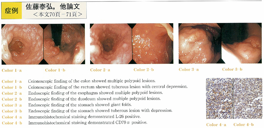
    A 80-year-old male patient referred to our hospital because of constipation and fresh bloody stool. On admission, superficial lymph nodes were palpable in the inguinal resions, and spleno megaly was detected by CT scan examination. Endoscopic examination revealed multiple polypoid lesions in the whole colon, esophagus and duodenum, and tuberous lesions with central depression in the stomach, cecum and rectum. Histopathological examinations of biopsy specimens showed diffuse proliferation of abnormal lymphocytes. Immunohistochemical staining demonstrated that these consisted of L-26, CD-79α positive, and of UCHL1, DFT1, CD3 negative. Consequently this case was diagnosed as B-cell malignant lymphoma, diffusemedium sized cell type. We think that it should be appropriate to deal lymphomatous tumors forming multiple polypoid lesions with distant lymph node involvement as an advanced type of MLP, even if they can not be confirmed as gastrointestinal origin. Further discussion should be raised on diagnostic criteria of MLP.
    Download PDF (936K)
  • Miho Tabata, Yoshihiro Furuichi, Kenji Ueda, Manabu Nishimaki, Toshio ...
    2001 Volume 59 Issue 2 Pages 72-73
    Published: December 05, 2001
    Released on J-STAGE: May 22, 2014
    JOURNAL FREE ACCESS
    The patient was a 59-year-old woman with the chief complaints of abdominal pain and fever. Upon palpation of a tumor the size of an egg accompanied by precordial tenderness, she was hospitalized. Leukocytosis and positive CRP were observed, and abdominal US and CT revealed a tumor measuring 40 mm by 60 mm protruding out of the stomach wall. Endoscopic examination of the upper gastrointestinal tract disclosed a smooth-surfaced hemispheric elevation near the posterior wall of the antrum, and a white pus-like exudate was seen oozing from part of the mass. As she was suspected of having gastric abscess, an antibiotic was administered to control IVH. The symptoms and inflammatory reaction improved, and the tumor was reduced in size on the scan. On the 26th hospital day, however, subtotal gastrectomy and cholecystectomy were performed. At surgery, a tumor measuring 30 mm by 53 mm was seen adhering to the ligament of the transverse colon near the posterior wall of the antrum. The gallbladder wall was prominently thickened, and the gallbladder was found to contain many bilirubin stones. Considering that the gallbladder showed notable adhesion to the surrounding organs, gastric abscess seemed to have developed from pericholecystitis.
    Download PDF (370K)
  • Hiroaki Hagiwara, Teruo Yoshinaga, Tsugio Higuchi, Tatsuya Higuchi, Na ...
    2001 Volume 59 Issue 2 Pages 74-75
    Published: December 05, 2001
    Released on J-STAGE: May 22, 2014
    JOURNAL FREE ACCESS
    A hyperplastic foveolar polyp (about 2cm in diameter) on greater curvature of antrum was found in a 56 year-old-man who had gastro-duodenal ulcers. H. pylori infection was detected by histological examination and cultures of biopsied spiecimens taken from greater curvature of antrum and upper body. H. pylori IgG antibody was positive in serological examination. As the initial H. pylori eradication by 1-week triple therapy (rabeprazole 20mg, amoxicillin 1500mg and clarithromycin 600mg) was not succeeded, he was re-treated by 1 week course of lansoprazole 60mg, amoxicillin 1500mg and metronidazole 750mg 3 months later. 6 months after re-treatment, the polyp was disappeared endoscopically and histologically and H. pylori eradication was confirmed by 13C-urea breath test.
    Eradication of H. pylori may be an effective treatment in cases with hyperplastic foveolar polyp of stomach.
    Download PDF (425K)
  • Minako Yoshida, Keiji Koshibu, Shinsuke Tokoi, Hideaki Fujimoto, Yuhsu ...
    2001 Volume 59 Issue 2 Pages 76-77
    Published: December 05, 2001
    Released on J-STAGE: May 22, 2014
    JOURNAL FREE ACCESS
    Before surgery, precise diagnosis of submucosal tumors in the gastrointestinal tract remains quite difficult. A 75-year-old female was endoscopically pointed out a gastric submucosal tumor, less than 1 cm in diameter, on the greater curvature of the antrum at a health checks in 1985. Subsequent endoscopic examinations were followed up periodically for 15 years. In 2000, the lesion enlarged to 2 cm. The internal echo pattern on endoscopic ultrasonography (EUS) was almost homogenous but partially hyperechoic. On EUS guided fine needle aspiration biopsy, it was diagnosed as leiomyoma. Because the malignancy could not be excluded, it was resected by laparoscopic approach. Histopathologically, it was mainly composed of uniform myogenic cells, but included abnormal cells in localized area. By immunohistochemical staining for α-smooth muscle actin, malignant cell infiltration to lamina propria was confirmed.
    Download PDF (909K)
  • Noriaki Kameyama, Nobutaka Yasui, Atsushi Toizumi, Yoichiro Tamura, Ta ...
    2001 Volume 59 Issue 2 Pages 78-79
    Published: December 05, 2001
    Released on J-STAGE: May 22, 2014
    JOURNAL FREE ACCESS
    We report a case of hand assisted laparoscopic surgery (HLS) for gastric schwannoma. The patient was a 65-year-old woman, who had been asymptomatically diagnosed to have gastric extramural tumor, and gallbladder polyp. Because of its growing in 8 months, she was hospitalized for surgical treatment. We started the operation under the open method of laparoscopic surgery using the intraabdominal pressure of 8 mmHg. Three ports were inserted into the abdomen as follows ; upper umblication (15mm) , below the xiphoid process on the median line (5mm) , and the right abdomen (11.5mm) . The tumor of the maximum diameter of 7 cm was observed on the greater curvature without any adhesion to other organs. After I enlarged the wound of the port below the xiphoid process on the median line to 5cm, I put my right hand into the abdomen using LAP-DISK and performed the partial resection of the stomach by Endo-GIA. The cholecystectomy underwent simultaneously performed for the gallbladder polyp. The size of the gastric tumor was 6.5cm×4.3cm×4.0cm. The tumor was diagnosed as gastrointestinal stromal tumor by frozen sections, but the added immunohistochemical staining of the tumor was positive only for S-100 protein. It was eventually diagnosed as gastric schwannoma.
    Download PDF (927K)
  • Yoshiaki Osaka, Shigeru Sato, Yu Takagi, Kazuhiko Tamura, Ryosuke Okad ...
    2001 Volume 59 Issue 2 Pages 80-81
    Published: December 05, 2001
    Released on J-STAGE: May 22, 2014
    JOURNAL FREE ACCESS
    A 90-year-old man was admitted to our hospital in March of 2000 because of the difficulty of oral ingestion. He underwent distal gastrectomy with the reconstruction of Billroth I method due to the gastric cancer in 1994. Gastrointestinal endoscopy showed the stenosis due to the cancerous recurrence at the oral side of anastomosis, in which the endoscope could not be passed through it. Pathological specimen showed the poorly differentiated adenocarcinoma. Instead of reoperation, the stent (Ultra-Flex®, 10cm in length) was placed because the patient was too old and his general condition was also poor. Concerning on the technique of stent placement, endoscope was first inserted with leaving the delivery system in the stomach, because it could not be passed through the stenosis due to bending. Then, the delivery system was grasped with the forceps and was endoscopically placed in the stenosis. After the stent placement, the patient could ingest the rice gruel and leave the hospital.
    The stent placement for the pyloric or duodenal stenosis is relatively difficult. Our technique is thought to be one of the useful methods for these diseases.
    Download PDF (928K)
  • Junichi Saito, Eiji Kurihara, Jun Yamamoto, Kenji Iwata
    2001 Volume 59 Issue 2 Pages 82-83
    Published: December 05, 2001
    Released on J-STAGE: May 22, 2014
    JOURNAL FREE ACCESS
    A 58-year-old male complaining of swallowing disturbance was admitted to our hospital. Exploratory laparotomy for cardiac gastric cancer performed because of peritoneal dissemination and invasion to phren of the cancer. Seven months later, he was diagnosed as cardiac stricture by progression of the cancer. Non-covered stent was applied to improve the stricture. Five months later, the oral portion of the stent developed stenosis due to tumor growth, and placement of a covered stent through the first stent was performed to obtain the patency. Two months later, the oral portion and the anal portion of these stents developed stenosis due to tumor growth, and additional placement of each two covered stents was done at both oral and anal side of formed treatment. As a result, he could take semi-solid food for about eleven months after cardiac stenosis till cancer death. Our case showed problem in placing stent in stent without full stent expansion. Inadequate expansion maked food impaction and dead space between esophageal wall and stent. So far, we think that expandable metaric stent theraphy for gastric stenosis is useful to the inprovement of quality of life and hope more development of expandable metaric stent.
    Download PDF (810K)
  • Takaaki Kobayashi, Takeshi Omura, Hiroaki Nozawa, Hiroyoshi Morita, Ta ...
    2001 Volume 59 Issue 2 Pages 84-85
    Published: December 05, 2001
    Released on J-STAGE: May 22, 2014
    JOURNAL FREE ACCESS
    A 60-year-woman was admitted to our hospital complaining of the anastomotic reccurence tumor of rectum. She underwent sigmoidectomy for sigmoid colon cancer, extended left hemicolectomy for descending colon cancer and endoscopic-mucosal-resection for ascending colon cancer.
    Endoscopic examination showed two lesions in the stomach, one was IIa in greater curvature of lower body and the other was depressed lesion in lesser curvature of antrum. The biopsy specimen of the former lesion was pathologically diagnosed as well~modalately differenciated adenocarcinoma.
    Distalgastorectomy and super low anterior resection was performed. Three lesions were detected in the resected stomach. All three lesions pathologically showed carcinoma, IIa in the lower body showed tub1 (m) , IIc in the antrum showed papillary adenocarcinoma (m) , IIc in the prepyloric lesion showed tub1 (sm) .
    Microsatellite instability (MSI) of all lesions and nomal mucosa of the stomach were positive. Rectal tumor was well differenciated adenocarcinoma and MSI of tumor and normal mucosa of the rectum was negative.
    MSI of multiple cancer shows higher positive rate than that of single cancer. We think that MSI may be usefull for genetic diagnosis of gastric cancer as well as hereditary nonpolyposis colorectal cancer.
    Download PDF (589K)
  • Naomi Kakushima, Mitsuhiro Fujishiro, Naohisa Yahagi, Shotaro Enomoto, ...
    2001 Volume 59 Issue 2 Pages 86-87
    Published: December 05, 2001
    Released on J-STAGE: May 22, 2014
    JOURNAL FREE ACCESS
    A protruded tumor on the lesser curve of lower gastric body was detected by a barium meal for medical check-up in a 64-year-old man. Endoscopically, it was a doom-like protruded lesion, 3 cm in diameter, and biopsy revealed a well differentiated adenocarcinoma. Endoscopic ultrasonography showed that it was semi-pedunculated and the depth seemed to be limited in mucosal or shallower submucosal layer. Therefore, the tumor was treated endoscopically and pathological diagnosis revealed a mucosal cancer with lymphatic vessel infiltration. Furthermore, beneath it, remarkable lymph follicle formation was observed in the mucosal muscle layer and component of moderately to poorly differentiated adenocarcinoma invaded those lymph follicles. Because lymphatic vessel infiltration and component of poorly differentiated adenocarcinoma were observed, the patient underwent an additional surgery, but no residual tumor or lymph node metastasis was found. This case was well worth reporting, because of the rare macroscopic shape and remarkable lymph follicle formation in the mucosal muscle layer accompanied with the tumor invasion.
    Download PDF (904K)
  • Yoshifumi Takenaka, Masataka Yamazaki, Ryouhei Miyata, Osahiro Nitori, ...
    2001 Volume 59 Issue 2 Pages 88-89
    Published: December 05, 2001
    Released on J-STAGE: May 22, 2014
    JOURNAL FREE ACCESS
    A 77-year-old male was admitted with appetite loss and abdominal tumor. His tumor markers were of high levels : CEA 176.2 ng/ml and CA 19-9 115.8 U/ml. Examinations such as gastrofiberscopy, CT, MRI, ultrasonography, and angiography revealed a gastric carcinoma with extraluminal growth. A distal gastrectomy was performed palliatively, as well as a resection of the infiltrated transverse colon. The operative findings were : T4, N3, H1, P0, CYX, M1 (LYM) , and Stage IV. The pathological finding of the intragastric specimen was well-differentiated adenocarcinoma (4×3.5×2.5 cm) , but one of the extraluminal growth specimens showed mucinous carcinoma (11×9×4.5 cm) . In between the gastric carcinoma and the extraluminal area, a malignant cystic area was discovered, but the connective area was not pointed out. Four months after the surgical operation, the patient died of peritonitis carcinomatosae.
    We found only 79 relative case reports in the Japanese literature, with patient ages ranging from 25 to 87 with average of 60.2 years of age. The primary tumors, as per our case, originated in the greater curvature side of the antrum in most of the reported cases. Their prognoses were poor due to infiltration, or metastasis, and exceptionally, only 10 patients survived over one year. We present an extremely rare type of gastric carcinoma with extraluminal growth.
    Download PDF (1112K)
  • Wen San Ji, Koichiro Kumai, Hiroyuki Imaeda, Koichi Aiura, Haruhiko Og ...
    2001 Volume 59 Issue 2 Pages 90-91
    Published: December 05, 2001
    Released on J-STAGE: May 22, 2014
    JOURNAL FREE ACCESS
    A 67-year old man underwent endoscopic examination because of abnormal findings in upper GI series at the other hospital. Multiple erosions were observed at the lower gastric body and the antrum. And histological findings of the biopsied specimen at the anterior wall of the lower gastric body showed signet-ring cell carcinoma. Although endoscopic findings at our hospital showed the same as ones there, however histological findings of the biopsied specimen could not reveal malignancy. After 1.5 and 3 months, the patient underwent endoscopic examination again, however malignancy was not detected. Nine months after primary examination, slightly depressed lesion with small redness (0'IIc type) was obseved at the greater curvature of the lower gastric body in the fourth endoscopy, and histological findings showed signet-ring cell carcinoma. The best treatment among gastrectomy, local resection of the stomach and endoscopic mucosal resection was discussed. As histological typing was signet-ring cell carcinoma, 9 months had been already passed from the first diagnosis, and it was difficult to detect the site of this lesion, distal gastrectomy was selected. Hisotological findings of resected specimen showed minute signet-ring cell carcinoma (0IIb type) 3 mm in diameter in the lamina propriae mucosae without vascular invasion and lymph node metastasis. Strict diagnosis and decision of the treatment were difficult in this case.
    Download PDF (648K)
  • Hiroshi Shiozaki, Toshiyuki Tahara, Yoshiyuki Yamagishi, Isao Nakata
    2001 Volume 59 Issue 2 Pages 92-93
    Published: December 05, 2001
    Released on J-STAGE: May 22, 2014
    JOURNAL FREE ACCESS
    A 20-year-old female was admitted to our hospital because of bilateral lower limb edema for 2 months. Her symptom developed one month after the birth of her first child. Laboratory tests on admission showed hypoproteinemia (TP4.6g/dl, Alb2.4g/dl) without proteinuria or liver function disorder. Serum anti-nuclear antibody was negative. Her cardiac finding and thyroid function were normal.
    Esophagogastroduodenoscopy showed scattered white spots in the second portion of duodenum. Biopsy specimen of the lesion demonstrated dilated lymphatics in the lamina propria microscopically. Abdominal image 5 minutes after intravenous injection of 111In-transferrin revealed a leak of radioisotope into the upper gastrointestinal tract, indicating the presence of protein-losing gastroenteropathy. Other examinations showed neither neoplasm nor inflammatory disease, so she was diagnosed as primary intestinal lymphangiectasia. Elemental enteral alimentation with low fat diet led to increase her serum protein (TP6.0g/dl, Alb3.1g/dl) . Although a few other cases were reported, which presented protein-losing gastroenteropathy due to primary intestinal lymphangiectasia after delivery, the pathogenesis remains unclear.
    Download PDF (461K)
  • Yuichiro Eguchi, Ken Higuchi, Toru Kakinuma, Shin Arai, Kenji Fujimori ...
    2001 Volume 59 Issue 2 Pages 94-95
    Published: December 05, 2001
    Released on J-STAGE: May 22, 2014
    JOURNAL FREE ACCESS
    A 55-year-old woman with alcoholic liver cirrhosis was admitted to our hospital because of hemorrhagic shock after repeated episode of tarry stool. Endoscopic examination disclosed a solitary duodenal varix at the third portion of the duodenum with white plug on the surface. EVL was performed for the varix, and she recovered from hemorrhagic shock. Additional therapy was not done, because both trans-superior mesenteric arterial portography and magnetic resonance angiography revealed no collateral vessels toward the esophagus, stomach and duodenum. At 4 months later, tarry stool recurred. On endoscopic examination, there was a newly developing duodenal varix at anal side of the third portion of the duodenum, which was treated with EVL. However, intestinal endoscopic examination performed 7 days later revealed another small varix at the same side, but this varix was not treated. She was followed up only by endoscopical observation thereafter, and was quite well for 8 months despite of the remaining varix. It is concluded that EVL may be useful as an emergent therapy for massive hemorrhage from ruptured duodenal varices, but from the prognostic aspect, its efficacy must be evaluated precisely in future.
    Download PDF (422K)
  • Kiyoshi Terai, Hirofumi Gonda, Yuji Fujii, Eiichiro Seki, Yoh Sekine, ...
    2001 Volume 59 Issue 2 Pages 96-97
    Published: December 05, 2001
    Released on J-STAGE: May 22, 2014
    JOURNAL FREE ACCESS
    A 56-year-old man was admitted to our hospital because of diabetes mellitus, meanwhile endoscopic examination revealed a elevated lesion at the second portion of the duodenum. The biopsy specimen showed adenoma with severe dysplasia, Endoscopic mucosal resection (EMR) was performed safely with a detacable snare and a bipolar snare. Histological examination revealed a well differentiated tubular adenocarcinoma with depth of invasion into the mucosal layer, and the resected margin were tumor-free.
    Download PDF (968K)
  • Koji Asai, Hironobu Umezawa, Hironobu Ooyanagi, Tokuhiko Saito, Yoshit ...
    2001 Volume 59 Issue 2 Pages 98-99
    Published: December 05, 2001
    Released on J-STAGE: May 22, 2014
    JOURNAL FREE ACCESS
    A 60-year-old male visited our medical center for presenting positive reaction in stool occult blood test. Barium enema showed a gigantic tumor, which stalk was on terminal ileum and which head was into ascending colon. Colonoscopy revealed that it was a cushion-sign-positive submucosal tumor that was smooth in surface and good in mobility. On the same day, snare excision was performed combining with snare holding method. The specimen was 75×38×30mm in size. Histology proved the tumor to be lipoma. Among our experience of 5 small intestinal lipoma cases treated by endoscopic resection, the size of this case was largest.
    Download PDF (914K)
  • Yoshiki Takashima, Takeshi Matsuda, Yuji Okihama, Yuji Nagashima, Yosh ...
    2001 Volume 59 Issue 2 Pages 100-101
    Published: December 05, 2001
    Released on J-STAGE: May 22, 2014
    JOURNAL FREE ACCESS
  • Takeshi Matsuda, Yuji Nagashima, Yoshihiro Hiramoto, Yoshiki Takashima ...
    2001 Volume 59 Issue 2 Pages 102-103
    Published: December 05, 2001
    Released on J-STAGE: May 22, 2014
    JOURNAL FREE ACCESS
  • Kazuo Maruta, Shingo Miyaguchi, Kenji Nukaga, Aritoshi Yasuda, Mitsuhi ...
    2001 Volume 59 Issue 2 Pages 104-105
    Published: December 05, 2001
    Released on J-STAGE: May 22, 2014
    JOURNAL FREE ACCESS
    The patient was a 70-year-old female. She noticed abdominal pain, which was followed by diarrhea and hematochezia on the night of April 1, 2000. She was taken to the emergency department and hospitalized for further examination. Colonoscopy revealed ulceration complicated by edema in sigmoid colon. The ulcerous lesion had spread over the entire circumference of the deeper portion of sigmoid colon. A tumorous lesion of 20 mm in diameter was detected remaining in the normal mucosa within the inflamed mucosa. The tumorous lesion was biopsied and a diagnosis of colon cancer was made. In this case, ischemic colitis was found to accompany colon cancer. The patient was placed on dialytic therapy because of chronic renal failure. A CT scan and X-ray examination revealed marked arteriosclerosis, which was assumed to be responsible for ischemia. A review of literature yielded many case reports on so-called obstructive colitis, ischemic colitis that occurred on the oral side of colorectal cancer. There appeared to be a limited number of cases of colorectal cancer detected at colonoscopy performed to examine ischemic colitis. This rare case was reported in the present study.
    Download PDF (983K)
  • Hidejiro Kawahara, Fumiaki Yano, Isao Tabei, Takemasa Cho, Hideyuki Ka ...
    2001 Volume 59 Issue 2 Pages 106-107
    Published: December 05, 2001
    Released on J-STAGE: May 22, 2014
    JOURNAL FREE ACCESS
    51 year-old male who had undergone total gastrectomy with splenectomy 4 years ago was admitted to our hospital for examination and treatment for melena. Type 4 tumors were found in the vermiform appendix and the rectum by endoscopic examination. Histopathological fingings showed signet ring cell carcinoma similar to that of the gastric cancer. Therefore it was diagnosed as metastatic diffuse infiltrative type 4 carcinoma of the appendix and rectum. Lymphangio-metastasis and peritoneal dissemination of the gastric cancer was determined negative in relation by pre- and opelative examination. Therefore hematogenous metastasis of the signet ring cell carcinoma from the original gastric cancer through SMA for the appendix and IMA for the rectal lesions were most suspected for the route of metastasis.
    A rare case of gastric cancer with hematogenous metastasis to the appendix and rectum is reported.
    Download PDF (1032K)
  • Kohei Kawakami, Ichirou Sudoh, Tsuneto Oda, Daisuke Ishizuka, Kazuo Ma ...
    2001 Volume 59 Issue 2 Pages 108-109
    Published: December 05, 2001
    Released on J-STAGE: May 22, 2014
    JOURNAL FREE ACCESS
    The patient was a 38-year-old male suffering from acute myelocytic leukemia. He had been treated in the department of hematology at this hospital since November 1998. On May 30, 2000, he was admitted to this hospital for surgical treatment. He underwent a bone marrow transplant on June 22. From July 19, he became febrile and erythema was present all over the body. A skin biopsy specimen was collected from the inside of the upper arm and a diagnosis of acute graft versus host (GVHD) was made on the basis of the results obtained. He had frequent diarrher from July 28, and was transferred to this department for further examination on August 7. He underwent colonoscopy on August 8, and the large intestine was endoscopically observe up to the sigmoid colon. Edematous mucosa was observed and irregular rubefactions and erosions were occasionally detected among severe edemas. There findings and his clinical symptoms were suggestive of GVHD of the intestinal tract or of TMA. In the treatment of patient suffering from repeated episodes of diarrhea after bone marrow transplantation, the possibility of GVHD of the intestinal tract or TMA must be born in mind and implementation of colonoscopy should generally be considered.
    Download PDF (266K)
  • Hirotoshi Ebinuma, Hiroyuki Imaeda, Haruhiko Ogata, Junichi Kaburagi, ...
    2001 Volume 59 Issue 2 Pages 110-111
    Published: December 05, 2001
    Released on J-STAGE: May 22, 2014
    JOURNAL FREE ACCESS
    A 58-year-old woman visited our hospital complained of right lower abdominal pain. The ultrasound sonography and computed tomography examination showed intussusception in the ileocecal lesion. Barium enema examination revealed an elevated lesion in the ostium of appendix and the contrast barium was not fully filled in the appendix. Endoscopic examination also revealed an elevated lesion with redness in the ostium and obstruction of the hole, which is called volcano sign. These findings suggested that the mucocele in appendix caused ileocecal intussusception. During her admission the lower abdominal pain due to ileocecal intussusception was frequently occurred and therefore ileocecal resection was performed. Histological findings showed mucinous cystadenoma in the distal appendix and fibrous change in the appendix wall. It was suggested that this fibrous mucosa caused intussusception in the cecum. Mucinous cystadenoma in the appendix causing intussusception is rare, and in most of the cases preoperative diagnosis has not been made because emergency operation has been performed. Here we report a rare case of mucinous cystadenoma causing intussusception which was preoperatively suggested from the findings of the barium enema and endoscopic examination.
    Download PDF (593K)
  • Toshio Katakami, Miho Tabata, Yoshihiro Furuichi, Kenji Ueda, Michio H ...
    2001 Volume 59 Issue 2 Pages 112-113
    Published: December 05, 2001
    Released on J-STAGE: May 22, 2014
    JOURNAL FREE ACCESS
    The patient was a 59-year-old man. He visited a neighborhood clinic because of diarrhea. He had alopecia at the top of his head and swelling of finger tips around that time. As barium enema examination revealed multiple polyps in the entire colon, he visited our hospital. Endoscopic examination disclosed numerous red polyps of irregular size, and histologic examination revealed cyst-like dilation of the ducts, interstitial edema, and cellular infiltration which were consistent with hamartomatous polyps. Endoscopic examination of the upper gastrointestinal tract similarly disclosed multiple polyps. Nail exfoliation and pigmentation were also observed. The patient was diagnosed as having Cronkhite-Canada syndrome and was started on corticosteroid therapy. The subsequent course was uneventful and favorable.
    Download PDF (658K)
  • Tadashi Teramoto, Kaku Egami, Nobutoshi Hagiwara, Hidehiro Watanabe, H ...
    2001 Volume 59 Issue 2 Pages 114-115
    Published: December 05, 2001
    Released on J-STAGE: May 22, 2014
    JOURNAL FREE ACCESS
    Therapeutic balloon dilation was performed colonoscopically, using a balloon dilatation catheter, on a patient who had a stricture of the terminal ileum resulting from Crohn's disease. The patient was a 53-year-old man who had undergone 4 abdominal operations for Crohn's disease between 1990 and 1995. He had been treated with medical and nutritional therapy, and had obtained symptomatic relief.
    The patient consulted our hospital complaining of lower abdominal pain and loss of appetite, and was admitted with a diagnosis of ileus on August 5, 2000. Contrast study of the small intestine showed a 6-cm-long stenosis of the terminal ileum. Conservative therapy for 2 months did not relieve the symptoms. Endoscopic treatment was considered preferable to surgery because the patient's multiple resections in the past increased the risk of short bowel syndrome. Balloon dilation was performed 3 times using a commercially available balloon dilatation catheter (CRE, Boston Scientific) , and sustained improvement was observed during a 9-month follow-up period.
    The present authors believe that endoscopic balloon dilation is a safe and effective therapeutic option for patients with intestinal strictures resulting from Crohn's disease, which has a high rate of symptomatic recurrence and re-operation.
    Download PDF (672K)
  • Shuya Yoshinaga, Jun Tashiro, Hitohiko Koizuka, Toshiki Nikami, Tomoo ...
    2001 Volume 59 Issue 2 Pages 116-117
    Published: December 05, 2001
    Released on J-STAGE: May 22, 2014
    JOURNAL FREE ACCESS
    The use of a metal stent in malignant colorectal obstruction is not well established in Japan. The traditional method of managing inoperable colonic obstruction has been palliative colostomy or insertion of long tube. However, these methods were invasive and/or umcomfortable for patients. We report two cases of malignant rectal obstruction which was treated with expandable metal stent. Case 1 is a 65-year-old female diagnosed to have advanced gastric cancer with Schnitzler metastasis. Barium enema and colonoscopy showed severe stenosis due to extraluminal compression. Case 2 is an 89-year-old female diagnosed to have rectal cancer. They were inoperable and developed abdominal pain due to ileus during hospitalization. EMS with the inner diameter of 18mm (Ultraflex prosthesis) was inserted for their rectal obstruction. Stent expansion brought about immediate decompression of the obstructed bowel. After the treatment, their abdominal symptom were relieved and no complications were developed in both cases. In our opinion, the treatment could maintain patient's QOL until their death compared with traditional method. Stent decompression could be palliative treatment for inoperable malignant colonic obstruction.
    Download PDF (837K)
  • Yousuke Otake, Tsuyoshi Abe, Akihiko Ohta, Koichiro Sato, Takaaki Tama ...
    2001 Volume 59 Issue 2 Pages 118-119
    Published: December 05, 2001
    Released on J-STAGE: May 22, 2014
    JOURNAL FREE ACCESS
    A 63-year-woman visited our hospital for further evaluation of submucosal tumor in the sigmoid colon. The lesion was hard and was approximately 10mm in diameter. Endoscopic ultrasonography demonstrated a hypoechoic lesion located mainly in the shallow part of the third layer without swelling of the lymph nodes. We strongly suspected it to be a carcinoid. Endoscopic resection with aspiration and ligation was performed. By this procedure, the lesion was completely resected with submucosal tissue between tumor and resected surface. Microscopic examination including immunohistological study of the specimen diagnosed a benign schwannoma.
    Endoscopic resection with aspiration and ligation was considered to be an effective and safety procedure for diagnosis and therapy of submucosal tumor when the lesion was comparatively small (less than 10mm in diameter) and located in the shallow part of submucosa without swelling of the lymph nodes.
    Download PDF (434K)
  • Ayako Watanabe, Susumu Yamakado, Isao Shinozawa, Toshihiko Nagai, Yume ...
    2001 Volume 59 Issue 2 Pages 120-121
    Published: December 05, 2001
    Released on J-STAGE: May 22, 2014
    JOURNAL FREE ACCESS
    A 89 year-old woman visited to our hospital with a complaint of right lower abdominal pain. Colonoscopic examination revealed a Isp type protruding lesion in 7mm diameter in lower rectum. We performed endoscopic mucosal resection to this lesion for accurate diagnosis because biopsy specimen revealed the atypical lymphocyte infiltration. Pathological findings of the resected tissue showed marginal zone B-cell lymphoma of mucosa-associated lymphoid tissue type (MALToma) . Because of her age was high and the lesion located in the lower rectum without any other lymphoma lesions, we followed up this ulcer lesion with endoscopic examination. As Helicobacter pylori (HP) infection to her stomach was positive, we eradicated HP infection by triple therapy. After EMR to this rectal MALToma, she has not a relapse until today.
    We suggested to be able to treat with EMR for gastrointestinal primary MALToma when its lesion was the indication for EMR and patient was senile and/or had any inoperable complications.
    Download PDF (226K)
  • Kazunori Miyata, Hiroshi Abe, Hiroyuki Machino, Yuichi Handa, Kyoya Sa ...
    2001 Volume 59 Issue 2 Pages 122-123
    Published: December 05, 2001
    Released on J-STAGE: May 22, 2014
    JOURNAL FREE ACCESS
    A 38-year-old male was admitted to our hospital because of jaundice. 18 months before admission, he was diagnosed as insulin-dependent diabetes mellitus. Then we could detect no abnormalities of abdominal CT scan.
    On this admission abdominal US revealed hypoechoic swelling of pancreas. ERCP demonstrated focal narrowing of the main pancreatic duct and constriction of the common bile duct in the pancreas with dilatation of upstream.
    Pathological finding of pancreas was infiltration of inflammatory cells (lymphocytes, eosinophils and plasma cells) and fibrosis. Pathological findings of gall bladder and lymphnode were similar to that of pancreas. We diagnosed his illness as autoimmune pancreatitis. After surgery jaundice improved but swelling of pancreas continued. Though we recommended steroid therapy, he refused it. We are carefully following.
    Download PDF (784K)
  • Koichi Haruyama, Takayoshi Nishino, Fumitake Toki, Shoko Hisada, Yuji ...
    2001 Volume 59 Issue 2 Pages 124-125
    Published: December 05, 2001
    Released on J-STAGE: May 22, 2014
    JOURNAL FREE ACCESS
    Glucagon has been shown to depress the gut motility and pancreatic secretion and it has also been reported that glucagon has a beneficial effect on experimental pancreatitis in mice.
    The present study investigates whether glucagon administration decreases duodenal moltility and prevents the increase of serum pancreatic enzymes after endoscopic retrograde chlangiopancreatography (ERCP) . ERCP was performed in 29 consecutive cases (18 males and 11 females) , which were divided into two groups. Scopolamine butylbromide was given at a dose of 20 mg to Group B, and glucagon was given at a dose of 1 mg to Group G. Each drug was administered by intramuscular injection before ERCP, and then duodenal motility was evaluated by modified Niwa's criteria : (1 : complete supression to 5 : no supression) . Serum pancreatic enzymes were determined to be suppresed at 0h, 4h and 24h after ERCP.
    Result : 1. The glucagon treatment suppressed duodenal motility slightly more than the scopolamine butylbromide treatment (1.5±0.7 vs 2.0±0.4) . 2.Serum lipase levels at 4h after ERCP in patients in Group G were slightly lower than in patients in Group B ; however, there was no differece at 24h between two groups.
    In conclusions, the glucagon treatment suppressed duodenal motility sufficiently in ERCP. Further research is needed to determine whether the incidence of postcannulation pancreatitis can be reduced by glucagon.
    Download PDF (291K)
  • Tsunao Imamura, Fumihiko Nozu, Masashi Sakamoto, Akihisa Ishikawa, Kou ...
    2001 Volume 59 Issue 2 Pages 126-127
    Published: December 05, 2001
    Released on J-STAGE: May 22, 2014
    JOURNAL FREE ACCESS
    A 76-year-old woman was admitted to our hospital for severe acute biliary pancreatitis. The patient was evaluated as“severe”according to the severity criteria of the Japanese Ministry of Health and Welfare Study Group. The patient was treated by continuous regional arterial infusion using nafamostat mesilate and imipenem for 7 days. Symptoms of pancreastitis improved but high fever appeared on 15th hospital day. Liver abscess was suspected on the findings of the abdominal computed tomography. Antibiotic was intravenously administered but fever persisted. We judged that the patient has a indication of percutaneous abscess drainage, but we could not performed this because of bleeding tendency of idiopathic thrombocytopenic purpura. We performed endoscopic naso-biliary drainage (ENBD) for treatment of liver abscess. Endoscopic retrograde cholangiography visualized the dilated bile duct and connection between this duct and the liver abscess. A 7.5Fr. ENBD tube was placed in the abscess through the dilated bile duct. The clinical symptoms improved dramatically. Our experience suggests that ENBD is one of the effective treatment for liver abscess after biliary tract infection.
    Download PDF (566K)
  • Tadashi Ohshima, Takeshi Ishihara, Taketo Yamaguchi, Genichiro Kadono, ...
    2001 Volume 59 Issue 2 Pages 128-129
    Published: December 05, 2001
    Released on J-STAGE: May 22, 2014
    JOURNAL FREE ACCESS
    A twenty-year-old male had failed with the conservative treatment of a traumatic pancreatic pseudocyst and had documented cyst persistence for over four weeks. The size of the pseudocyst was 110mm in diameter and CT scans showed apposition of the pseudocyst in the pancreatic body to tail against the stomach wall. We performed EUS-guided transgastric puncture of the pseudocyst with a 19 gauge ultrasound needle (ECHO TIP. Wilson-Cook Medical) , then a 0.035 inch guide wire was passed into the pseudocyst after with drawn of a stylet and a 7-F stent coaxially inserted into the pseudocyst. There were no complications associated with the procedure. The pseudocyst had collapsed in five days after the drainage. The pseudocyst had completely resolved at follow-up of six months after the drainage.
    Download PDF (674K)
  • Terumi Kamisawa, Takashi Fujiwara, Mizuka Suzuki, Kozue Amemiya, Naoto ...
    2001 Volume 59 Issue 2 Pages 130-131
    Published: December 05, 2001
    Released on J-STAGE: May 22, 2014
    JOURNAL FREE ACCESS
    A case who had pancreatic cancer during the course of the chronic pancreatitis is reported. A man of heavy drinker was diagnosed as pancreatolithiasis in 33 years old. He continued drinking. Abdominal CT and ERCP demonstrated typical findings of chronic pancreatitis in 50 years old. He was admitted to our hospital again complaining of back pain in 55 years old. Abdominal CT showed irregular mass in the uncus of the pancreas. ERCP showed stenosis of the main pancreatic duct in the head and body of the pancreas. In laparotomy, a 7cm-sized mass was detected in the head and body of the pancreas. Histological examination was moderately differentiated adenocarcinoma. This is the first case who had pancreatic cancer in the course of chronic pancreatitis, in 130 patients with chronic pancreatitis in our hospital.
    Download PDF (1232K)
feedback
Top