
Volume 82, Issue 1
Displaying 1-50 of 81 articles from this issue
-
2013Volume 82Issue 1 Pages 1-22
Published: 2013
Released on J-STAGE: July 05, 2013
Download PDF (9253K)
-
2013Volume 82Issue 1 Pages 45-48
Published: June 14, 2013
Released on J-STAGE: July 05, 2013
 Download PDF (447K)
 -
2013Volume 82Issue 1 Pages 49-52
Published: June 14, 2013
Released on J-STAGE: July 05, 2013
Download PDF (401K) -
2013Volume 82Issue 1 Pages 53-55
Published: June 14, 2013
Released on J-STAGE: July 05, 2013
Download PDF (348K)
-
2013Volume 82Issue 1 Pages 56-59
Published: June 14, 2013
Released on J-STAGE: July 05, 2013
Download PDF (263K) -
2013Volume 82Issue 1 Pages 60-63
Published: June 14, 2013
Released on J-STAGE: July 05, 2013
Download PDF (358K) -
2013Volume 82Issue 1 Pages 64-67
Published: June 14, 2013
Released on J-STAGE: July 05, 2013
 Download PDF (234K)
 -
2013Volume 82Issue 1 Pages 68-71
Published: June 14, 2013
Released on J-STAGE: July 05, 2013
 Download PDF (330K)
 -
2013Volume 82Issue 1 Pages 72-76
Published: June 14, 2013
Released on J-STAGE: July 05, 2013
Download PDF (527K) -
2013Volume 82Issue 1 Pages 77-81
Published: June 14, 2013
Released on J-STAGE: July 05, 2013
Download PDF (608K) -
2013Volume 82Issue 1 Pages 82-86
Published: June 14, 2013
Released on J-STAGE: July 05, 2013
Download PDF (682K)
-
2013Volume 82Issue 1 Pages 87-89
Published: June 14, 2013
Released on J-STAGE: July 05, 2013
 Download PDF (293K)
 -
2013Volume 82Issue 1 Pages 90-91
Published: June 14, 2013
Released on J-STAGE: July 05, 2013
 Download PDF (362K)
 -
2013Volume 82Issue 1 Pages 92-93
Published: June 14, 2013
Released on J-STAGE: July 05, 2013
 Download PDF (418K)
 -
2013Volume 82Issue 1 Pages 94-95
Published: June 14, 2013
Released on J-STAGE: July 05, 2013
 Download PDF (261K)
 -
2013Volume 82Issue 1 Pages 96-97
Published: June 14, 2013
Released on J-STAGE: July 05, 2013
 Download PDF (339K)
 -
2013Volume 82Issue 1 Pages 98-99
Published: June 14, 2013
Released on J-STAGE: July 05, 2013
 Download PDF (409K)
 -
2013Volume 82Issue 1 Pages 100-101
Published: June 14, 2013
Released on J-STAGE: July 05, 2013
 Download PDF (398K)
 -
2013Volume 82Issue 1 Pages 102-103
Published: June 14, 2013
Released on J-STAGE: July 05, 2013
 Download PDF (544K)
 -
2013Volume 82Issue 1 Pages 104-105
Published: June 14, 2013
Released on J-STAGE: July 05, 2013
 Download PDF (373K)
 -
2013Volume 82Issue 1 Pages 106-107
Published: June 14, 2013
Released on J-STAGE: July 05, 2013
 Download PDF (267K)
 -
2013Volume 82Issue 1 Pages 108-109
Published: June 14, 2013
Released on J-STAGE: July 05, 2013
 Download PDF (351K)
 -
2013Volume 82Issue 1 Pages 110-111
Published: June 14, 2013
Released on J-STAGE: July 05, 2013
 Download PDF (332K)
 -
2013Volume 82Issue 1 Pages 112-113
Published: June 14, 2013
Released on J-STAGE: July 05, 2013
 Download PDF (416K)
 -
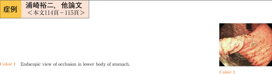
2013Volume 82Issue 1 Pages 114-115
Published: June 14, 2013
Released on J-STAGE: July 05, 2013
 Download PDF (597K)
 -
2013Volume 82Issue 1 Pages 116-117
Published: June 14, 2013
Released on J-STAGE: July 05, 2013
 Download PDF (419K)
 -
2013Volume 82Issue 1 Pages 118-119
Published: June 14, 2013
Released on J-STAGE: July 05, 2013
 Download PDF (301K)
 -
2013Volume 82Issue 1 Pages 120-121
Published: June 14, 2013
Released on J-STAGE: July 05, 2013
 Download PDF (276K)
 -
2013Volume 82Issue 1 Pages 122-123
Published: June 14, 2013
Released on J-STAGE: July 05, 2013
 Download PDF (386K)
 -
2013Volume 82Issue 1 Pages 124-125
Published: June 14, 2013
Released on J-STAGE: July 05, 2013
 Download PDF (466K)
 -
2013Volume 82Issue 1 Pages 126-127
Published: June 14, 2013
Released on J-STAGE: July 05, 2013
 Download PDF (275K)
 -
2013Volume 82Issue 1 Pages 128-129
Published: June 14, 2013
Released on J-STAGE: July 05, 2013
 Download PDF (480K)
 -
2013Volume 82Issue 1 Pages 130-131
Published: June 14, 2013
Released on J-STAGE: July 05, 2013
 Download PDF (319K)
 -
2013Volume 82Issue 1 Pages 132-133
Published: June 14, 2013
Released on J-STAGE: July 05, 2013
 Download PDF (214K)
 -
2013Volume 82Issue 1 Pages 134-135
Published: June 14, 2013
Released on J-STAGE: July 05, 2013
 Download PDF (498K)
 -
2013Volume 82Issue 1 Pages 136-137
Published: June 14, 2013
Released on J-STAGE: July 05, 2013
 Download PDF (618K)
 -
2013Volume 82Issue 1 Pages 138-139
Published: June 14, 2013
Released on J-STAGE: July 05, 2013
 Download PDF (374K)
 -
2013Volume 82Issue 1 Pages 140-141
Published: June 14, 2013
Released on J-STAGE: July 05, 2013
 Download PDF (452K)
 -
2013Volume 82Issue 1 Pages 142-143
Published: June 14, 2013
Released on J-STAGE: July 05, 2013
 Download PDF (338K)
 -
2013Volume 82Issue 1 Pages 144-145
Published: June 14, 2013
Released on J-STAGE: July 05, 2013
 Download PDF (374K)
 -
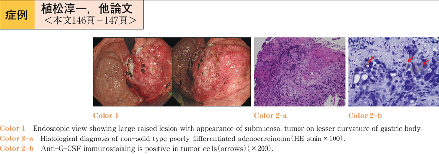
2013Volume 82Issue 1 Pages 146-147
Published: June 14, 2013
Released on J-STAGE: July 05, 2013
 Download PDF (306K)
 -
2013Volume 82Issue 1 Pages 148-149
Published: June 14, 2013
Released on J-STAGE: July 05, 2013
 Download PDF (249K)
 -
2013Volume 82Issue 1 Pages 150-151
Published: June 14, 2013
Released on J-STAGE: July 05, 2013
 Download PDF (409K)
 -
2013Volume 82Issue 1 Pages 152-153
Published: June 14, 2013
Released on J-STAGE: July 05, 2013
Download PDF (507K) -
2013Volume 82Issue 1 Pages 154-155
Published: June 14, 2013
Released on J-STAGE: July 05, 2013
 Download PDF (358K)
 -
A case of early duodenal cancer observed during follow-up of intraductal papillary mucinous neoplasm2013Volume 82Issue 1 Pages 156-157
Published: June 14, 2013
Released on J-STAGE: July 05, 2013
 Download PDF (377K)
 -
2013Volume 82Issue 1 Pages 158-159
Published: June 14, 2013
Released on J-STAGE: July 05, 2013
 Download PDF (288K)
 -
2013Volume 82Issue 1 Pages 160-161
Published: June 14, 2013
Released on J-STAGE: July 05, 2013
 Download PDF (300K)
 -
2013Volume 82Issue 1 Pages 162-163
Published: June 14, 2013
Released on J-STAGE: July 05, 2013
 Download PDF (365K)
 -
2013Volume 82Issue 1 Pages 164-165
Published: June 14, 2013
Released on J-STAGE: July 05, 2013
 Download PDF (338K)