Progress of Digestive Endoscopy
Online ISSN : 2187-4999
Print ISSN : 1348-9844
ISSN-L : 1348-9844
84 巻, 1 号
選択された号の論文の78件中51~78を表示しています
症例
  • 彌勒寺 紀栄, 西村 誠, 大村 卓也, 田村 優子, 中嶋 研一朗, 佐々木 美奈, 上垣 佐登子, 小宮 陽仁, 杉原 毅彦, 新井 冨 ...
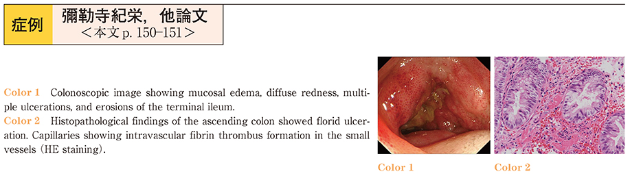
    2014 年 84 巻 1 号 p. 150-151
    発行日: 2014/06/14
    公開日: 2014/06/21
    ジャーナル フリー
    A 35-year-old man presented to our emergency department with a 4-day history of intermittent abdominal pain, watery diarrhea and fever. Physical examination revealed a flat and soft abdomen with marked tenderness of the umbilical region, but no abdominal rebound tenderness or guarding. Abdominal computed tomography showed ascites and edematous thickening of the wall of the ascending colon and part of the small intestine. Colonoscopy revealed mucosal edema, diffuse reddening, multiple ulcerations, and erosions in the terminal ileum. These findings and the pathological examination results strongly suggested the diagnosis of lupus mesenteric vasculitis. Treatment was commenced with high-dose prednisolone (40 mg/day, intravenous) , cyclophosphamide (1000 mg/day, intravenous) , and dalteparin sodium (5000 IU/day, intravenous) . The patient achieved gradual and complete control of the illness, which allowed tapering of the prednisolone dose and warfarin therapy. Post-treatment, colonoscopy revealed normal mucosa and normalization of most of the serological parameters.
  • 並河 健, 高田 康裕, 志村 裕人, 武藤 英知, 木村 祐, 石田 康男, 山中 太郎, 有泉 泰, 前田 一郎, 保阪 政樹
    2014 年 84 巻 1 号 p. 152-153
    発行日: 2014/06/14
    公開日: 2014/06/21
    ジャーナル フリー
    A 77-year-old man with a history of asthma presented with a 1-month-history of numbness of both lower thighs. In July 2013, the patient was hospitalized because of diarrhea and abdominal pain. Laboratory examination revealed leucocytosis with eosinophilia, increased serum immunoglobulin E levels, and presence of rheumatoid factor. The test for myeloperoxidase-antineutrophil cytoplasmic antibodies was negative. Computed tomography showed wall thickening of the duodenum. Although upper gastrointestinal endoscopy revealed gastric ulcers and evidence of duodenitis, histopathologic examination did not reveal any granulomas or vasculitis. The patient developed purpura on both feet and reported bloody stools, therefore, colonoscopy was performed. There were multiple ulcers throughout the large intestine. These clinical findings were consistent with a diagnosis of Churg-Strauss syndrome. Colonoscopic biopsy revealed vasculitis with eosinophilic infiltration, and the diagnosis of allergic granulomatous angiitis was made. The patient was started on steroid treatment, following which his symptoms rapidly resolved and the eosinophil count decreased. The disease was well-controlled by systemic steroid therapy, and follow-up endoscopies showed improvement of both the mucosal and pathological manifestations.
  • 芦谷 啓吾, 米野 和明, 山岡 稔, 筋野 智久, 大庫 秀樹, 菅野 龍, 野口 哲, 小林 威仁, 有馬 博, 木下 俊介, 飯田 慎 ...
    2014 年 84 巻 1 号 p. 154-155
    発行日: 2014/06/14
    公開日: 2014/06/21
    ジャーナル フリー
    Tacrolimus, a calcineurin inhibitor, has come to be well-known as effective therapy for steroid-refractory and steroid-dependent ulcerative colitis (UC) . It is necessary to achieve and maintain a trough level of tacrolimus in the blood of 10─15 ng/ml for effective induction of remission. We encountered 2 UC cases in which such target trough levels were rapidly achieved. Case 1 was a 48-year-old man with ulcerative colitis in whom remission could not be achieved in spite of mesalazinesteroid and adsorptive granulocyte and monocyte apheresis treatments. We started to administer 3 mg of tacrolimus daily and adjusted the daily dosage by checking the trough levels daily. The target trough level could be achieved by four days later after the start of treatment, and was effective to induce remission. Case 2 was a 38-year-old man with chronic continuous-type UC. He was under treatment with mesalazine, a steroid and azathioprine, however, his symptoms continued to worsen. We started treatment with 6 mg of tacrolimus daily, and the target trough level was achieved the very next day, with prompt induction of remission. Oral tacrolimus was discontinued three months later and oral azathioprine was used to maintain the remission in both patients, neither of whom showed any recurrence. The initial dose of tacrolimus for UC is usually 0.05 mg/kg/day, and the dose must be adjusted based on the trough levels. We have reported herein that an initial high dose of tacrolimus and daily measurement of the trough levels are effective for achieving the desired trough levels and prompt induction of remission.
  • 葛西 豊高, 川辺 晃一, 村松 誠司, 山田 拓郎, 宮原 庸介, 福田 裕昭, 石川 文彦, 新田 宙, 藤田 昌久, 釜田 茂幸, 山 ...
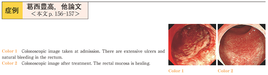
    2014 年 84 巻 1 号 p. 156-157
    発行日: 2014/06/14
    公開日: 2014/06/21
    ジャーナル フリー
    An 18-year-old woman was admitted to our hospital with fever, abdominal pain and bloody stool. Colonoscopy revealed severe ulcerative colitis. She was treated with prednisolone (70 mg/day) and sessions of granulocyte/monocyte apheresis (GMA) , which induced remission. However, the symptoms returned following the start of oral intake. Prednisolone and GMA treatment were not effective this time, and the patient was started on treatment with tacrolimus, which successfully induced remission. During treatment with tacrolimus, blood trough level control is important. By using rapid induction therapy and adjusting the tacrolimus dose according to the formula in the package insert, we were able to treat the intractable ulcerative colitis safely. Tacrolimus is thought to be an effective treatment agent for intractable ulcerative colitis in ordinary hospitals like ours.
  • 中下 学, 大場 彬博, 関 由喜, 中嶋 緑郎, 伊藤 高章, 松本 由華, 岡沢 啓, 水城 啓, 永田 博司
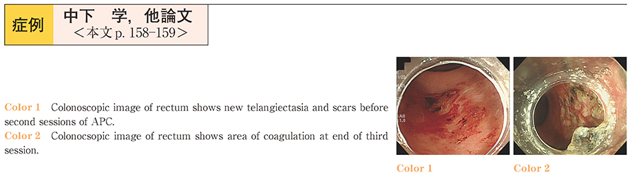
    2014 年 84 巻 1 号 p. 158-159
    発行日: 2014/06/14
    公開日: 2014/06/21
    ジャーナル フリー
    Chronic radiation proctitis (CRP) occurs in 2%─20% of patients undergoing pelvic radiotherapy. The symptoms of CRP include diarrhea, tenesmus, rectal pain and persistent rectal bleeding, rectal bleeding being the most common symptom. While most standard treatments for CRP, including medical, surgical and endoscopic treatments, remain unsatisfactory, endoscopic argon plasma coagulation (APC) is reported as an effective, efficient and reasonably safe non-contact method for the treatment of hemorrhagic CRP. While APC is reported to be associated with limited complications, major ulcers, strictures and fistulas have been reported previously, and careful monitoring and attention are necessary. We report the case of a 78-year-old man who presented with rectal bleeding after having undergone radiation therapy for prostatic cancer six months earlier. Endoscopic examination revealed hemorrhages and multiple rectal telangiectasias, and the patient was diagnosed as having CRP. Three sessions of brief pulse APC under the power setting of 30 W and argon flow rate of 0.6 l/min were performed, and complete hemostasis was achieved with no complications. Post-procedure follow-up endoscopy performed two months later revealed no telangiectasias or complications. In conclusion, plural therapy of brief pulse APC for CRP using a low power setting and low argon flow rate seems to be useful to reduce the complications of APC for the treatment of CRP.
  • 助川 誠, 藤田 晃司, 天田 塩, 内 雄介, 一坂 俊介, 森 克昭, 石川 啓一, 堂脇 昌一, 菊永 裕行, 熊井 浩一郎, 皆川 ...
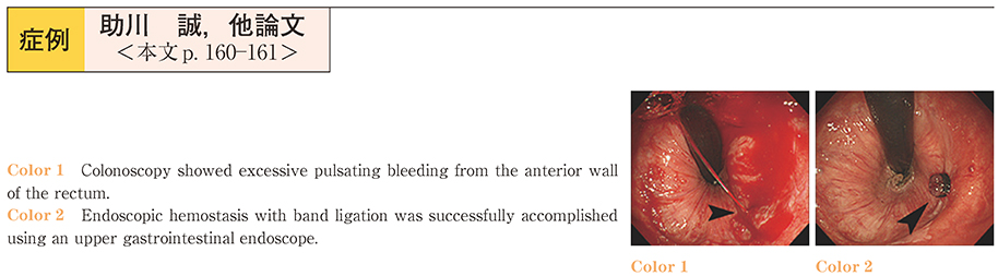
    2014 年 84 巻 1 号 p. 160-161
    発行日: 2014/06/14
    公開日: 2014/06/21
    ジャーナル フリー
    A man in his eighties on antiplatelet therapy was admitted to our hospital with hematochezia. He had undergone a transrectal prostatic biopsy 14 days earlier. Emergency colonoscopy showed hemorrhage and a massive clot in the rectum below the peritoneal reflection (2 cm oral to the anal verge) . Excessive pulsating bleeding from the anterior wall of the rectum was also observed. Endoscopic hemostasis with band ligation was accomplished successfully using an upper gastrointestinal endoscope. He could resume oral intake from Day 5 and was discharged from the hospital on Day 13. Rectal bleeding is recognized as one of the acute complications of transrectal prostatic biopsy; however, there is only one report of the late onset of bleeding in the Japanese literature from 2005-2012. Endoscopic hemostasis by clipping is commonly performed for rectal bleeding. However, our case suggests the effectiveness of endoscopic hemostasis with band ligation for the control of rectal bleeding.
  • 加藤 孝征, 小山 茂, 金崎 峰雄, 新倉 利啓, 藤井 徹朗, 梅沢 翔太郎, 城野 文武, 秋本 恵子, 高畑 彩子, 北條 裕美子, ...
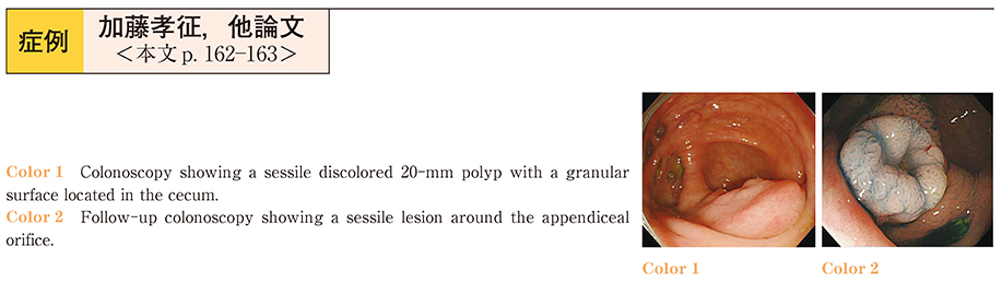
    2014 年 84 巻 1 号 p. 162-163
    発行日: 2014/06/14
    公開日: 2014/06/21
    ジャーナル フリー
    A 61-year-old man presented to our hospital with abdominal pain and hematochezia. No abnormal findings were noted on laboratory examination, while total colonoscopy revealed a sessile discolored 20-mm polyp with a granular surface in the cecum. Endoscopic piecemeal mucosal resection (EPMR) of the polyp was performed. Pathological examination indicated findings consistent with a sessile serrated adenoma/polyp (SSA/P) . A follow-up colonoscopy performed after 1 year revealed a sessile lesion around the appendiceal orifice and a biopsy examination indicated findings consistent with a SSA/P, similar to the EPMR specimen. The lesion was judged to be endoscopically unresectable, therefore, laparoscopic excision was performed. The patient exhibited a good clinical course after the surgery, and showed no furtherrecurrence. Thus, we report a rare case of relatively early recurrence of an SSA/P surrounding the appendiceal orifice.
  • 木村 麻衣子, 小泉 浩一, 桑田 剛, 田畑 拓久, 堀口 慎一郎, 比島 恒和, 大橋 一輝
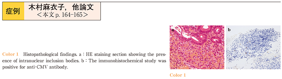
    2014 年 84 巻 1 号 p. 164-165
    発行日: 2014/06/14
    公開日: 2014/06/21
    ジャーナル フリー
    A case of acute hemorrhagic rectal ulcer (AHRU) with cytomegalovirus (CMV) enteritis is reported. AHRU is characterized by sudden painless onset of massive hemorrhage from rectal ulcer (s) in patients with serious underlying illnesses. A 70-year-old woman with multiple myeloma was referred to our division for fresh blood in the stool. Endoscopic examination revealed two deep ulcers, one in the lower rectum and the other in the ascending colon. The initial endoscopic diagnosis was AHRU, however, histopathological examination of the biopsy specimen from both ulcers showed evidence of CMV infection. The patient died 22 days later from progression of the multiple myeloma. We reviewed the data of 17 cases (including our case) reported previously from Japan. In conclusion, CMV infection can be a cause or a risk factor for AHRUs.
  • 守屋 まりこ, 倉持 みずき, 櫛田 幸, 玉井 恒憲, 島田 哲也, 細田 泰雄, 関塚 永一
    2014 年 84 巻 1 号 p. 166-167
    発行日: 2014/06/14
    公開日: 2014/06/21
    ジャーナル フリー
    A 76-year-old woman visited our hospital complaining of diarrhea and left lower abdominal pain. Colonoscopy showed erythema and edema of the mucosa. Non-specific enteritis was suspected and the patient was observed with supportive treatment. However, as the patient’s symptoms continued to deteriorate, further investigation was conducted. A complete blood count showed eosinophilia, which led to the suspicion of eosinophilic gastroenteritis. Therefore, prednisolone was started, both for diagnostic and therapeutic purposes. However, the symptoms did not improve. Findings of a repeat colonoscopic examination suggested cap polyposis (CP) . After eradication of H.pylori infection with antibiotic therapy, her clinical condition and endoscopic findings improved remarkably, consistent with previous reports.
  • 山本 洋輔, 田原 利行
    2014 年 84 巻 1 号 p. 168-169
    発行日: 2014/06/14
    公開日: 2014/06/21
    ジャーナル フリー
    A 47-year-old man visited our hospital with the chief complaints of diarrhea, dysgeusia, alopecia and onychodystrophy. Endoscopic examinations revealed an edematous and reddish mucosa and many polypoid lesions in the stomach and colon. Cronkhite-Canada syndrome was diagnosed and the patient was treated with intravenous prednisolone. The symptoms improved gradually, however, remission was not achieved. At one month after treatment, the serum albumin level still persisted. We then decided to administer steroid pulse therapy. With this therapy, the symptoms improved dramatically, and steroid maintenance therapy was initiated with betamethasone, which has a longer half-life than prednisolone. The patient’s condition improved, and repeat endoscopy revealed improvement of the polypoid lesions and mucosal inflammation. This case indicates that steroid pulse therapy may be useful for at least a proportion of patients with Cronkhite Canada syndrome, and may also be useful for helping in deciding the treatment policy.
  • 水野 達人, 岡村 幸重, 伊倉 顕彦, 山本 悠太, 小林 真介, 片山 正, 白石 貴久, 上原 淳, 佐伯 恵太, 寺元 研, 東澤 ...
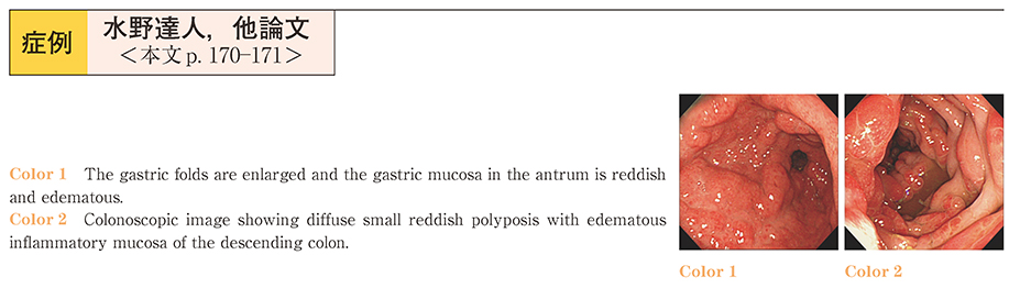
    2014 年 84 巻 1 号 p. 170-171
    発行日: 2014/06/14
    公開日: 2014/06/21
    ジャーナル フリー
    The patient was a 66-year-old male, with the chief complaint of diarrhea. He began experiencing diarrhea at the beginning of September 2012, with the symptom worsening in October, along with the onset of pedal edema and dysgeusia. He was hospitalized in November 2012. Blood biochemistry at the time of hospitalization confirmed hypoalbuminemia, and a colonoscopic examination revealed edematous change of the mucous membrane of the entire large intestine and mucosal hyperpigmentation. Upper gastrointestinal endoscopy revealed the presence of hypertrophic adenomatous polyps from the stomach to the duodenum. Histopathology confirmed severe edema of the mucous membrane of both the upper and lower GI tract, mild to moderate chronic inflammatory cell infiltration and ductal dilatation, consistent with the diagnosis of Cronkhite-Canada syndrome. The patient was then treated with PSL 50 mg/day, and the clinical symptoms and endoscopic findings improved rapidly after the treatment initiation.
  • 河村 貴広, 浅川 剛人, 金城 美幸, 高浦 健太, 西尾 匡史, 勝倉 暢洋, 小橋 健一郎, 橋口 真子, 先田 信哉, 有村 明彦, ...
    2014 年 84 巻 1 号 p. 172-173
    発行日: 2014/06/14
    公開日: 2014/06/21
    ジャーナル フリー
    Pneumatosis cystoides intestinalis (PCI) is a relatively rare disease characterized by multiple gas-filled cysts in the bowel wall. We report a rare case of recurrent PCI in a patient with alcoholic cirrhosis, who was successfully treated by oxygen supplementation via a nasal cannula. A 57-year-old female who was being followed up at our hospital for alcoholic cirrhosis presented with persistent abdominal pain. CT showed pneumatosis in the large intestinal wall, and PCI was diagnosed. Although her symptoms were mild at onset, they deteriorated 10 months later. The patient was admitted and treated by oxygen supplementation at 5 L/minute via a nasal cannula for one week, and her symptoms and intestinal pneumatosis disappeared. Although the PCI recurred four times in the subsequent 14 months and sometimes intraabdominal free air was seen, the oxygen supplementation via a nasal cannula (5 L/min×1 week) was always successful. Within one week, her symptoms, intestinal pneumatosis, and intraabdominal free air disappeared. Because oxygen supplementation via a nasal cannula is easy, safe and useful, it should be considered as the treatment of first choice for PCI. There are no reports yet of PCI in patients with alcoholic cirrhosis, and the relation between the PCI and liver disease in this patient remains uncertain.
  • 南雲 大暢, 安達 哲史, 江川 優子, 市原 広太郎, 齋藤 訓永, 多田 正弘, 風間 博正
    2014 年 84 巻 1 号 p. 174-175
    発行日: 2014/06/14
    公開日: 2014/06/21
    ジャーナル フリー
    A 45-year-old woman presented with a 2-month history of diarrhea and lower abdominal pain.The patient began to suffer from bloody diarrhea, and was referred to hospital.
    Colonoscopy revealed elevated lesions like a submucosal tumor at two locations in the sigmoid colon and rectum.
    The overlying mucosa was edematous and showed small red nodules at the center of the lesion with white spots and a scar.
    Intestinal endometriosis was suspected, and biopsy specimens were obtained from the nodules.
    On histopathological examination, the submucosal layer through the lamina propria showed a structure similar to the uterine stroma and glands.
    Immunostaining revealed that the lesion was estrogen-receptor positive, therefore, the lesions were diagnosed as intestinal endometriosis.
  • 柿本 年春, 辻 忠男, 桂 英之, 金田 浩幸, 篠崎 博志, 水谷 友美, 三浦 邦治, 宮永 亮一, 松波 幸寿
    2014 年 84 巻 1 号 p. 176-177
    発行日: 2014/06/14
    公開日: 2014/06/21
    ジャーナル フリー
    A 58-year-old man was admitted to our hospital with a seven-day history of right lower quadrant abdominal pain. Contrast-enhanced CT showed multiple diverticula and pericolic inflammation in the ascending colon. The patient was diagnosed as having colonic diverticulitis and treated with antibiotics. After hospitalization for 10 days, the patient improved and was discharged.
    A month later, total colonoscopy was performed, which showed multiple colonic diverticula in the ascending colon and a polypoid lesion at the same site. The polypoid lesion was covered with normal colonic mucosa and the top of the lesion was reddish and slightly depressed. We obtained a biopsy specimen and noticed some fatty tissue at the site of the biopsy. We finally diagnosed the polypoid lesion as an inverted colonic diverticulum and immediately closed the biopsy site with endoclips. The patient was admitted and showed no complaints. He was discharged on day 4 of admission.
    Inverted colonic diverticulum is a rare disease. It resembles a colonic polyp or submucosal tumor and may be misdiagnosed. Therefore, the possibility of this condition must be borne in mind when polypoid lesions are encountered, to avoid unnecessary biopsy or polypectomy, especially in patients with diverticular disease.
  • 柳沼 行宏, 呉 一眞, 盧 尚志, 青木 順, 岡澤 裕, 高橋 里奈, 水越 幸輔, 嵩原 一裕, 高橋 玄, 小島 豊, 五藤 倫敏, ...
    2014 年 84 巻 1 号 p. 178-179
    発行日: 2014/06/14
    公開日: 2014/06/21
    ジャーナル フリー
    A 86 year-old male who visited a local physician with the chief complaint of blood in the stool. At the initial colonoscopy, a polyp with a partially disrupted top was found in the sigmoid colon. Histopathological examination of a biopsy specimen obtained from the tumor base showed moderately differentiated adenocarcinoma. A second colonoscopy was performed 10 days later, which revealed that the tumor top was almost completely disrupted. The patient was referred to our hospital and admitted for endoscopic therapy 35 days after the initial colonoscopy. A repeat colonoscopy confirmed an increase of the tumor volume and progression of the lesion to a sessile-type cancer. Consequently, the lesion was diagnosed as a submucosal cancer and endoscopic submucosal dissection (ESD) was performed. Histopathological examination showed that the tumor was a well to moderately well differentiated adenocarcinoma, SM 5,000 μm. We recommended radical resection; however, the patient was kept under follow-up because he refused surgery. We present this case as an example of early colon cancer in which the tumor top was rapidly disrupted and morphological change was observed.
  • 大津 威一郎, 鎮西 亮, 笹島 圭太, 土井 浩達, 高橋 正憲, 熊谷 純一郎, 塩屋 雄史, 大島 忠, 甲嶋 洋平, 登内 昭彦
    2014 年 84 巻 1 号 p. 180-181
    発行日: 2014/06/14
    公開日: 2014/06/21
    ジャーナル フリー
    A 47-year-old man was admitted to our hospital complaining of anal pain and difficulty in defecation. Gastrografin enema showed marked narrowing of the lumen and spicula of the lower rectum. Colonoscopy revealed circumferential stenosis with edematous mucosa, a coarse cobblestone like appearance, but no ulcer in the lower rectum. Histological findings of biopsy specimens obtained from the first colonoscopy showed non-neoplastic cells. However, the imaging findings and history of present illness strongly suggested the possibility of type 4 colorectal cancer. Therefore, we performed colonoscopy three times until a definitive diagnosis was obtained. A total of 51 biopsy specimens were examined until a signet ring cell carcinoma was detected, which was detected in a specimen only obtained from an erosion. The resected specimen showed marked thickening of the rectal wall. Histological examination revealed transmural invasion by poorly differentiated adenocarcinoma and a signet ring cell carcinoma associated with severe desmoplastic reaction. Type 4 colorectal cancer is an uncommon morphological type of colon cancer. Even many biopsy specimens obtained from non-erosive areas can rarely provide cancer cells, because of the invasive configuration of the cancer. Therefore, multiple biopsies, especially from erosions, are necessary to obtain a definitive histological diagnosis.
  • 別府 加寿子, 坂本 直人, 武田 武史, 磯野 彩, 山本 悠子, 石井 重登, 小谷 知弘, 水井 智和, 猪狩 功遺, 折笠 英紀, ...
    2014 年 84 巻 1 号 p. 182-183
    発行日: 2014/06/14
    公開日: 2014/06/21
    ジャーナル フリー
    A 74-year-old woman was admitted to our hospital complaining of a sense of abdominal fullness. Abdominal CT with a radiocontrast agent showed marked ascites, opacification of the mesenteric fat, and several enhancing lesions, including swollen lymph nodes in the right groin area, a small nodule in the pouch of Douglas, and nodules in the anorectal area. Ascitic cell block cytology showed tumor cells containing blackish brown granules. Based on these findings, we made a preliminary diagnosis of malignant melanoma. Colonoscopy revealed black elevated lesions in the anorectal area, and histology of biopsy specimens obtained from the lesions showed tumor cells containing blackish brown granules. Based on these findings, a final diagnosis of stage IV malignant anorectal melanoma was made.
    A definitive diagnosis of malignant anorectal melanoma can be challenging, because the colonoscopic findings of anal polyps, anal canal cancer and malignant lymphoma can be similar. Careful examination for detecting the colonic elevated lesions and obtaining biopsies from these lesions may allow early diagnosis of anorectal melanoma at a higher frequency.
  • 山宮 知, 北村 勝哉, 石井 優, 佐藤 悦基, 岩田 朋之, 野本 朋宏, 吉田 仁 
    2014 年 84 巻 1 号 p. 184-185
    発行日: 2014/06/14
    公開日: 2014/06/21
    ジャーナル フリー
    A 79-year-old woman was admitted to our hospital with weight loss and appetite loss. Physical examination revealed no abnormal findings, and laboratory data, including the serum levels of tumor makers, were normal. A whole-body CT showed a tumor extending from the left kidney to the pelvic cavity. EUS revealed an unclear hyperechoic tumor measuring 2 cm in size near the left kidney. EUS-FNA was performed, and the samples revealed malignancy of cytology (Class V) and histology (poorly differentiated adenocarcinoma) . However, immunostaining did not yield a diagnosis of the primary cancer.
    Based on the above, the patient was diagnosed as having cancer of unknown primary. EUS-FNA performed around the kidney such as this case is very rare, and there has been only one case report of cancer of unknown primary. EUS-FNA is useful not only for the diagnosis of pancreatic, biliary and digestive tract diseases, but also for that of mediastinal, abdominal and pelvic lesions, and ascites. EUS-FNA is a safe and minimally invasive procedure with a high diagnostic value.
  • 千嶋 巌, 矢吹 拓, 千嶋 さやか, 北岡 吉民, 上原 慶太, 中山 成一
    2014 年 84 巻 1 号 p. 186-187
    発行日: 2014/06/14
    公開日: 2014/06/21
    ジャーナル フリー
    A 51-year-old man who drank about 3 L/day of alcohol was admitted to our hospital with abdominal distention. The serum amylase was 367 IU/l, serum lipase was 165 IU/l, and the ascitic cell count and amylase level were 1,764IU/l and 8,732 IU/l, respectively. Contrast-enhanced CT and MRCP revealed a massive pleural effusion and ascites, and dilatation of the pancreatic duct branches with an additional parenchymal pseudocyst at the pancreatic head. ERCP showed communication between the pancreatic duct and the pseudocyst. A 5F, 5-cm pancreatic stent was placed, and the symptom gradually improved. Subsequent follow-up CT and X-ray revealed marked improvement. On the 45th day, the patient was discharged from the hospital without any symptoms or complications.
    Decompression of the pancreatic duct by pancreatic stent placement may be useful for the treatment of pancreatic ascites and pleural effusion.
  • 井上 健太郎, 相浦 浩一, 市東 昌也, 星本 相淳, 壁島 康郎, 小柳 和夫, 有泉 健, 高木 英恵, 玉井 博修, 入江 理恵, ...
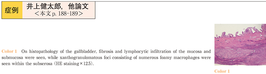
    2014 年 84 巻 1 号 p. 188-189
    発行日: 2014/06/14
    公開日: 2014/06/21
    ジャーナル フリー
    A 71-year-old man presented with a 5-day history of abdominal distention. Abdominal ultrasonography showed diffuse thickening of the gallbladder wall and an 18 mm-sized gallstone in the neck. Magnetic resonance cholangiopancreatography showed a stricture, 18 mm in length, at the proximal portion of the extrahepatic biliary duct and dilatation of the intrahepatic biliary duct. Dynamic computed tomography revealed gradual enhancement of the thickened wall at the neck of the gallbladder, and the border between the gallbladder and the liver was indistinct. EUS showed a well-circumscribed hypoechoic mass at the neck of the gallbladder interrupting the extrahepatic biliary tract, suggesting gallbladder carcinoma with bile duct invasion. However, ERC and intraductal ultrasonography yielded conflicting results. Endoscopic retrograde cholangiography revealed a smooth stricture of the biliary tract ; furthermore, intraductal ultrasonography showed homogeneous thickening of the submucosal layer of the gallbladder with a smooth mucosal layer, and no evidence of direct invasion of the bile duct. These findings were indicative of xanthogranulomatous cholecystitis with Mirizzi syndrome. In open surgery, there were no findings suggestive of malignancy and cholecystectomy was performed. On histopathology, fibrosis and lymphocytic infiltration with xanthogranulomatous foci were present at the thickened wall of the gallbladder neck. The final pathological diagnosis was chronic cholecystitis with xanthogranulomatous change, which was consistent with the results obtained by intraductal ultrasonography. Intraductal ultrasonography was useful for differentiating between gallbladder carcinoma and chronic cholecystitis.
  • 三枝 陽一, 美原 静香, 加藤 彩, 竹下 知親, 西元寺 克禮, 大井田 正人
    2014 年 84 巻 1 号 p. 190-191
    発行日: 2014/06/14
    公開日: 2014/06/21
    ジャーナル フリー
    A 77-year-old man gave a history of passing brown urine 1 month earlier. The patient presented with jaundice 2 weeks ago, and was admitted to our hospital. Blood tests revealed evidence of obstructive jaundice. Abdominal ultrasonography, computed tomography (CT) , and magnetic resonance cholangiopancreatography (MRCP) showed dilatation of the intrahepatic bile ducts and thickening of the common bile duct. Endoscopic retrograde cholangiopancreatography (ERCP) revealed an unnatural stricture of the common bile duct. Strictures of the intrahepatic bile ducts were sporadically noted, and immunoglobulin G (IgG) 4-related sclerosing cholangitis was suspected. Additional blood tests revealed significant elevation of the serum IgG and IgG4 levels to 3,830 mg/dl and 1,390 mg/dl, respectively. IgG4-related sclerosing cholangitis was diagnosed based on the findings. The patient was treated with oral ursodeoxycholic acid (600 mg/day) and prednisolone (30 mg/day) . The liver function improved, and ERCP, MRCP and CT showed marked improvement in the stricture of the common bile duct. The dose of prednisolone was reduced, and there has been no recurrence as at the time of this writing. Because IgG4-related sclerosing cholangitis is a relatively uncommon disease, diagnosis and treatment are often challenging. We describe our experience of a patient diagnosed as having IgG4-related sclerosing cholangitis.
  • 乾山 光子, 岸本 有為, 岡野 直樹, 原 精一, 宅間 健介, 三村 享彦, 伊藤 謙, 塩沢 一恵, 渡邉 学, 大久保 陽一郎, 渋 ...
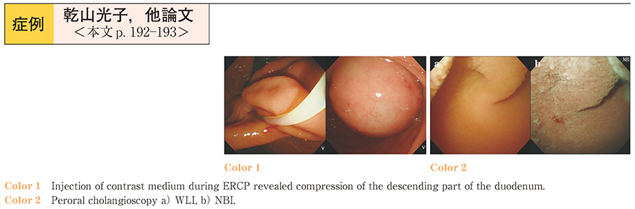
    2014 年 84 巻 1 号 p. 192-193
    発行日: 2014/06/14
    公開日: 2014/06/21
    ジャーナル フリー
    A 62-year-old woman referred to us for the management of suspected acute cholecystitis was hospitalized. Abdominal CT showed choledocholithiasis and a duodenal diverticulum. Esophagogastroduodenoscopy (EGD) revealed compression of the descending part of the duodenum in the same region as the diverticulum on the abdominal CT. ERCP was performed for the choledocholithiasis. Numerous stones were seen in the cystic lesion upon injection of contrast medium, and continuous injection revealed a cystic lesion measuring 22 mm in diameter. A diagnosis of choledochocele was made. The major duodenal papilla could not be identified on the EGD because of the ballooned choledochocele filled with contrast medium. A needle knife precut was made, and the choledochocele subsequently collapsed after the CBD stones were removed. Endoscopic hemostasis using coagulation and hemoclip was performed to stop oozing from the site of the precut. ERCP was performed again 1 week later. The major duodenal papilla was identified and EST was performed, followed by CBD stone removal. The level of pancreatic amylase in the bile was high at the first ERCP and decreased after the precut. Functional pancreaticobiliary maljunction was diagnosed, which disappeared after the precut and EST.
  • 小谷 知弘, 石井 重登, 別府 加寿子, 磯野 彩, 山本 悠子, 水井 智和, 猪狩 功遺, 中谷 晃典, 二川 俊二, 折笠 英紀
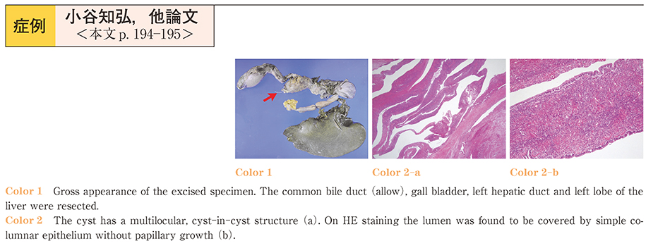
    2014 年 84 巻 1 号 p. 194-195
    発行日: 2014/06/14
    公開日: 2014/06/21
    ジャーナル フリー
    A 28-year-old woman visited our hospital with the complaints of itching and jaundice. Blood examination showed elevation of the hepatobiliary enzymes and bilirubin. Abdominal US, CT and MRI revealed cystic lesions with septae spreading contiguously to the left lobe, left hepatic duct and common bile duct. Endoscopic retrograde cholangiography (ERC) demonstrated translucent components in the bile duct, and CT immediately after the ERC suggested that one partial cystic lumen filled with contrast medium was communicating with the bile duct. Therefore, we made the diagnosis of intraductal papillary neoplasm of the bile duct (IPNB) preoperatively. However, histological examination of the resected specimens after a left hepatectomy revealed the diagnosis of mucinous cystic neoplasm (MCN) , because the ovarian-like stroma showed positive immunostaining for the estrogen receptor and progesterone receptor. At present, 10 months after the surgery, there is no evidence of tumor recurrence.
    Hepatobilialy cystic neoplasms are classified into MCNs, which contain an ovarian-like stroma, and IPNB, which do not contain an ovarian-likes stroma. Although tens of cases of MCN have been reported, the reason for the existence of the stromal cells is still unclear. Our case was atypical in respect of its macroscopic appearance and its communication to the bile duct. Herein, we report this case as we believe that it will contribute to elucidation of the pathogenesis.
  • 山田 俊哉, 丸山 秀樹, 古谷 健介, 鏑木 大輔, 待木 雄一
    2014 年 84 巻 1 号 p. 196-197
    発行日: 2014/06/14
    公開日: 2014/06/21
    ジャーナル フリー
    A 55-year-old woman was diagnosed as having Gardner syndrome’s at the age of 28. Blood chemistry analysis showed repeated episodes of liver dysfunction and inflammatory worsening from early 2013. MRCP revealed intrahepatic bile duct dilatation. Abdominal CT revealed a low-density lesion in the porta hepatis causing biliary obstruction. Further investigation by ERCP revealed a post-papilloplasty state and a sessile lesion in the lower bile duct. We obtained biopsy specimens from the two different lesions and placed a drainage tube at the portal hepatis. Transnasal endoscopy with a slim endoscope allowed us to visualize the irregular lesion in the bile duct at the hilum. There was obstruction of the bile duct at the porta hepatis and a type IIa lesion in the lower bile duct. Endoscopic biopsy of these separate lesions showed identical features, those of an adenocarcinoma. Surgery was performed. Histopathological examination of the resected specimens revealed papillary adenocarcinoma of the bile duct, pat BlcsiA, m/s (-) , pHinf0, pGinf0, pPanc0, pDU0, pPV0, pA0, pN0, pHM0, pDM0, pEM0, ly0, v0, ne0, fStage I. The two lesions were histopathologically connected.
    We report a rare case of superficial widely spreading carcinoma of the bile duct occurring 27 years after the diagnosis of Gardner’s syndrome.
  • 竹内 千尋, 鴨志田 敏郎, 佐々木 翔一, 浜野 由花子, 綿引 隆久, 大河原 悠, 大河原 敦, 柿木 信重, 平井 信二, 岡 裕爾 ...
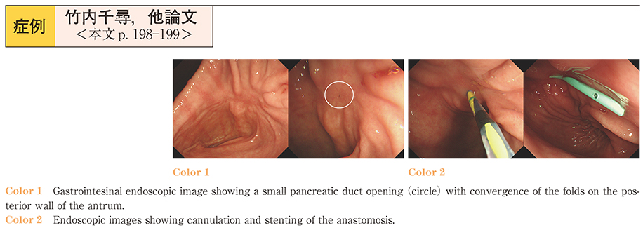
    2014 年 84 巻 1 号 p. 198-199
    発行日: 2014/06/14
    公開日: 2014/06/21
    ジャーナル フリー
    A 65-year-old male patient was admitted to the hospital complaining of epigastralgia. He was suffering from chronic pancreatitis associated with alcoholism and had undergone partial pancreatectomy with pancreaticogastrostomy because of pancreatic tumor, for which cancer could not be ruled out; the operative specimen revealed tumor-forming pancreatitis. On admission, hematologic examination revealed no elevation of the pancreatic enzymes and a contrast-enhanced CT showed no evidence of active pancreatitis. Despite intravenous hydration and opiate administration, the patient continued to suffer from epigastric pain, therefore, we attempted endoscopic therapy. Upper GI endoscopy showed a small pancreatic duct opening with convergence of the folds on the posterior wall of the antrum, and stenting was performed through the anastomotic opening. Thereafter, the patient’s symptom disappeared and there has been no recurrence.
    Endoscopic treatment for abdominal pain associated with chronic pancreatitis has been reported to yield high success rates of symptom relief without major complications, but also a high recurrence rate during follow-up. We describe an unusual case of endoscopic stenting performed through a pancreaticogastrostomy anastomosis. Owing to its low invasiveness, endoscopic therapy can be offered as first−line treatment in such cases, although careful follow-up is required.
  • 伊藤 裕幸, 今井 仁, 矢崎 利典, 市川 仁志, 永田 順子, 小嶋 清一郎, 高清水 真二, 白井 孝之, 渡辺 勲史, 山田 美鈴, ...
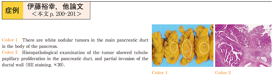
    2014 年 84 巻 1 号 p. 200-201
    発行日: 2014/06/14
    公開日: 2014/06/21
    ジャーナル フリー
    A 75-year-old man had been followed up after treatments of lung cancer, retroperitoneal fibrosis and malignant lymphoma at our hospital. Abdominal CT in February 2013 revealed dilatation of the main pancreatic duct in the body and the tail. MRCP and ERCP showed a nodular mass, 18 × 8 mm in size, in the pancreatic body, as well as dilatation of the distal pancreatic duct. Distal panreatectomy and splenectomy were performed under the preoperative diagnosis of early-stage pancreatic ductal carcinoma. However, the pathological diagnosis of the resected pancreatic mass was intraductal tubulopapillary neoplasm (ITPN) . Histochemical examinations showed positive results for MUC1 on the luminal side of the pancreatic ducts, and negative results for MUC2 and MUC5AC. Acinar cell carcinoma and malignant lymphoma were excluded on the basis of the negative staining results for trypsin and Bcl-10. ITPN is a rare and relatively newly described clinical type of pancreatic neoplasm. This case serves to emphasize that we must bear in mind the possibility of ITPN in the differential diagnosis of pancreatic tumors.
  • 石田 奈々, 矢野 貴史, 三島 孝仁, 山根 敬子, 長谷川 力也, 中目 哲平, 石黒 康浩, 村田 東, 荒木 正雄, 風間 暁男, ...
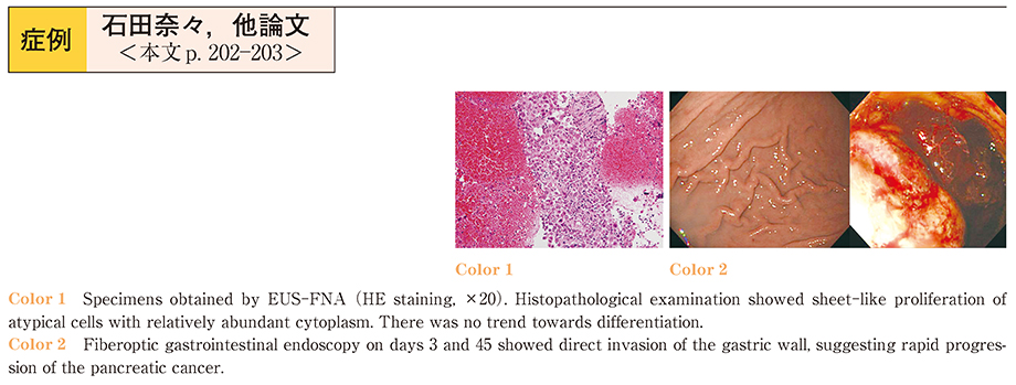
    2014 年 84 巻 1 号 p. 202-203
    発行日: 2014/06/14
    公開日: 2014/06/21
    ジャーナル フリー
    A 68-year-old man presented at our hospital with abnormal results of blood tests performed by a local physician. Computed tomography revealed a tumor in the tail of the pancreas with multiple liver tumors, splenic invasion, and gastric-wall invasion, and the patient was admitted. Endoscopic ultrasonography-guided fine-needle aspiration (EUS-FNA) was subsequently performed, and anaplastic carcinoma of the pancreas was diagnosed. The patient was started on combination chemotherapy with gemcitabine and S-1. On day 4 of treatment, he developed fever and altered consciousness, and the chemotherapy was discontinued. On day 45, the patient vomited blood, presumably because of direct invasion of the stomach by the pancreatic ductal carcinoma. Subsequently, multiple organ failure developed, and the patient died on day 56.
    Anaplastic carcinoma is an extremely rare type of pancreatic cancer that is characterized by extremely rapid progression and poor outcomes as compared to the usual pancreatic ductal carcinomas. Most anaplastic carcinomas are diagnosed at surgery or autopsy. We describe our experience of a rapidly progressive case of anaplastic carcinoma of the pancreas with gastric invasion that was diagnosed by EUS-FNA.
お詫びと訂正
feedback
Top