Journal of Spine Research
Online ISSN : 2435-1563
Print ISSN : 1884-7137
Volume 12, Issue 11
Displaying 1-12 of 12 articles from this issue
Editorial
Review Article
  • Atsuhiro Yoshida, Tsutomu Akazawa, Yoshiaki Torii, Jun Ueno, Tasuku Um ...
    2021 Volume 12 Issue 11 Pages 1278-1286
    Published: November 20, 2021
    Released on J-STAGE: November 20, 2021
    JOURNAL FREE ACCESS

    Background: Although it has been concluded that the natural course of adolescent idiopathic scoliosis (AIS) is not highly detrimental to later health even if untreated, there are few data with long-term follow-up, and there is some variation among researchers in their interpretation. However, long-term follow-up data are scarce and interpretation of such data varies among researchers.

    Purpose: To conduct a literature review of the long-term natural history of AIS, to reevaluate cases in which therapeutic intervention should be performed after bone maturation, and to identify problems with previous studies.

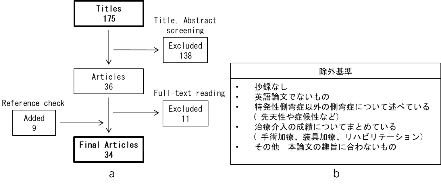
    Methods: We conducted a literature search using PubMed, checked the contents of 175 papers, and finally adopted 34 papers.

    Results: Curve progression is more likely to occur in the thoracic curve, especially in the Cobb angle of 50-75°, with less than 1° progression per year. Lung function is related to Cobb's angle, with shortness of breath in daily life occurring at angles of 80° or more, and a greater impact on cardiac function at angles of 110° or more. Low back pain is not related to Cobb's angle, but its frequency and severity tend to be higher than in the healthy group. Mortality, prevalence of psychiatric disorders, and social functioning are not inferior to normal subjects. Problems with previous studies include lack of clarification of etiology, lack of data on long-term natural history, and uncertainty of Onset.

    Conclusion: Surgical intervention should be considered for thoracic curves with Cobb angle >50 degrees, otherwise it should be considered multifaceted.

    Download PDF (1145K)
Original Article
  • Sunao Tanaka, Kanichiro Wada, Gentaro Kumagai, Toru Asari, Yasuyuki Is ...
    2021 Volume 12 Issue 11 Pages 1287-1293
    Published: November 20, 2021
    Released on J-STAGE: November 20, 2021
    JOURNAL FREE ACCESS

    Introduction: The aim of this study was to clarify the risk factors of distal adding-on (DAO) after selective thoracic fusion surgery without osteotomy for patients with Lenke types 1A-R and 2A-R adolescent idiopathic scoliosis.

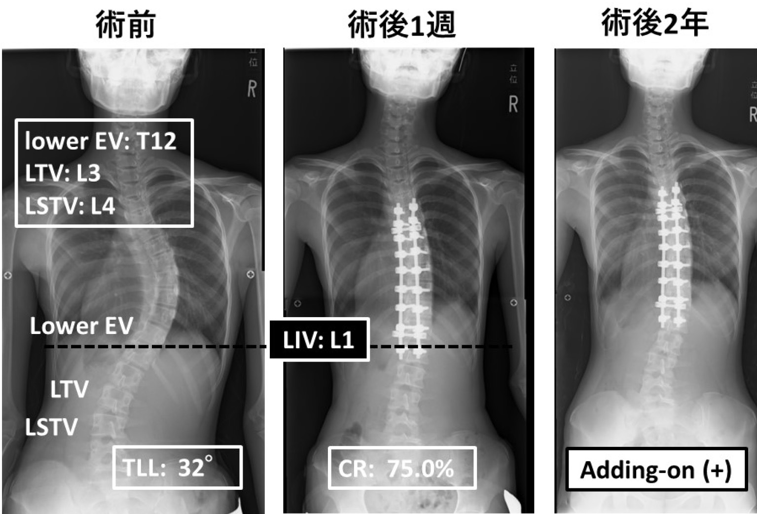
    Methods: Eleven Lenke 1A-R (10 females, average 14.6 years old), and 11 Lenke 2A-R (9 females, average 13.6 years old) patients were investigated retrospectively. Patients were classified to two groups based on the occurrence of DAO until 2 years after surgery: DAO group and non-DAO group. Standing posteroanterior spine radiographs were obtained at three times: preoperatively, 1 week after surgery and 2 years after surgery. In preoperative X-ray, lowest end vertebra (EV), last touching vertebra (LTV) and last substantially touching vertebra (LSTV) were identified. Cobb angles of the proximal thoracic curves, the main thoracic (MT) curves, and the thoracolumbar/lumbar (TLL) curves were measured before and after surgery and correction rates were obtained in each curve. The risk factors associated with DAO were analyzed by univariate analysis and logistic regression analysis.

    Results: Twelve patients (8 of Lenke 1A, 4 of 2A) were placed in the DAO group. There were significant differences in preoperative MT curve (P = 0.040), TLL curve at 1 week after surgery (P=0.025), and correction rates of TLL curve (P = 0.002) between the DAO and the non-DAO group. Logistic regression analysis revealed high correction rate of TLL curve was a risk factor of DAO (Odds ratio was 1.14, P = 0.019). There were 9 patients whose TLL correction rates were more than 75%, 4 were in DAO group and 5 were non-DAO group. In all patients with DAO whose TLL correction rates were more than 75%, LIV were cranial to LTV and LSTV. On the other hand, two of three patients with non-DAO cases, LIV were caudal to every three landmarks, lower EV, LTV and LSTV.

    Conclusions: Our results suggested we should choose LIV as the same or more distal as the lowest EV, LTV and LSTV when correction rates of TLL curves are estimated to be more than 75 percent.

    Download PDF (1564K)
  • Ayato Nohara, Noriaki Kawakami, Tetsuya Ohara, Toshiki Saito, Ryoji Ta ...
    2021 Volume 12 Issue 11 Pages 1294-1299
    Published: November 20, 2021
    Released on J-STAGE: November 20, 2021
    JOURNAL FREE ACCESS

    Introduction: The purpose of this study is to evaluate the long-term postoperative results of patients with idiopathic scoliosis (AIS) with lumbar modifier C and LIV selected as L3, and to determine whether DD could be predicted from preoperative X-ray parameters.

    Methods: A total of 44 patients with AIS met the inclusion criteria: 1) female diagnosed with AIS with lumbar modifier C, 2) PSF or Anterior and PSF, 3) L3 as the LIV, 4) 11-20 years of age at the time of surgery, and 5) MRI and X-ray images at 10 years postoperatively.

    Results: DD at unfused lumbar intervertebral discs was identified in 48% of the patients at 10 years postoperatively. The selected patients were classified into 2 groups based on DD occurrence. Preoperative and postoperative 10 years L3/4 disc angle (DA) exhibited significant differences between two groups. The incidence of DD markedly increased at the preoperative L3/4DA greater than 5°.

    Conclusion: In the AIS patients with lumbar modifier C and LIV at L3, it was suggested that not only postoperative but also preoperative wedge formation of the L3/4 disc was closely related to the occurrence of disc degeneration in the long-term postoperative period.

    Download PDF (821K)
  • Taro Okumura, Takehide Katogi, Toshiaki Kotani, Tsuyoshi Sakuma, Keita ...
    2021 Volume 12 Issue 11 Pages 1300-1305
    Published: November 20, 2021
    Released on J-STAGE: November 20, 2021
    JOURNAL FREE ACCESS

    Introduction: We investigated the effect of exercise habits on abdominal muscle strength in postoperative adolescent idiopathic scoliosis (AIS) patients.

    Methods: Fifty-nine female patients were evaluated quantitatively for abdominal muscle strength before and 1 and 2 years after surgery using sits-up according to the Japanese National Physical Fitness Test. The number of sits-up times in 30 seconds was recorded and the rate of change (%) at 2 year after surgery was calculated. The patients were divided into athletic and non-athletic groups based on their preoperative club affiliation. Statistical analyses were performed to compare the numbers of sits-up performed preoperatively and two years after surgery and correlations between the rate of change (%) at 2 year after surgery and postoperative Cobb angle, Cobb angle correction rate, number of vertebral bodies fixed, Lowest Instrumented Vertebra (LIV), and forward bending were examined within each group.

    Results: The athletic group showed improvement in sits-up to the same level as preoperative at 2 years postoperatively, while the non-athletic group did not recover.

    Conclusions: The abdominal muscle strength of patients with exercise habits recovers to the preoperative level 2 years after surgery.

    Download PDF (965K)
  • Norihiro Oku, Satoru Demura, Satoshi Kato, Kazuya Shinmura, Noriaki Yo ...
    2021 Volume 12 Issue 11 Pages 1306-1310
    Published: November 20, 2021
    Released on J-STAGE: November 20, 2021
    JOURNAL FREE ACCESS

    Introduction: Brace treatment is generally applied as a conservative treatment for pediatric spine deformity, and it is reported to be effective in preventing the progression of scoliosis. On the other hand, it has been also reported that the strength of abdominal trunk muscles is reduced by wearing the brace. In addition, there are no reports on prospective studies of the relationship between brace treatment and abdominal trunk muscle strength (ATMS) in pediatric scoliosis. In this study, we prospectively measured ATMS in pediatric scoliosis patients who started brace treatment, and examined the effect of brace treatment on ATMS and the relationship between ATMS and scoliosis progression.

    Methods: 20 patients with pediatric scoliosis who started on brace treatment were included in this study. Physical measurements (height, weight, BMI, grip strength, knee extension strength) were performed before and six months after the start of treatment. ATMS was measured using an exercise device for the abdominal trunk muscles that also measures muscle strength. We also measured Cobb angle before and six months, and classified those of improved or unchanged main curve into "stable group (SG) " and those of advanced main curve into "progressive group (PG) ". In each group, the measurement items were examined using the paired t-test. In addition, the correlation between ATMS, Cobb angle, and brace wearing time in each group was examined using Pearson and Spearman's correlation coefficient.

    Results: There were 12 cases in SG, and 8 cases in PG. Height, weight, BMI and ATMS were significantly increased in SG. On the other hand, in PG, height, weight and BMI were significantly increased, but there was no significant increase in ATMS. There was no significant correlation between ATMS, Cobb angle, and brace wearing time.

    Discussion and conclusion: There were no cases of decrease of ATMS in pediatric scoliosis patients who started brace treatment. There was a significant increase in ATMS in SG, but not in the PG. It was suggested that the change of ATMS might be related to the progression of scoliosis.

    Download PDF (730K)
  • Satoshi Baba, Noriaki Kawakami, Tetsuya Ohara, Toshiki Saito, Ryoji Ta ...
    2021 Volume 12 Issue 11 Pages 1311-1318
    Published: November 20, 2021
    Released on J-STAGE: November 20, 2021
    JOURNAL FREE ACCESS

    Introduction: The Growth process after growth-friendly surgery for early onset scoliosis (EOS) has not been clarified. The purpose of this study was to investigate the growth process of EOS patients who underwent the traditional Growing-rod (TGR) procedure from initial placement to 2 years after final fusion.

    Methods: To evaluate the growth process in 10 EOS patients who underwent the TGR procedure, physical and radiographic changes were measured from initial placement to 2 years after final fusion, and surgery-related complications during the course of the treatment were investigated.

    Results: On average, the Cobb angle improved by 39° at initial placement, progressed by 4° during the growth sparing surgery period, improved by 16° at final fusion, and progressed by 3° 2 years after final fusion. Spinal height increased significantly at initial placement and during the growth-sparing surgery period, with little change at final fusion or at 2 years after final fusion. Surgery-related complications were observed in 40% of the patients.

    Conclusions: In growth-sparing surgery for EOS, initial placement was shown to have the greatest contribution to the correction of the main Cobb angle and gain in spinal height during the entire treatment process.

    Download PDF (1559K)
  • Teppei Suzuki, Koki Uno, Kohei Kawakita, Masaaki Ito
    2021 Volume 12 Issue 11 Pages 1319-1325
    Published: November 20, 2021
    Released on J-STAGE: November 20, 2021
    JOURNAL FREE ACCESS

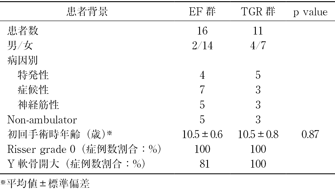
    Introduction: In juveniles with progressive curves, there is a controversy regarding the indication of early definitive fusion (EF) vs. traditional growing rod (TGR). The purpose of this study is to compare radiographic outcomes and complications of EF vs. TGR surgery for immature patients at a single institution.

    Material: There were 16 patients treated by EF who met the following criteria: (1) diagnosis before 10 years old; (2) grade 0 of Risser sign; (3) had definitive spinal fusion at the age of 9-11 years; (4) excluding congenital scoliosis, neurofibromatosis. To compare the outcomes, 11 patients treated by TGR who underwent the initial surgery at the age of 9-11 years were included in this study. Average age at the initial surgery were 10.5±0.6, 10.5±0.8 years respectively (p=0.87). The magnitude of the major curve was 98±16, 92±23 degree respectively (p=0.42).

    Results: Four patients in the group EF underwent staged surgeries. The average number of fused segments were 14±2, 15±1, respectively (p=0.18). Initial curve correction in the group EF was significantly greater than that in the group TGR after the initial surgery (60% vs. 40%; p<0.01). There was not significant difference in the overall correction rate of major curve (58% vs. 44%; p=0.12). The group EF maintained the thoracic kyphosis at latest follow-up better than the group TGR (34±15 vs. 56±29 degree; p=0.01). T1-12 height at latest follow-up were similar between the group EF and TGR (215±24 vs. 223±27 mm; p=0.42). Number of surgeries in the group EF was significantly lower than those in the group TGR (1.4±0.5 vs. 8.6±3.2; p<0.01).

    Conclusion: While similar spinal growth was obtained in both groups, the number of operations could be reduced by EF. Early definitive spinal fusion might be a surgery of choice for 9- to 11-year-old children with EOS.

    Download PDF (1334K)
  • Masaaki Iizuka, Ayato Nohara, Shunichi Kishida, Satoshi Baba, Naoki Sh ...
    2021 Volume 12 Issue 11 Pages 1326-1331
    Published: November 20, 2021
    Released on J-STAGE: November 20, 2021
    JOURNAL FREE ACCESS

    Background: In our center we treat early-onset scoliosis with corrective cast and brace from 2017. The main treatment for Early-onset scoliosis is the growth friendly spine surgery. Few centers provide corrective cast treatment so we report our short-term outcome.

    Methods: A review of a single center's experience with corrective casting was performed from 2017 to 2020. Patients were included who treated with corrective cast more than 4 times. Radiographic evaluation was performed.

    Results: There were 18 patients included. Average age at first corrective cast was 4.9 years. 2 were congenital, 11 were symptomatic and 5 were idiopathic scoliosis. The main thoracic Cobb angle before casting was 69.4±23.4 degrees, which corrected to 41.3±19.0 degrees in a cast. And correction rate was 39.2%. Cobb angle was 53.3±25.6 degrees in brace, and after cast removal was 59.3±26.8 degrees. Average corrective cast was performed 7times. There was no serious complication. Only one patient undergone surgery.

    Conclusions: Corrective cast can correct the scoliotic spine more effectively than bracing. Corrective cast may help delay surgical intervention. We will continue report these patients.

    Download PDF (1208K)
  • Masayuki Miyagi, Mitsuyoshi Matsumoto, Yusuke Mimura, Wataru Saito, Ta ...
    2021 Volume 12 Issue 11 Pages 1332-1337
    Published: November 20, 2021
    Released on J-STAGE: November 20, 2021
    JOURNAL FREE ACCESS

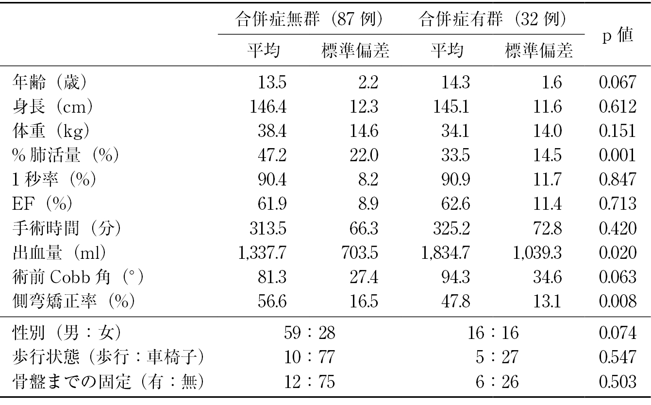
    Introduction: High perioperative complication rate in neuromuscular scoliosis (NMS) had been reported. The aim of the current study was to elucidate the perioperative complications in NMS surgery and the risk factors of the perioperative complications.

    Methods: In the current study, 126 NMS patients who were underwent posterior fusion surgery for scoliosis were included. We reviewed perioperative complications, age at operation, body height, body weight, pre-operative %VC and FEV1.0 (%) for pulmonary function, and pre-operative ejection fraction (EF) for cardiac function, ambulation status, preoperative Cobb angle, operation time, blood loss, fusion to pelvis and correction rate of scoliosis.

    Results: There were 39 (31.0%) complications including 7 complications due to surgical technique, 12 pulmonary complications, and 5 cardiovascular complications. When we divided into complication (+) group and complication (−) group, the pre-operative %VC and correction rate of scoliosis in complication (+) group were significantly lower than that in complication (−) group. (p<0.05) In addition, the blood loss at operation in complication (+) group were significantly higher than that in complication (−) group. (p<0.05) In the multiple logistic regression analysis, the pre-operative %VC was significant independent risk factor for perioperative complications.

    Conclusions: Severe preoperative restrictive ventilatory impairment, higher blood loss, and worse correction rate of scoliosis might affect the incidence of perioperative complications in posterior spinal fusion surgery for NMS.

    Download PDF (851K)
  • Ryoji Tauchi, Noriaki Kawakami, Tetsuya Ohara, Toshiki Saito, Ippei Ya ...
    2021 Volume 12 Issue 11 Pages 1338-1342
    Published: November 20, 2021
    Released on J-STAGE: November 20, 2021
    JOURNAL FREE ACCESS

    Introduction: Although there have been several studies about surgical outcomes in dystrophic type scoliosis of neurofibromatosis type I (NF1), there have been no reports on the thoracic cage deformity in this disease. The aim of this study was to investigate the characteristics of thoracic cage deformity and pulmonary function in NF1 dystrophic type scoliosis.

    Methods: Thirty-eight patients with dystrophic type scoliosis due to NF1 who had undergone corrective surgery were included in this study. There were 22 males and 16 females with a mean age of 12.6 years old at 1st surgery. The mean follow-up period was 13.1 years. We evaluated main curve Cobb angle, thoracic cage deformity, site of thoracic deformity, and the relationship with rib penciling. Pulmonary function was assessed at FU.

    Results: Main curve Cobb angle was 69.6° preoperatively, 27.5° at final follow-up. Deformity of the thoracic cage was observed in 11 (29%) at final follow-up. The sites of thoracic cage deformity were at convex side in 10 and at concave side in 1. There was a significant difference in the mean age at surgery between the group with and without thoracic deformity (8.9 years vs. 14.1 years, p=0.004). The T1-T12 height in the group with thoracic cage deformity was significantly smaller than in the group without thoracic cage deformity (21.3 cm vs. 23.6 cm, p=0.01). The numbers of rib penciling in the group with thoracic cage deformity was significantly higher than in the group without thoracic cage deformity (6.1 vs. 1.9 ribs, p<0.001). Pulmonary function (FVC) was lower in the group with thoracic cage deformity (1,880 ml vs. 2,705 ml, p<0.001). There was no significant difference in FEV1.0% between the two groups.

    Conclusions: In dystrophic type scoliosis of NF1, thoracic cage deformity was present in 29%, mostly on the convex side. We should pay attention to not only spinal deformity but also pulmonary function during follow-up for thoracic cage deformity cases.

    Download PDF (1194K)
  • Takashi Terao, Yoshinobu Yamanouchi, Takuto Kurakawa, Masao Ryu, Tomoy ...
    2021 Volume 12 Issue 11 Pages 1343-1348
    Published: November 20, 2021
    Released on J-STAGE: November 20, 2021
    JOURNAL FREE ACCESS

    Introduction: In activity of daily living (ADL), changing underwear is one of the problem after adult spinal deformity (ASD) surgery. However, few reports have evaluated the factors. We performed this study to investigate the relationship between activity of changing underwear and spinopelvic parameters after surgery.

    Methods: We reviewed 111 patients (75 females, average 70-year at the surgery) who underwent fixation surgery from lower thoracic spine down to pelvis for ASD. Patients were divided into independent group and requiring assistance group, according to the degree of self-reliance of dressing activities after surgery. X-ray parameters (SVA, TK, LL, PT, SS, PI, PT-LL) and preoperative ROM of hip joint were compared between two groups.

    Results: UIV were T10 in 95 patients, S2 alar-iliac screws were used in all cases, and interbody fusion at L5/S1 were performed in 88 cases. Mean spinopelvic parameters (independent group/requiring assistance group) which SVA (28 mm/34 mm), PT (25°/39°) preoperatively, and PT (18°/24°) postoperatively were significantly different between two groups (p <0.01). However, preoperative ROM of hip joint was not significantly different.

    Conclusions: Postoperative pelvic tilt was significantly smaller in patients with independent activity of changing underwear than in patients requiring assistance, that suggested flexion of the hip joint might allow body trunk to lean forward more deeply. Preoperative intervention to improve mobility of hip joint will be required to rehabilitation for adult spinal deformity patients, especially with severe deformity.

    Download PDF (983K)
feedback
Top