Journal of Spine Research
Online ISSN : 2435-1563
Print ISSN : 1884-7137
Volume 14, Issue 8
Displaying 1-14 of 14 articles from this issue
Original Article
  • Kei Takagi, Kazuya Kishima, Keishi Maruo, Fumihiro Arizumi, Norichika ...
    2023 Volume 14 Issue 8 Pages 1080-1085
    Published: August 20, 2023
    Released on J-STAGE: August 20, 2023
    JOURNAL FREE ACCESS

    Introduction: Full-endoscopic spine surgery (FESS) using transforaminal (TF) approach can be performed through an 8 mm skin incision under local anesthesia and is considered a minimally invasive spine surgery. The minimal invasiveness of FESS was evaluated using pre- and postoperative blood data.

    Material and Methods: The subjects of this study were 68 patients (39 males and 29 females) who underwent TF-FESS (FESS group), 24 patients (18 males and 6 females) who underwent laminectomy (L group), and 76 patients (38 males and 38 females) who underwent posterior interbody fusion (F group) for lumbar disc herniation and lumbar spinal canal stenosis between 1 vertebra from April 2021 to July 2022. The mean age of TF-FESS, laminectomy, and lumbar interbody fusion were 53.6, 73.2, and 71.0 years old. Preoperative and postoperative day 1 blood data were used.

    The items examined were creatinine kinase (CK), C-reactive protein (CRP), white blood cell count (WBC), and hemoglobin (Hb). The amount of change in each item in the FESS group was determined and compared with the L and F groups.

    Results: CK did not differ pre- and postoperatively in the FESS group (130 U/L vs 114 U/L, P=0.06), but was significantly increased postoperatively in the L group (158 U/L vs 269 U/L, P<0.05) and F groups (128 U/L vs 584 U/L, P<0.05). CRP did not differ pre- and postoperatively in the FESS group (0.44 mg/dL vs 0.51 mg/dL, P=0.35) and the L group (0.08 mg/dL vs 0.75 mg/dL, P=0.08), but was significantly increased postoperatively in the F group (0.34 mg/dL vs 1.24 mg/dL, P<0.05). WBC did not differ pre- and postoperatively in the FESS group (6,757/μL vs 6,982/μL, P=0.22), but was significantly increased postoperatively in the L (6,188/μL vs 9,263/μL, P<0.05) and F groups (6,137/μL vs 10,108/μL, P<0.05). Hb was significantly decreased postoperatively in the FESS group (13.9 g/dL vs 13.5 g/dL, P<0.05), L group (13.9 g/dL vs 12.3 g/dL, P<0.05) and F group (13.1 g/dL vs 11.2 g/dL, P<0.05). Comparison of pre- and postoperative changes showed significant differences among the three groups for all items.

    Conclusion: The results of this study show that TF-FESS is a minimally invasive spine surgery in terms of blood data.

    Download PDF (975K)
  • Tomowaki Nakagawa, Masako Tokunaga, Eiji Takahashi, Ken Hoshikawa, Hir ...
    2023 Volume 14 Issue 8 Pages 1086-1090
    Published: August 20, 2023
    Released on J-STAGE: August 20, 2023
    JOURNAL FREE ACCESS

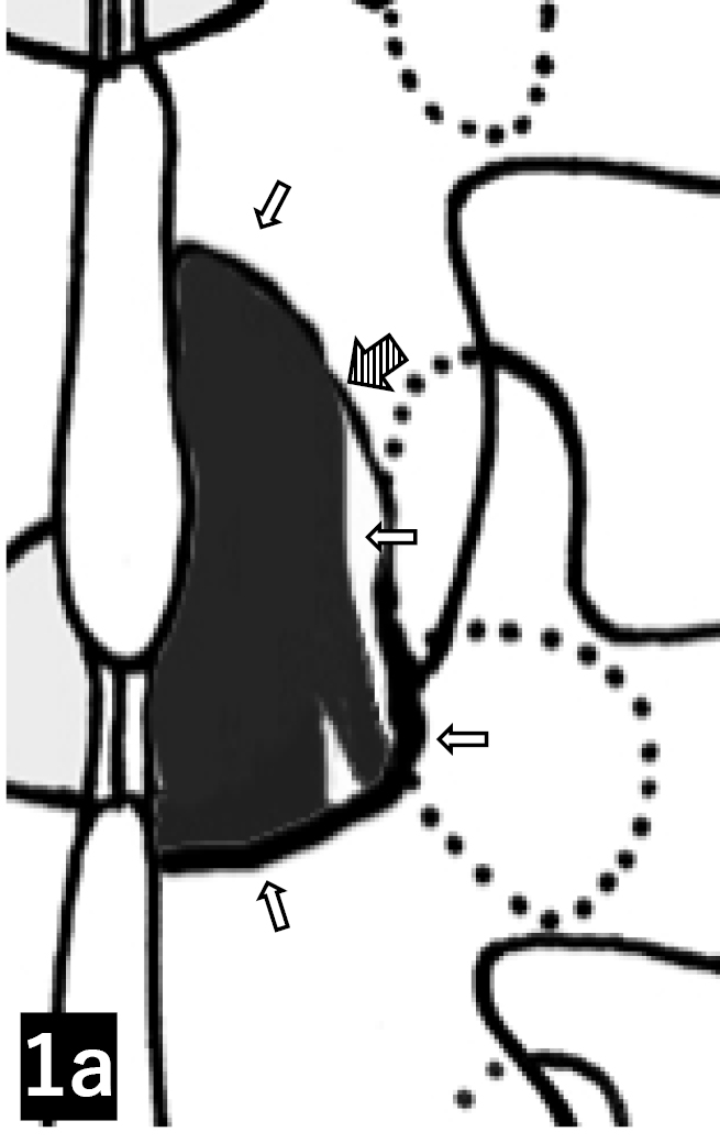
    The basic surgical treatment for spinal canal lesion of lumbar spinal canal stenosis is decompression surgery. However, the endpoints of decompression surgery are unclear in many procedure manuals. The purpose of this study was to identify the location of spinal canal stenosis, posterior compression factors, and their relationship to the endpoints of the surgical procedure. The patients were 100 cases, 167 vertebrae, who underwent endoscopic laminectomy for spinal canal stenosis at our institution. Lumbar degenerative spondylolisthesis was found in 47 vertebrae (28%). Nerve compression sites were classified as high in the intervertebral disc, cephalad, or caudal to the intervertebral disc. Dorsal compression factors were also identified. Results. Nerve compression sites included cephalad to intervertebral disc in 3%, intervertebral disc only in 81%, intervertebral disc to caudal in 13%, cephalad to caudal in 1%, and caudal only in 2%. 98% of the cases included intervertebral disc. Posterior compression factors were ligamentum flavum in 91% and lipomatosis in 8%. Decompression surgery for lumbar spinal canal stenosis should be performed using the intervertebral disc as a guide for decompression and removal of the ligamentum flavum and lipomatosis at the same site.

    Download PDF (911K)
  • Tomowaki Nakagawa, Masako Tokunaga, Eiji Takahashi, Ken Hoshikawa, Hir ...
    2023 Volume 14 Issue 8 Pages 1091-1098
    Published: August 20, 2023
    Released on J-STAGE: August 20, 2023
    JOURNAL FREE ACCESS

    For lumbar spinal canal stenosis, we only perform minimally invasive decompression surgery in all patients with or without degenerative spondylolisthesis. The aim of this study was to examine and validate the 1-year results of patients who underwent decompression surgery only, focusing on the presence or absence of degenerative spondylolisthesis.

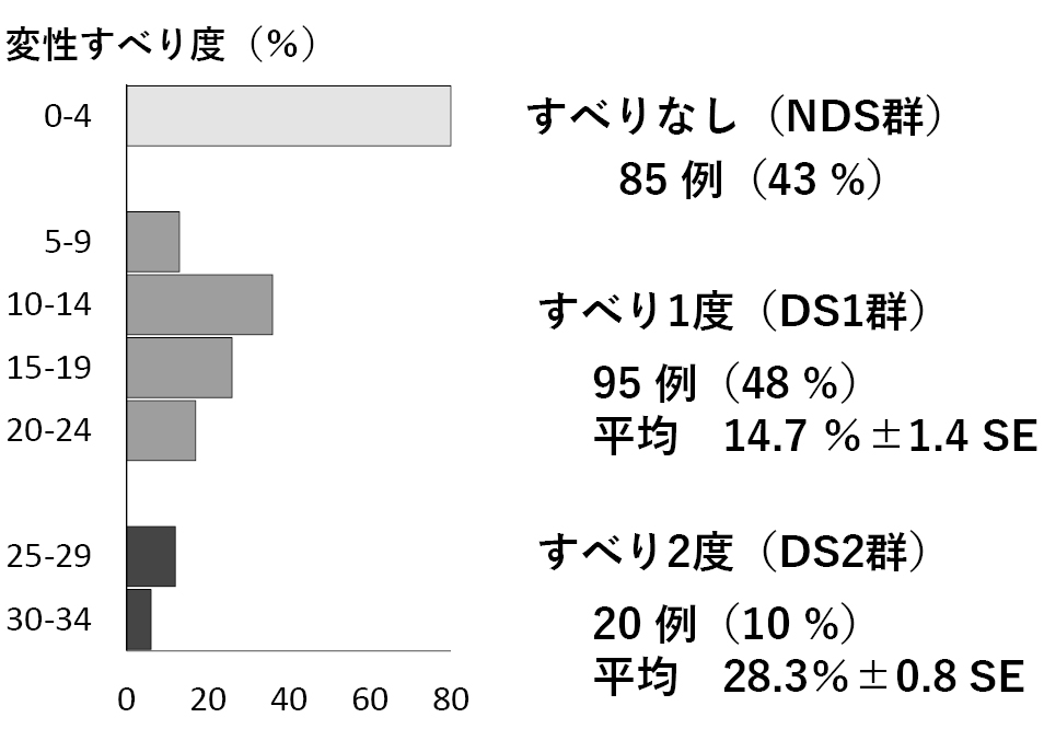
    Methods: 200 patients with lumbar spinal canal stenosis operated on only the L4/5 vertebrae were divided into 85 patients in the non-degenerative spondylolisthesis group (NDS group), 95 patients in the Meyerding classification 1st degree group (DS1 group) and 20 patients in the 2nd degree group (DS2 group). Operative technique, operative time, blood loss, perioperative complications, reoperation rate, JOA score, ODI, JOABPEQ and Lumbago/lower limb pain NRS were compared.

    Results: There were no differences in operative time or blood loss between the three groups. There was one dural injury in the DS1 group and one re-operation in the NDS group; JOA score and JOABPEQ did not differ between the three groups; DS2 group had inferior results in postoperative ODI and lower limb NRS; DS2 group had some inferior results. However, the DS2 group was considered to have a good surgical outcome based on the reoperation rate, the improvement in ODI and the significant improvement in lower limb pain NRS. The presence or absence of a spondylolisthesis was less involved in the short-term results and did not suggest the need for fusion surgery.

    Download PDF (1118K)
  • Masayuki Ishihara, Shinichiro Taniguchi, Takashi Adachi, Masaaki Paku, ...
    2023 Volume 14 Issue 8 Pages 1099-1108
    Published: August 20, 2023
    Released on J-STAGE: August 20, 2023
    JOURNAL FREE ACCESS

    Objective: To investigate the clinical results of lateral access corpectomy combined with release of anterior longitudinal ligament deep layer (LACADR) for kyphotic deformity after osteoporotic vertebral fractures (KOVF).

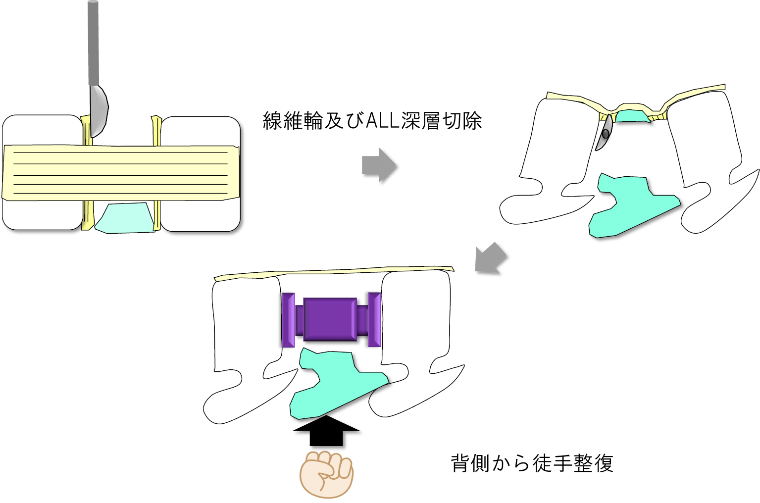
    Subjects and methods: KOVF who underwent surgery using LACADR, lateral lumbar interbody fusion (LLIF), and percutaneous pedicle screw (PPS) at our hospital and was able to follow up for more than 2 years. The subjects were 12 patients with the mean age was 74.6±10.1 years, and the mean follow-up period was 47.1±10.1 months. Fractured vertebral body, blood loss and operative time, low back pain VAS, anterior interbody distance (AID) and posterior interbody distance (PID), spinal canal occupancy rate of bone fragment (ORBF), local kyphosis angle (LK), Various spinopelvic parameters, bone fusion rate, and complications were investigated. The surgical procedure is to perform LACADR and vertebral body replacement using an extreme lateral interbody fusion (XLIF®) retractor in the lateral decubitus position, then place the patient in the prone position and perform posterior fusion using PPS. Regarding LACADR, the annulus ligament and the deep layer of ALL on either craniocaudal side of the fractured vertebral body are bluntly dissected with a hump, manual reduction is performed from the dorsal side, and the X-core® is installed while maintaining that state.

    Results: The fractured vertebral body was T11 in 1 case, T12 in 10 cases, L1 in 4 cases, and L2 in 1 cases. Bleeding volume was 365±150 ml, operation time was 207±92 minutes, AID improved from 19.5±5.0 mm preoperatively to final 35.5±5.9 mm, and PID improved from 25.0±3.3 mm preoperatively to final 35.1±4.5 mm. ORBF was 44.3±13.1% before surgery, 34.6±10.4% immediately after surgery, and 29.3±8.9% at the end, showing significant improvement. LK was 37.4±4.5° before surgery, 3.6±2.3° after LACADR, 3.9±2.2° after PPS, and 4.6±3.7° at the end, and was significantly improved after surgery. In spinopelvic parameters, SVA was 56.7±48.5 mm preoperatively, 33.0±26.2 mm postoperatively, final 38.0±28.1 mm (pre. vs post. p< 0.05), TK was 44.3±14.5 mm preoperatively, 32.6±12.1°postoperatively, final 35.6±18.3° (preop. vs post. p< 0.05), LLL was 28.2±19.2° preoperatively, 22.3±17.2°postoperatively, final 23.8±18.3° (preop. vs postop. p< 0.05). LL was 34.1±24.0° preoperatively, 36.1±9.4° postoperatively, and final 35.1±11.4° (preop. vs. postop. p=0.37), and no significant improvement was observed. The bone fusion rate was 85% at 12 months, and 92% at the end. As for the morphology of bone fusion, 83% were bridging type or both bridging and interbody fusion type, and 17% were intra-cage fusion type. Complications included intraoperative endplate injury in 1 case, cage subsidence in 2 cases, proximal junctional kyphosis in 1 case, and parietal pleural injury in 4 cases.

    Conclusion: The radiographical results of LACADR for KOVF was investigated. Sufficient correction and indirect decompression effects and bone fusion were confirmed.

    Download PDF (2269K)
  • Yoichi Tani, Takahiro Tanaka, Koki Kawashima, Kohei Masada, Masaaki Pa ...
    2023 Volume 14 Issue 8 Pages 1109-1116
    Published: August 20, 2023
    Released on J-STAGE: August 20, 2023
    JOURNAL FREE ACCESS

    Introduction: Although previous studies reported that robot-assisted spine surgery have helped improve accuracy of pedicle screw placement, it still remains undetermined whether such assistant devices can be alternatives to spine surgery experts. This study has verified if the accuracy varies among three groups of operators with different years of experience in this field: expertly trained spine surgeons (group A), spine surgeons less than 3 years of experience (group B), and inexperienced trainees (group C).

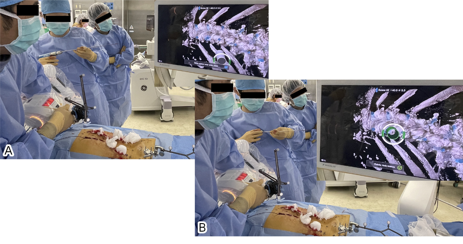
    Methods: Forty patients (21 men), aged 12 to 89 (mean, 72.5) years underwent a total of 264 percutaneous pedicle screw (PPS) placements (40 each for cervical and thoracic spines and 184 for lumbosacral spines and pelvis) using the Cirq robotic arm system coupled with intraoperative CT-based navigation. With the patient positioned and prepared on the operating table, we attached the reference array to the two 3 mm-diameter K-wires inserted into the iliac bone unilaterally followed by registration CT scanning for the thoracic/lumbosacral PPS and S2 alar-iliac screw placements. For the cervical PPS placements, we affixed the reference array to the C2 spinous process or the Mayfield tongs. When carrying out PPS insertion, the surgeon located the entry area with blunt finger dissection through a 2-cm incision first, then he/she moved a robot arm thereto manually. Subsequently, the robot arm, when activated, automatically positioned to align to the preplanned pedicle trajectory. The arm locked in place, with a drill-stabilizing metallic tube assembled manually at its end, allowed a desired drilling and guidewire insertion. In the cervical vertebrae, we employed a posterolateral intermuscular approach through a small incision described by Tokioka et al.

    Results: Considering no pedicle wall breach for cervical PPSs and both no breach and breach < 2 mm for thoracic/lumbosacral and iliac PPSs as successful placements, overall success rate accounted for 93.6% (247 of 264 screws): 95.0% for cervical, 85.0% for thoracic, and 95.1% for lumbosacral PPS and alar-iliac screw placements. No patients required revision surgery for repositioning or replacing the PPSs.

    The success rates showed no significant difference among three groups of operators: 94.4% for group A, 92.7% for group B, and 93.9% for group C.

    Conclusions: The surgeon-controlled robotic arm with intraoperative CT-based navigation served well for PPS placements at any spinal level including cervical spine. By using this device, safety and accuracy of PPS placements did not significantly vary depending on surgeon's years of experience in spinal surgery.

    Download PDF (1655K)
  • Shuntaro Tsuchida, Yoshihiro Kitahama
    2023 Volume 14 Issue 8 Pages 1117-1127
    Published: August 20, 2023
    Released on J-STAGE: August 20, 2023
    JOURNAL FREE ACCESS

    We present two cases of late palsy that occurred after an osteoporotic vertebral fracture, which were treated using transforaminal full-endoscopic removal of bone fragments. The criteria for selection were as follows:

    The vertebral fracture has already healed.

    The primary cause of palsy was static compression by bone fragments.

    Both cases showed improvement in palsy and leg pain after procedure. This method is technically demanding, but it offers advantage of minimally invasive direct removal of the bone fragments.

    Download PDF (2586K)
  • Reiko Yoneyama, Kazuo Ohmori, Deokcheol Lee, Yasuhiro Endo
    2023 Volume 14 Issue 8 Pages 1128-1132
    Published: August 20, 2023
    Released on J-STAGE: August 20, 2023
    JOURNAL FREE ACCESS

    Introduction: Postoperative pain and neuropathy due to epidural hematoma is a complication after spinal endoscopic surgery. In this study, we examined the effect of the number of drains placed after full-endoscopic laminoplasty (FEL) on epidural hematoma.

    Methods: Twenty-four patients (10 males and 14 females, mean age 70.0 years) who underwent single level FEL for lumbar spinal canal stenosis from December 2021 to June 2022 were included in the study. Patients were alternately assigned to two groups: a single drain group (D1 group: 12 patients) and a double drain group (D2 group: 12 patients). The drain used was a NIPRO UK slim drain (10 Fr). The drain was negative pressure, removed on day 4, and the drainage volume was examined. The area of the epidural hematoma was measured using MRI horizontal images at 1 month postoperatively. The rate of improvement of JOA score was compared between the two groups.

    Results: No differences were found between the two groups in age, gender, preoperative antiplatelet medication, operative level, or operative time. Compared to the D1 group, the D2 group had a significantly higher drainage volume (D1 group; 161.6 ml vs. D2 group; 218.8 ml, p=0.026). Epidural hematoma area at 1 month postoperatively also tended to be larger in D2 group than in D1 group (D1 99.0 mm2, D2 186.5 mm2, p = 0.09). But no significant difference in JOA score improvement between the two groups (D1 75.0%, D2 85.7%, p =0.33).

    Conclusion: The drain drainage volume and hematoma area were greater with double drains placed than with single drain. The increased negative pressure caused by the drains may have contributed to posterior hemorrhage from the epidural space and bone resection and inhibited hemostasis. However, no difference in postoperative clinical outcomes was observed between the D1 and D2 groups, and from an invasive standpoint, a single post-FEL drain would be preferable.

    Download PDF (1239K)
  • Takeshi Hori, Kiyoshi Sakai, Ayano Tokunaga, Ryo Ueshima, Masatoshi Sa ...
    2023 Volume 14 Issue 8 Pages 1133-1137
    Published: August 20, 2023
    Released on J-STAGE: August 20, 2023
    JOURNAL FREE ACCESS

    Introduction: The purpose of this study was to investigate whether degeneration of the paravertebral muscles occurs after full-endoscopic spine surgery (FESS).

    Methods: A total of 39 examinations in 30 patients who underwent MRI after FESS surgery were included. There were 21 males and 9 females, with an average age of 67.4 years. The surgical approaches were interlaminar approach (IL) in 18 cases, transforaminal approach (TF) in 10 cases, and posterolateral approach (PL) in 2 cases. Average time to postoperative MRI was 245 days. Reasons for postoperative MRI examination included recurrence of leg pain in 12 cases, residual symptoms in 12 cases, and occurrence of contralateral leg pain in 9 cases. Degeneration of the paravertebral muscles was assessed by applying the Goutallier classification.

    Results: Preoperative MRI showed that 9 cases of stage 1, 19 cases of stage 2, 1 case of stage 3, and 1 case of stage 4 degeneration of the paravertebral muscles at the level of the operation. Progressive degeneration of the paravertebral muscles after surgery was observed in 3 cases, all of which were TF cases, and degeneration was observed in the paravertebral muscles caudal to the surgical site.

    Conclusions: During the TF approach, it was suggested that the degeneration of the caudal paravertebral muscle progressed more than at the surgical level due to damage to the posterior nerve root branch at the intervertebral foramina. On the other hand, no change was observed in the paravertebral muscle of the invasion route.

    Download PDF (1025K)
  • Shigeki Urayama, Kiyoshi Iida, Naoya Kashiwagi, Sae Shibashiro, Shiko ...
    2023 Volume 14 Issue 8 Pages 1138-1143
    Published: August 20, 2023
    Released on J-STAGE: August 20, 2023
    JOURNAL FREE ACCESS

    The purpose of this study was to retrospectively investigate the effects and complications of DPEL scope.

    Fifty patients were included in this study group. Forty patients suffered from lumbar spinal canal stenosis, and the other 10 suffered from large extruded lumbar intervertebral disc herniation which migrated cranially or caudally. Follow-up period was 12.1 (2-36) months after operation.

    JOA score was 14.6 (5-19) points preoperatively and improved to 26.5 (19-29) points at the final follow up (P<0.0001). Improvement ratio was 82.6%. Complications occurred in 4 patients (8%) who had bilateral decompression surgery for spinal canal stenosis. Injury of a nerve root in the opposite side occurred in one patient who had extensive removal of the yellow ligament, which recovered within one year after operation. Dural tear occurred in the other three patients during removal of the yellow ligament. Dural tears in these 3 patients were treated by using 2-3 layers of combination of PGA (Polyglycolic acid) sheet with free fat tissue graft. This technique was effective in 2 patients. They did not complain of symptom of cerebrospinal fluid leakage and discharged from hospital as scheduled.

    DPEL scope was effective for laminectomy of lumbar spinal canal stenosis and large lumbar intervertebral disc herniation. However, dural tear occurred during removal of the yellow ligament in three patients (6%).

    Download PDF (1253K)
  • Katsuhiko Kikuchi, Koichi Yoshikane
    2023 Volume 14 Issue 8 Pages 1144-1148
    Published: August 20, 2023
    Released on J-STAGE: August 20, 2023
    JOURNAL FREE ACCESS

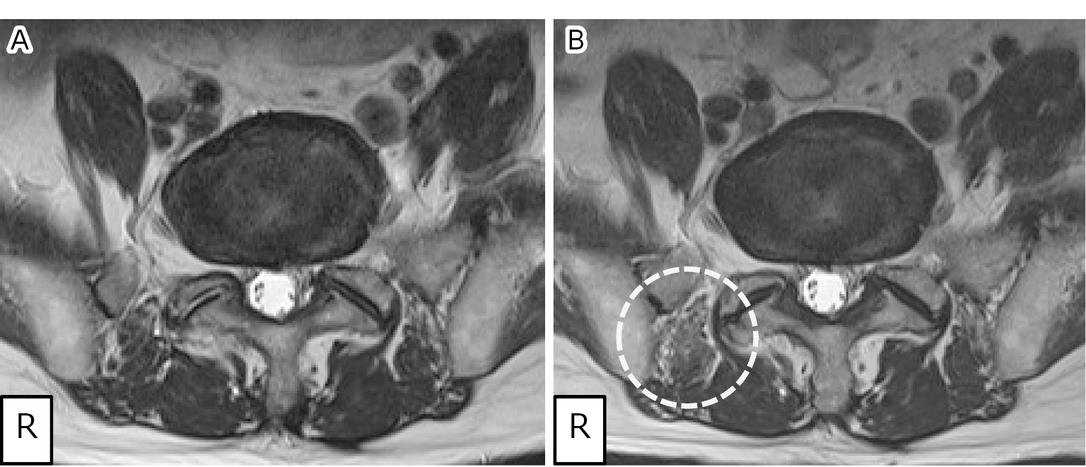
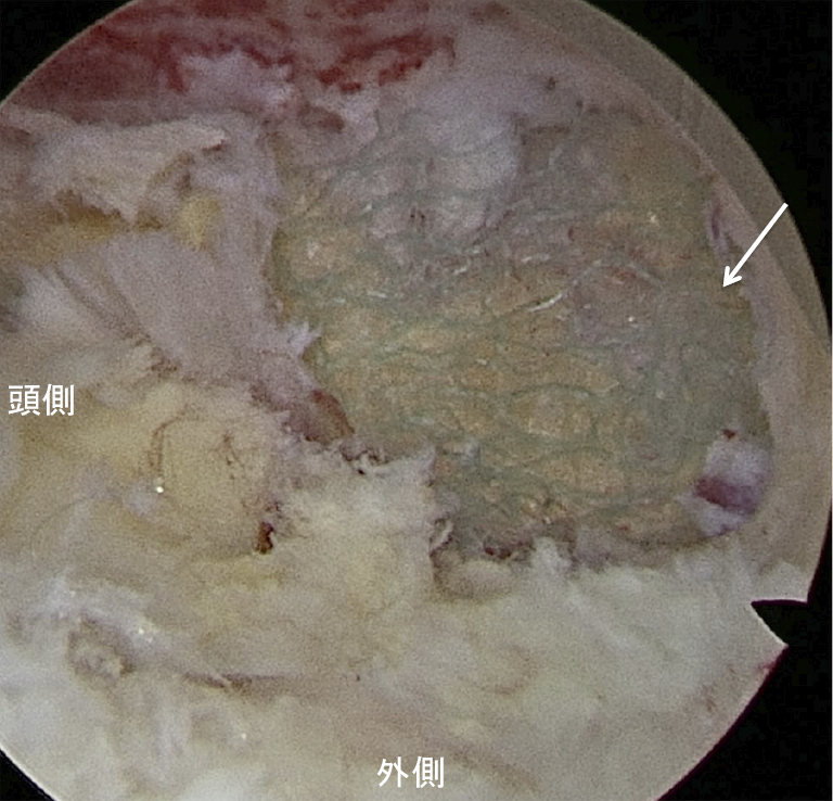
    Introduction: With the increasing use of full-endoscopic spine surgery (FELD) in lumbar disc herniation (LDH), FELD has been introduced to the treatment of LDH in teenagers. FELD by transforaminal approach (FELD TF) can be performed under local anesthesia. Although FELD TF is good indication for LDH in teenagers because it is minimum invasive and enable patients to return to school early, surgeons may have some concern about whether teenagers can understand requests from surgeons intraoperatively or not.

    Purpose: To evaluate surgical outcomes of FELD TF at lumbar spine under local anesthesia for teenagers.

    Subjects and methods: We evaluated the mean operation time, perioperative complications, VAS and the improvement of VAS for teenagers who was performed FELD TF under local anesthesia from April 2014 to March 2021.

    Results: 14 male and 6 female were included in this study. The mean age was 16.9 years. The mean operation time was 34.5 minutes. Intraoperative complication was nothing, but a discal cyst was occurred.

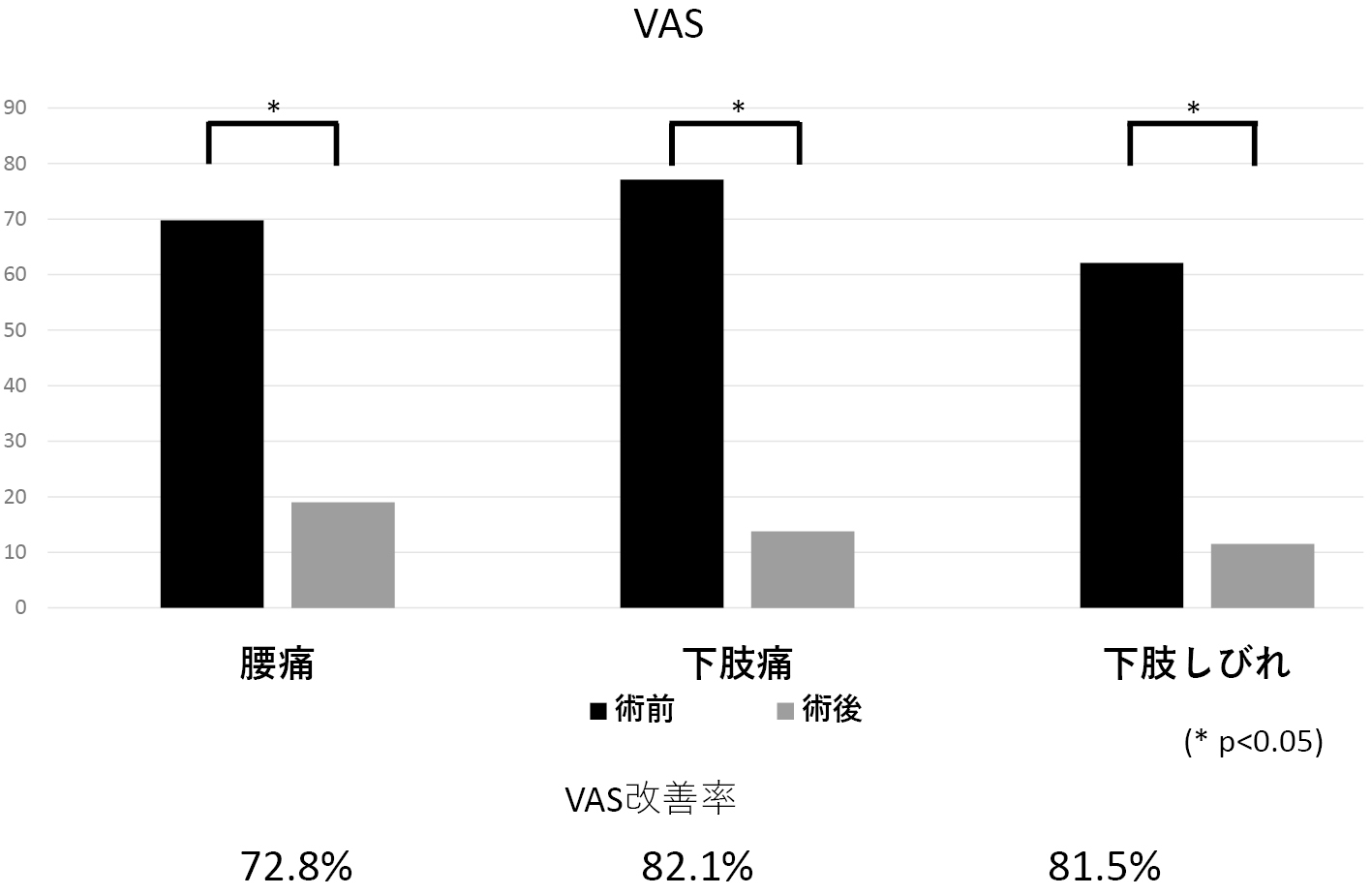
    Preoperative VAS in LBP, leg pain and numbness was 69.8, 77.1 and 62.1. Postoperative VAS was 19, 13.8 and 11.5. The improvement of VAS was 72.8, 82.1 and 81.5%.

    Conclusions: FELD TF at lumbar spine under local anesthesia for teenagers was useful and safe procedure.

    Download PDF (914K)
  • Yoshio Enyo, Yukihiro Nakagawa, Takahiro Maeda, Teiji Harada, Hidenobu ...
    2023 Volume 14 Issue 8 Pages 1149-1156
    Published: August 20, 2023
    Released on J-STAGE: August 20, 2023
    JOURNAL FREE ACCESS

    Purpose: The purpose of this study is to evaluate the clinical outcomes of midline spinous process-splitting cervical laminoplasty with titanium lamina plates.

    Materials and methods: 13 patients (Group C) with cervical myelopathy were treated by midline spinous process-splitting laminoplasty with titanium lamina plates and they were able to follow up for 6 months. The investigating items were operation time, blood loss, recovery rate at JOA score (6 months after surgery), lamina enlargement rate immediately after surgery, lamina closure at 6 months after surgery, presence or absence of bony union in the lateral gutter. 12 patients (Group H) with cervical myelopathy were treated by midline spinous process-splitting laminoplasty with HA spacers. Group C and Group H were compared by the investigating items.

    Results: Operation time and blood loss were significantly more in Group C than Group H. Recovery rate at JOA score was 45.6% in both groups. There was no significant difference in laminar closure between the two groups, but there was some tendency to have more lamina closures in Group H. The rates of bony union in the lateral gutter at six months after surgery were 74.2% in the group C and 14.9% in the group H.

    Conclusion: 1. There was no significant difference between titanium lamina plates and HA spacers in the clinical outcome of midline spinous process-splitting cervical laminoplasty. 2. Midline spinous process-splitting cervical laminoplasty with titanium lamina plates may be able to achieve early bony union in the lateral gutter with strong fixation.

    Download PDF (1833K)
  • Yoshio Enyo, Yukihiro Nakagawa, Takahiro Maeda, Teiji Harada, Hidenobu ...
    2023 Volume 14 Issue 8 Pages 1157-1164
    Published: August 20, 2023
    Released on J-STAGE: August 20, 2023
    JOURNAL FREE ACCESS

    Purpose: We compared the clinical results of vertebral body stenting (VBS) and balloon kyphoplasty (BKP) for osteoporotic vertebral fracture (OVF).

    Materials and Methods: 51 patients were treated by VBS for OVF at our hospital and affiliated hospitals. The examination items were operation time, blood loss, Numerical Rating Scale (NRS) in low back pain, amount of cement filling, presence or absence of cement leakage from the vertebral body, and presence or absence of subsequent vertebral fractures. The anterior and posterior height of the vertebral body, the wedge angle of the fractured vertebral body, and the local kyphosis angle were measured. We compared VBS patients with 234 patients who were treated by BKP for OVF.

    Results: Operation time was 42.5 minutes for VBS and 34.7 minutes for BKP, blood loss was 11.1 ml for VBS and 4.6 ml for BKP. There was no significant difference in NRS between VBS and BKP before, after, and 3 months after surgery. The amount of cement filling was 7.6 ml for VBS and 8.7 ml for BKP. The rate of cement leakage was found in 13.7% (7/51) for VBS and in 19.7% (46/234) for BKP. There were no significant differences in the anterior height of the fractured vertebral body, the posterior height of the fractured vertebral body, the wedge angle of the fractured vertebral body, and the local kyphosis angle before surgery, after surgery, at 3 and 6 months after surgery. Subsequent vertebral fractures were found in 9.8% (5/51) for VBS and in 13.7% (32/234) for BKP with no significant difference.

    Conclusion: 1. VBS showed no difference in pain relief compared to BKP, but the amount of cement filling and cement leakage outside the vertebral body were less in VBS than in BKP.

    2. There was no difference between VBS and BKP in vertebral body correction and correction loss.

    Download PDF (1829K)
  • Shu Nakamura, Fujio Ito, Yasushi Miura, Motohide Shibayama, Zenya Ito, ...
    2023 Volume 14 Issue 8 Pages 1165-1172
    Published: August 20, 2023
    Released on J-STAGE: August 20, 2023
    JOURNAL FREE ACCESS

    Foraminal stenosis is usually approached from the posterolateral side on the affected side, but when the angle of approach from the posterolateral side is restricted, such as at the L5-S1 level, and spinal stenosis is complicated at the same level, the contralateral interlaminar approach may be more advantageous. We compared percutaneous full-endoscopic lumbar contralateral interlaminar foraminoplasty (PfELCIF) with conventional percutaneous full-endoscopic lumbar foraminoplasty (PfELF).

    There were no cases of postoperative dysesthesia in either group. Enlargement ratio of the intervertebral foramina was significantly higher in PfELCIF at medial area, and significantly higher in PfELF at lateral area.

    The contralateral interlaminar foraminoplasty is performed in a considerable deep area from the bone surface, which is possible with percutaneous full-endoscope. PfELCIF was as effective as PfELF in clinical results. We consider that PfELCIF is a useful option for patients with lumbar spinal and foraminal stenosis at the L5-S1 level, especially when the main site of the foraminal stenosis is center to medial.

    Download PDF (1767K)
  • Koki Kawashima, Masayuki Ishihara, Shinichiro Taniguchi, Takashi Adach ...
    2023 Volume 14 Issue 8 Pages 1173-1180
    Published: August 20, 2023
    Released on J-STAGE: August 20, 2023
    JOURNAL FREE ACCESS

    Purpose: We investigated the bone fusion rate, segmental lordosis, and risk factors for bone union failure at L5/S1 in multi-level lumbosacral interbody fusion.

    Methods: This retrospective study included 35 patients who underwent lumbosacral interbody fusion at our hospital and were followed up for more than 2 years after surgery.Bone fusion rate according to number of interbody fusion, the cage material, and the number of cages, and segmental lordosis (SL) at L5/S1 for each cage lordotic angle were examined. Various parameters were compared between the group with bone fusion group at L5/S1 (U group) and the group without non-union at L5/S1 (N group).

    Results: The bone fusion rate at L5/S1 was 87% for 2 intervertebral fusion, 73% for 3 intervertebral fusion, and 62% for 4 intervertebral fusion. The bone union rate was the lowest in 4 intervertebral fusions. Preoperative PI, postoperative PI-LL, and postoperative SVA were significantly larger in the N group. Multivariate analysis detected preoperative PI as the risk factor of fusion failure at L5/S1, with a cutoff value of 51° and an area under the curve of 0.807. Mean SL improved from 5° preoperatively to 10° postoperatively. SL increased according to the lordotic angle of the cage, and the correlation coefficient was 0.73 (p<0.001), indicating a significant correlation.

    Conclusion: The results show that the higher the number of interbody fusion, the lower the rate of bone union. PI was a risk factor for bone fusion failure, with a cut-off value of 51°. Postoperateve SL was correlated with cage lordosis angle, but bone union rate was not correlated with cage material and cage number.

    Download PDF (2201K)
feedback
Top