Journal of Spine Research
Online ISSN : 2435-1563
Print ISSN : 1884-7137
Volume 12, Issue 9
Displaying 1-20 of 20 articles from this issue
Editorial
Review Article
  • Keiichi Katsumi, Takashi Hirai, Toshitaka Yoshii, Narihito Nagoshi, So ...
    2021 Volume 12 Issue 9 Pages 1087-1093
    Published: September 20, 2021
    Released on J-STAGE: September 20, 2021
    JOURNAL FREE ACCESS

    Ossification of the posterior longitudinal ligament (OPLL) is a progressive disease. The bridging of ossified lesions to the vertebral body gradually increases, thereby decreasing the mobility of the cervical spine; thus, cervical spine function may decrease over time. However, cervical spine function in patients with cervical OPLL has not been evaluated in large prospective studies. Therefore, we conducted a prospective multicenter study to clarify whether expansion of ossification can influence cervical spine function and quality of life (QOL) in patients with cervical OPLL. In total, 238 patients (162 men, 76 women; mean age, 63.9 years) were enrolled from 16 institutions in Japan. Each patient underwent whole spine computed tomography and was evaluated based on the Japanese Orthopaedic Association Cervical Myelopathy Evaluation Questionnaire (JOACMEQ). The JOACMEQ can evaluate cervical spine function using the cervical spine function domain (range 0-100 points; normal scale = 100 points). The mean score of cervical spine function was 65.9 points, and neck rotation was the most limited movement compared with flexion and extension. A higher neck VAS score and a larger number of bridge formations of OPLL in the whole spine were significant predictors of adverse outcomes related to cervical spine function. This is the first prospective multicenter study to reveal the impact of ossification expansion on cervical spine function. These findings are important for understanding the natural course of OPLL and can serve as controls when evaluating postoperative cervical spine function.

    Download PDF (1083K)
Original Article
  • Takashi Yamazaki, Nobuhiro Hara, Yusuke Sato
    2021 Volume 12 Issue 9 Pages 1094-1101
    Published: September 20, 2021
    Released on J-STAGE: September 20, 2021
    JOURNAL FREE ACCESS

    Introduction: There are few reports on iatrogenic nerve rupture in lumbar degenerative disease, which is an irreparable complication; thus, we examined cases of iatrogenic nerve rupture in department to obtain methods for its prevention.

    Methods: Cases of iatrogenic nerve rupture were extracted from the incident reports of 3,180 patients who underwent posterior surgery for lumbar degenerative disease, and the risk factors and causes were investigated.

    Results: There were 10 cases (0.31%) of iatrogenic nerve rupture. The risk factors were disc herniation as a disease, decompression, herniotomy, and laminectomy with Kerrison rongeurs in the presence of herniation as a surgical procedure. The cause was misidentification of the S2 nerve root as S1 in L5/S Love surgery (two cases), penetration of the medially moving drill tip into the epidural space, misidentification of nerve root compressed by a hernia, and dislodgement of Love retractor in L2/L3 hernia.

    Conclusions: Awareness of these risk factors can help reduce cases of iatrogenic nerve rupture.

    Download PDF (1223K)
  • Tomiya Matsumoto, Shinya Okuda, Yukitaka Nagamoto, Yoshifumi Takahashi ...
    2021 Volume 12 Issue 9 Pages 1102-1109
    Published: September 20, 2021
    Released on J-STAGE: September 20, 2021
    JOURNAL FREE ACCESS

    Introduction: This study aimed to investigate prevertebral soft tissue swelling (PSTS) after anterior cervical spine surgery over time and to compare the PSTS before and after extubation. This study also aimed to clarify whether the PSTS before extubation can be an index for predicting airway stenosis after extubation.

    Methods: In total, 28 patients (17 men and 11 women, mean age = 63 years) who underwent anterior cervical spine surgery and adapted our hospital's perioperative management protocol were included. The PSTS was measured at each level from C2 to C5 using cervical lateral radiographs. Radiological measurements were examined preoperatively, before extubation of POD1 (Pre-ex), after extubation of POD1 (Post-ex), 2 days after surgery (POD2), and 6 days after surgery (POD6). The rate of increase (ΔPSTS [%]) was measured at each postoperative period and at each level. Moreover, in POD2, bronchoscopy was performed. We examined (1) changes in ΔPSTS over time at each level, (2) correlation between pre-ex and post-ex, POD2, and POD6, and (3) comparison between ΔPSTS and bronchoscopic findings at POD2.

    Results: The temporal changes in ΔPSTS at each level (Pre-ex, Post-ex, POD2, POD6) were C2 (212±112,190±82,301±132,198±101[%]), C3 (154±76,188±81,289±137,216±79[%]), C4 (152±62,178±66,250±108,224±87[%]), C5 (127±33,150±42,163±51,140±118[%]). The ΔPSTS was higher at the upper cervical, and the ΔPSTS was highest at POD2 in each level. The respective correlation coefficient between pre-ex ΔPSTS and ΔPSTS (post-ex, POD2, and POD6) at each timing after extubation were C2 level (0.53, 0.43, and 0.17), C3 level (0.61, 0.45, and 0.47), C4 (0.58, 0.57, and 0.58), C5 (0.81, 0.71, and 0.76). Moderate correlations were observed up to the POD2 at each level, whereas especially strong correlations were noted up to POD6 at the mid-cervical spine. Mean ΔPSTS on POD2 showed no statistically significant differences between patients with bronchoscopic abnormal findings and those without. There were no cases of airway obstruction or reintubation caused by postoperative complications.

    Conclusions: The study outcomes showed that the PSTS before extubation is useful for predicting the deterioration of PSTS after extubation, and the evaluation of PSTS before extubation may help predict the occurrence of postoperative airway stenosis caused by PSTS.

    Download PDF (1736K)
  • Satoshi Nozawa, Chizuo Iwai, Kazunari Yamada, Kazunari Fushimi, Haruhi ...
    2021 Volume 12 Issue 9 Pages 1110-1116
    Published: September 20, 2021
    Released on J-STAGE: September 20, 2021
    JOURNAL FREE ACCESS
    Supplementary material

    Introduction: Although navigation in spinal surgery is effectively utilized, meticulous preoperative planning is still required for safe and successful surgery. We introduced a meticulous planning method using a three-dimensional (3D) image analysis system.

    Methods: Cases were analyzed using high-volume data, such as thin-slice CT images, using the software SYNAPS VINCENT (Fuji film). Multiplanar reconstruction (MPR), 3D image preparation, and virtual bone resection were conducted. Using the MPR images of patients with scoliosis, we measured the length and diameter of the pedicle screws (PS). The normally (Du) and carefully (Dm) measured diameters of 20 PS were compared statistically.

    Results: The average difference between Du and Dm was 0.69±0.09 (p < 0.01). The diameter of Dm was significantly increased compared to Du. Using VINCENT, we could accurately assess screw trajectory and bone condition before surgery. Additionally, we could effectively perform virtual laminectomy, foraminotomy, and PS insertion to correct a severely deformed spine.

    Conclusions: Based on careful planning using a 3D image analysis system, we could successfully perform a complex surgical procedure with little to no complications. This software is beneficial to spine surgeons and, with careful planning, has the potential to aid in reducing the intraoperative risk of vessel and nerve damage.

    Download PDF (1659K)
  • Tsunehiko Konomi, Takashi Asazuma, Masashi Saito, Shinjiro Kaneko, Yos ...
    2021 Volume 12 Issue 9 Pages 1117-1123
    Published: September 20, 2021
    Released on J-STAGE: September 20, 2021
    JOURNAL FREE ACCESS

    Introduction: The aim of this study was to clarify the radiological risk factors for a late revision surgery in patients with postoperative adolescent idiopathic scoliosis (AIS) with a long-term follow-up.

    Methods: A total of 47 adult patients who underwent spinal fusion surgery for the correction of scoliosis in adolescents with a minimum follow-up of 10 years were retrospectively evaluated. The radiographical parameters were compared between patients who underwent late (at least 10 years postoperatively) revision surgery and those who did not.

    Results: Revision surgeries were performed in nine patients with a mean time to revision of 21.9 years after the initial surgery. Postoperative radiological parameters before revision surgeries or at last follow-up, including pelvic tilt, lumbar lordosis, sagittal vertical axis, coronal balance, L3 and L4 tilt, and apical vertebral translation were 32.0°, 22.1°, 11.3 mm, 37.7 mm, 14.4°, 18.2°and 42.1 mm in patients with revision surgeries and 19.7°, 43.6°, 0.8 mm, 4.7 mm, 8.0°, 8.3°and 24.7 mm in those without revision surgeries, respectively, with statistical differences (P < 0.05).

    Conclusions: We demonstrated significant radiological risk factors for a late revision surgery, including increases in coronal off-balance, pelvic tilt, L3 and L4 tilt, and a decrease in lumbar lordosis. Maintaining a less lumbar tilt and well-balanced sagittal and coronal alignment should be considered for better long-term postoperative clinical outcomes in patients with AIS.

    Download PDF (1076K)
  • Tsubasa Sakai
    2021 Volume 12 Issue 9 Pages 1124-1129
    Published: September 20, 2021
    Released on J-STAGE: September 20, 2021
    JOURNAL FREE ACCESS

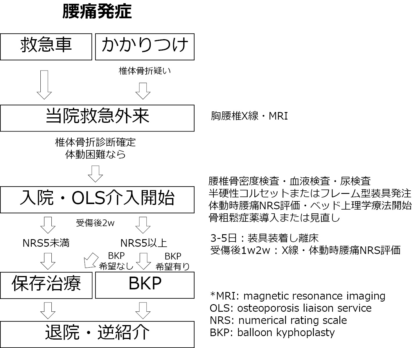
    Introduction: Previous studies have reported that conservative treatment for osteoporotic vertebral fractures (OVF) results in high bone union rates. However, other factors, including complications, decreased activities of daily living (ADL), and length of hospital stay, have not been fully evaluated.

    Methods: A total of 166 OVF patients who were conservatively treated in our hospital from April 2015 to March 2017 were included in this study. Among these, 16 cases with 5 or higher Numerical Rating Scale (NRS) of low back pain at the beginning of the movement 2 weeks postinjury were defined as the poor group. On the other hand, 150 cases with less than 5 NRS were defined as the poor group. Changes in NRS, length of hospital stay, walking ability decline rate, and complication rate were compared in both the groups.

    Results: Significant differences were found between the good and poor groups in terms of NRS (median [interquartile range]) at 1, 2, and 4 weeks postinjury (7 [6, 7.3] vs 6 [5, 7], 6.5 [6, 8] vs 2 [2, 3], 5.5 [4. 8, 6.3] vs 1 [1, 2], respectively), except at admission (8 [7, 9] vs 8 [8, 9]).

    Hospitalization days were 51 [44-61] days in the good group and 28 [24-32] days in the poor group, walking ability decline rates were 38% and 4%, the medical complication rates were 56% and 10%, and other adverse events were 38% and 8%, respectively, showing significant differences between the two groups.

    Conclusions: While treating OVF in the elderly, the evaluation of low back pain during movement 2 weeks postinjury can be a useful index to predict early recovery of ADL and extension of healthy life expectancy.

    Download PDF (951K)
  • Kenta Kurosu, Shin Oe, Tomohiko Hasegawa, Yu Yamato, Go Yoshida, Tomoh ...
    2021 Volume 12 Issue 9 Pages 1130-1134
    Published: September 20, 2021
    Released on J-STAGE: September 20, 2021
    JOURNAL FREE ACCESS

    Introduction: The preoperative nutritional status of patients undergoing thoracolumbar-lumbar surgery was evaluated using the prognostic nutritional index (PNI) for determining its effects on surgery and perioperative complications.

    Methods: Ninety-eight patients who had undergone thoracolumbar-lumbar surgery from November 2010 to July 2018 were recruited for the study (mean age 64.7±16.1 years). PNI was calculated using the following formula: serum albumin levels (g/dL) ×10+total lymphocyte count (/μL) ×0.005. Patients with a PNI of <50 and ≥50 were defined as the low PNI group and normal PNI group, respectively. Logistic regression analysis was performed by comparing the background, intraoperative/perioperative data, and complications of patients. The factors involved in the occurrence of complications were also examined.

    Results: While 43 patients were included in the low PNI group, 55 were included in the normal PNI group. A significant difference was observed in the mean age (70.8±12.0 vs. 59.9±17.3 years), length of hospital stay (31.2±16.1 vs. 21.6±12.6 days), home discharge rate (67.4% vs. 89.1%), and medical complication rate (18.6% vs. 0%) between the two groups (p < 0.05). Logistic regression analysis revealed that PNI was a predictor of medical complications.

    Conclusions: The present study showed that 43.9% of patients with low PNI had longer hospital stays, lower home discharge rates, and higher percentages of medical complications, thus suggesting that low PNI is a predictor of perioperative medical complications.

    Download PDF (798K)
  • Koki Kawashima, Masayuki Ishihara, Masaaki Paku, Yoichi Tani, Takashi ...
    2021 Volume 12 Issue 9 Pages 1135-1142
    Published: September 20, 2021
    Released on J-STAGE: September 20, 2021
    JOURNAL FREE ACCESS

    Introduction: Circumferential minimally invasive surgery (c-MIS) that employs lateral interbody fusion (LIF) and percutaneous pedicle screw (PPS) for adult spinal deformity (ASD) is becoming increasingly popular. However, studies that have thoroughly evaluated bone fusion rate and morphology, including those of the thoracic spine without bone grafting, are few. We examined the bone fusion process at the thoracic and lumbosacral levels of patients with ASD who underwent c-MIS using LIF and PPS.

    Methods: This retrospective study included 60 patients with ASD who underwent corrective surgery using LIF and PPS and were followed up for >24 months after surgery. Patients with anterior longitudinal ligament rupture were excluded. The average age and follow-up period of the patients were 73.5 years and 35.5 months, respectively. The fixation range was from the lower thoracic spine to the pelvis in all cases. Bone grafting was not performed in the thoracic spine. The bone fusion rate and morphology were examined for 191 thoracic, 240 lumbar, and 60 lumbosacral vertebrae. Moreover, implant-associated complications in the thoracic spine without bone grafting were examined and compared between the union and nonunion groups. The bone fusion morphology was classified into the bridging (type B), interbody fusion (type I), and posterior fusion (type P) types. The conditions of the preoperative disc/vertebral body were classified as follows: no degeneration (type N), diffuse idiopathic skeletal hyperostosis (DISH) (type D), and pre-DISH (type pre-D), which tends to form bridging.

    Results: The bone fusion rate at the thoracic level 2 years postoperatively was 52%; 17% of these patients had bone union preoperatively. The bone fusion rate at the lumbar and lumbosacral levels was 85%. Type B was the most common type of bone fusion. The preoperative disc/vertebral body conditions were type N in 36 cases, type D in 15 cases, and type pre-D in 9 cases, and the bone fusion rates were 27%, 93%, and 78%, respectively. The incidence of implant-associated complications in the thoracic spine was 38%. The proportion of male patients and type D cases were significantly higher and the proportion of type N cases was significantly lower in patients with union in the thoracic spine than in those without. Screw loosening was significantly more common in the nonunion group; however, no significant difference was observed in the incidence rate of postoperative implant-related complications between the two groups.

    Conclusions: Even without bone grafting, postoperative incidence of implant-related complications remains unaffected.

    Download PDF (2407K)
  • Yukitaka Nagamoto, Motoki Iwasaki, Shota Takenaka, Shinya Okuda, Tomiy ...
    2021 Volume 12 Issue 9 Pages 1143-1151
    Published: September 20, 2021
    Released on J-STAGE: September 20, 2021
    JOURNAL FREE ACCESS

    Introduction: Although surgery for adult spinal deformity (ASD) reportedly has good clinical outcomes, it also has unsolved issues. In this study, we conducted a satisfaction survey of patients who had undergone corrective surgery for ASD.

    Methods: Subjects included 55 patients who had undergone corrective surgery for ASD and were followed up for over 1 year. Background data, surgical data, radiographic parameter, SRS-22R, ODI, and an original questionnaire were obtained. The questionnaire included five-step evaluation of back pain, appearance, balance and appetite; five different ADL assessments for trunk stiffness; surgical expectations, achievement, and satisfaction. Patients were divided into two groups based on the degree of satisfaction. Univariate analysis followed by logistic regression analysis was performed to extract the satisfaction factor.

    Results: Surgical satisfaction was found to be 82%. As the satisfaction factor, improvement in back pain was selected by 55% of patients. As the dissatisfaction factor, trunk stiffness was selected by 45% of patients. Approximately 95% of patients were satisfied when preoperative expectations were achieved, whereas only 55% of patients were satisfied when preoperative expectations were not achieved. Improvement in back pain (OR = 4.58, p = 0.001), amount of change in lumbar lordosis before and after surgery (10 degree increment, OR = 2.34, p = 0.022), and amount of change in pelvic tilt before and after surgery (10 degree increment, OR = 2.79, p = 0.048) were extracted as the contributing factors.

    Conclusions: The factor that contributed the most to satisfaction was improvement in back pain. Surgically acquired LL and PT and long spinal fusion were believed to contribute to the improvement in back pain. Moreover, the achievement of preoperative expectations was shown to significantly improve satisfaction (p<0.001). It is important that the preoperative expectations regarding surgery are individually identified for post-operative satisfaction and that sufficient information is provided preoperatively regarding the achievement of these expectations.

    Download PDF (1169K)
  • Akira Miyauchi, Shin-ichi Saka, Hiroshi Terayama, Yoshifumi Fuse, Kobu ...
    2021 Volume 12 Issue 9 Pages 1152-1160
    Published: September 20, 2021
    Released on J-STAGE: September 20, 2021
    JOURNAL FREE ACCESS

    Introduction: Lumbar degenerative spondylolisthesis (DS), a self-limiting disorder, which is responsible for the efficacy of less invasive decompression for lumbar spinal stenosis (LSS) with DS. However, how and when the slip progression stops, its impact on clinical symptoms, and whether the female preponderance in DS affects the postdecompressive slip progression and clinical symptoms remain unknown. This study aimed to answer those questions and investigate the efficacy and limitations of decompression alone for LSS with DS.

    Methods: We enrolled 76 patients who had LSS with DS at L4/5 (≥10% slippage) and underwent microscopic decompression. A 2-year follow-up was performed. The Japanese Orthopedic Association (JOA) score, VAS for back pain (B-VAS), and VAS for leg symptoms (L-VAS) were used for clinical evaluation. The slipping rate (% slip) in the neutral, flexion, and extension positions, the absolute difference in the % slip between flexion and extension (Δ% slip), and the range of motion (ROM) were used for radiographic evaluation. A repeated-measures two-factor ANOVA was performed to determine differences in each parameter between the sexes and across three different time points (i.e., before, 1 year, and 2 years after surgery), and the interaction between the sex and the changes in each parameter across these time points.

    Results: Six patients (7.9%) required reoperation due to the degeneration at the decompressed site in four patients and LSS at L3/4 in two patients. In the remaining 70 patients, there were no interactions between sex and changes in parameters at any time point. The slip progression in each position stopped 1 year after decompression. Interestingly, the % slip at the neutral position decreased at 1-2 years after decompression. The Δ% slip did not change, and ROM tended to decrease. The JOA score increased up to 1 year after decompression, whereas B- and L-VAS scores decreased each year.

    Conclusions: Decompression alone is effective for LSS with DS, and the sex of the patient does not affect the postdecompressive course. The slip progression does not indicate secondary instability, which can stop 1 year after decompression, and clinical symptoms can improve even 1-2 years after decompression. However, this unique postoperative course reveals the intrinsic drawbacks of decompression alone.

    Download PDF (1469K)
  • Akinori Okuda, Hideki Shigematsu, Naoki Maegawa, Keisuke Masuda, Hiron ...
    2021 Volume 12 Issue 9 Pages 1161-1166
    Published: September 20, 2021
    Released on J-STAGE: September 20, 2021
    JOURNAL FREE ACCESS

    Introduction: Recently effectiveness of early surgical intervention (within 24 hours after trauma) for cervical spinal cord injury has been confirmed to achieve neurological improvement. However, the exact optimal time for surgical intervention is still unclear. We investigated the effects of early surgical intervention within 12 hours on severe traumatic cervical spinal cord injury patients with modified Frankel classification A or B on neurological prognosis and postoperative complications.

    Methods: The subjects were 42 patients with severe traumatic cervical spinal cord injuries who underwent surgical treatment, with Frankel A in 35 and B in 7. The patients who underwent surgery within 12 hours after injury constituted the early group, and the other patients constituted the late group. Patient background was compared between the two groups. Factors that influence surgical decisions within 12 hours were judged as confounding factors, and propensity scores were calculated for all patients. Adjustments were made using inverse weighting, and a linear regression model was used to provide data on neurological improvement at 1 month postoperatively, length of stay in ICU, respiratory complications, and cardiac arrest for analysis.

    Results: There was no significant difference between improvement of Frankel grade and neurological level of injury 1 month after surgery. There was a significant difference between the length of stay in ICU and respiratory complications, but no significant difference was found in cardiac arrest. Linear regression analysis using propensity scores showed no significant difference in improvement of Frankel grade 1 month after surgery but showed significantly less respiratory complications and cardiac arrest. The ICU stay tended to be shorter in the early group.

    Conclusions: Complications of postoperative respiratory distress and cardiac arrest can be reduced by early surgical intervention within 12 hours for severe traumatic cervical spinal cord injury.

    Download PDF (870K)
  • Keiichi Katsumi, Kei Watanabe, Toru Hirano, Masayuki Ohashi, Akiyoshi ...
    2021 Volume 12 Issue 9 Pages 1167-1173
    Published: September 20, 2021
    Released on J-STAGE: September 20, 2021
    JOURNAL FREE ACCESS

    Introduction: In the previous study, we established a method for three-dimensional analysis of computed tomography (CT) of cervical ossification of the posterior longitudinal ligament (OPLL) to identify the risk factors for the progression of ossification. The aim of this study was to establish biomarkers predicting progression of ossification by considering bone metabolism.

    Methods: In this study, 44 OPLL patients, who underwent imaging examinations and bone metabolism tests in our hospital or related facilities, were included. The subjects consisted of 26 men and 18 women with the mean age of 61 years.

    Bone metabolism was measured using complete blood count, biochemical parameters, electrolyte, 25-hydroxyvitamin D (25OHD), intact parathyroid hormone (PTH), fibroblast growth factor 23 (FGF-23), procollagen type 1 amino-terminal propeptide (P1NP), tartrate-resistant acid phosphatase 5b (TRACP-5b), sclerostin and Dickkopf-related protein 1 (Dkk-1), and bone mineral density. Progression of ossification was measured using the three-dimensional analysis of the volume of ossification to calculate the annual rate of increase. Based on the previous findings of the annual rate of increase (7.5%/year) in patients with cervical OPLL after laminoplasty, in the present study, the progression group (P group) was defined as those with an annual rate of increase of ≥8%, whereas the nonprogression group (N group) was defined as those with an annual rate of increase of <8%. Univariate and multivariate analyses were used to identify related factors.

    Results: The mean annual rate of increase was 5.0% per year. Univariate analysis identified age (P group, 50.0 years; N group, 63.9 years), BMI (P group, 30.4 kg/m2; N group, 24.8 kg/m2), serum phosphorus level (P group, 2.7 mg/dL; N group, 3.1 mg/dL), and TRACP-5b level (P group, 303.6 mU/dL; N group, 468.3 mU/dL) as significant factors (all, p < 0.05), whereas multivariate analysis identified age as the only significant factor (p < 0.05).

    Conclusions: In our previous studies, younger age and obesity were identified as significant risk factors for progression of ossification. Hence, the present study's results are consistent with our previous findings. In addition, univariate analysis also identified serum phosphorus and TRACP-5b as related factors. Both factors are closely related to bone metabolism and easy to measure. Future studies are required to evaluate the factors as possible biomarkers for the progression of OPLL ossification.

    Download PDF (1021K)
  • Kento Watanabe, Yutaka Nakamura, Masayoshi Kanai, Satoshi Asano
    2021 Volume 12 Issue 9 Pages 1174-1180
    Published: September 20, 2021
    Released on J-STAGE: September 20, 2021
    JOURNAL FREE ACCESS

    Introduction: Locomotive syndrome (LS) is a condition of reduced mobility due to the impairment of locomotive organs. The LS risk test is a comprehensive evaluation index of mobility and health-related quality of life (HRQOL). Spinopelvic abnormalities cause worsening in HRQOL. One of the causes of LS is osteoporosis. The purpose of this study was to clarify the relationship between spinopelvic parameters and LS in patients with osteoporosis.

    Methods: A total of 198 primary osteoporosis patients who visited the outpatient osteoporosis clinic at Higashi saitama general hospital underwent standing whole-spine sagittal X-rays and an LS risk test. Physical parameters, including age, height, weight, body mass index, and bone density of the lumbar spine were measured, and the patients were categorized by the LS risk test into four groups: LS1, LS2, LS3, and non-LS. Spinopelvic parameters, including thoracic kyphosis (TK), thoracolumbar kyphosis (TLK), lumbar lordosis (LL), sacral slope (SS), pelvic tilt (PT), pelvic incidence (PI), sagittal vertical axis (SVA), and pelvic incidence-lumbar lordosis (PI-LL) were measured on X-ray. The relationships between LS risk and spinopelvic parameters were analyzed.

    Results: In total, 84 patients were diagnosed as LS1, 46 were diagnosed as LS2, 51 were diagnosed as LS3, and 17 were diagnosed as non-LS. Spearman's rank correlation coefficient showed a significant correlation with LS risk for TLK (r = 0.23, p = 0.001), LL (r = −0.29, p < 0.001), SS (r = −0.26, p < 0.001), PT (r= 0.20, p = 0.004), SVA (r = 0.25, p < 0.001), and PI-LL (r = 0.26, p < 0.001). Stepwise multiple regression analysis showed that LL (odds ratio [OR]: 0.97, 95% confidence interval [CI] 0.95-0.98, p = 0.001) and age (OR: 1.08, 95% CI 1.03-1.13, p < 0.001) were related to LS risk.

    Conclusions: The present study demonstrated that older age and spinopelvic parameters abnormalities influence LS in osteoporosis patients. Hence, the spinopelvic parameters of LL are factors related to LS.

    Download PDF (924K)
  • Masashi Uehara, Toshimasa Futatsugi, Shota Ikegami, Shugo Kuraishi, Hi ...
    2021 Volume 12 Issue 9 Pages 1181-1187
    Published: September 20, 2021
    Released on J-STAGE: September 20, 2021
    JOURNAL FREE ACCESS

    Introduction: In patients with osteoporotic vertebral fractures (OVF) resulting in neurological deficit, it is controversial whether decompression should be added to posterior spinal fusion. The purpose of this study was to examine the necessity of concomitant decompression and spinal fusion surgery in OVF with delayed neurological deficit.

    Methods: Twenty-one patients who underwent posterior spinal fusion for OVF with delayed neurological deficit between 2011 and 2018 were retrospectively examined. Surgical invasiveness, complications, and postoperative outcomes were compared between the decompression group (n=10) in which decompression was added to fixation and the non-decompression group (n=11) with fixation alone.

    Results: There were no significant differences for age or gender between the groups, and preoperative cross-dural canal area and fragment occupancy rate were comparable. Operative time was significantly longer in the decompression group at 227 minutes versus 151 minutes in the non-decompression group (P=0.02). Bleeding volume was 325 ml in the decompression group and 260 ml in the non-decompression group, a nonsignificant difference (P=0.26). Postoperatively, Frankel classification improved to D2 or better in all patients of both groups.

    Conclusions: In posterior fixation for OVF with delayed neurological deficit, the addition of decompression to posterior spinal fusion was not associated with remarkably improved recovery of neurological symptoms.

    Download PDF (1325K)
  • Kazufumi Miyagishima, Kazuhiro Ishida, Eiki Tsushima, Takahiko Hyakuma ...
    2021 Volume 12 Issue 9 Pages 1188-1193
    Published: September 20, 2021
    Released on J-STAGE: September 20, 2021
    JOURNAL FREE ACCESS

    Introduction: The aim of this study was to investigate the effects of lumbar extension exercise performed immediately after lumbar discectomy.

    Methods: Overall, 32 patients who underwent lumbar discectomy were included. Lumbar extension exercises were performed on postoperative day 2-3. The patients were instructed to maintain lumbar extension in the prone position for 10 minutes.

    Results: Lumbar discectomy leads to improved symptoms (low back pain, lower extremity pain, and numbness). After 10 minutes of lumbar extension exercises, patients reported immediate relief from low back pain in eight (25.0%) cases and lower extremity pain and numbness in three (9.4%) cases. In one case, pain in the lower extremity narrowed from the posterior leg and foot to only the foot. Performing lumbar extension exercises for 10 minutes significantly decreased VAS for low back pain, lower extremity pain, and numbness (p < 0.05 for all). The score for patient satisfaction was 81 points per 100.

    Conclusions: Early postoperative lumbar extension exercises can provide immediate symptom relief with no worsening cases, indicating that exercises were safe and effective physical therapy.

    Download PDF (940K)
  • Kazuyuki Segami, Tomoya Asakura, Yusuke Kunieda, Syu Takahashi, Kentar ...
    2021 Volume 12 Issue 9 Pages 1194-1201
    Published: September 20, 2021
    Released on J-STAGE: September 20, 2021
    JOURNAL FREE ACCESS

    Introduction: Lung cancer is histologically divided into two main types: non-small cell lung cancer (NSCLC) and small cell lung cancer. NSCLC accounts for approximately 85%-90% of all lung cancers. NSCLC frequently shows spinal metastasis, most of which are osteolytic lesions. Therefore, therapeutic intervention by a spine surgeon for spinal metastasis of NSCLC is crucial to prevent the development of new skeletal-related events (SREs). Lung cancer treatment has rapidly advanced with molecularly targeted drugs. Long-term survival of patients with lung cancer in the advanced stage treated with molecularly targeted drugs can be expected. We investigated the effects of multidisciplinary therapy mainly comprising molecularly targeted drugs against spinal metastasis of NSCLC.

    Methods: Twenty-seven patients were diagnosed with NSCLC, of which 21, 5, and 1 patients were with adenocarcinoma, squamous cell carcinoma, and large cell carcinoma, respectively. Patients were divided into groups according to the methods of treatment against spinal metastasis of NSCLC. We investigated the image changes and frequency of SREs before and after treatment to identify the effect of molecularly targeted drugs on the lytic lesions of spinal metastasis of NSCLC.

    Results: Of all patients with spinal metastasis of NSCLC, 85.2% had osteolytic lesions and some SREs in the first medical examination. All patients who were treated with molecularly targeted drugs showed an outstanding osteoblastic change in lytic lesions 3 months after starting treatment. No patients showed new SREs.

    Conclusions: Molecularly targeted drugs exhibited a remarkable effect on spinal metastasis of NSCLC. It is considered that this effect contributed to a prevention of new SREs.

    Download PDF (1194K)
  • Shuhei Ueda, Kiyoshi Tarukado
    2021 Volume 12 Issue 9 Pages 1202-1209
    Published: September 20, 2021
    Released on J-STAGE: September 20, 2021
    JOURNAL FREE ACCESS

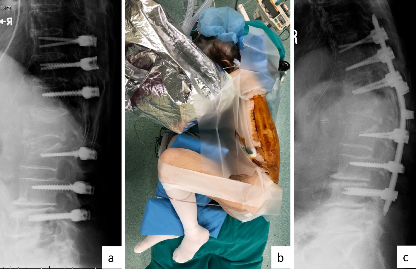
    Introduction: Vertebroplasty and posterior fusion for highly collapsed osteoporotic vertebral fractures have some complications such as implant failure due to postoperative correction loss. In contrast, because osteotomy and anterior fusion for super elderly over the age of 80 years are largely invasive, selecting them is difficult. Therefore, we performed surgery to fix the rod in the forward flexed lateral decubitus position to intentionally create kyphosis in anticipation of postoperative correction loss.

    Methods: The subjects were 4 patients (all females) who were over the age of 80 years with highly collapsed vertebra and severe angular instability from 25° to 30° or more between the supine position and sitting position in lateral Xp images. The surgical method involved the insertion of pedicle screws initially in the supine position. Next, wound closure was performed using a primary suture, and the position was changed to the forward flexed lateral decubitus position to create kyphosis. Finally, the 5.5 mm pure titan rod was fixed.

    Results: All patients had lower back pain visual analog scale of 0 mm at 6 months after the operation, and they went out on their own with little assistance. Six months after the operation, fusion was achieved in one patient and loosening of pedicle screws was observed in three patients. However, no symptoms were observed in the group of pedicle screw loosening.

    Conclusions: By intentionally creating kyphosis, we were able to prevent various problems caused by correction loss and achieved good clinical results in a short duration. It appears to be a useful technique if the patients were selected by appropriate adaptation.

    Download PDF (2312K)
  • Kiyonori Yo, Eiki Tsushima, Yosuke Oishi, Masaaki Murase, Yoko Matsuda
    2021 Volume 12 Issue 9 Pages 1210-1217
    Published: September 20, 2021
    Released on J-STAGE: September 20, 2021
    JOURNAL FREE ACCESS

    Introduction: The objective of the present study was to analyze factors associated with health-related quality of life (HRQoL) in patients with osteoporotic vertebral fracture (OVF) treated by balloon kyphoplasty (BKP) at the 1-year follow-up.

    Methods: Forty-seven patients in total (average age 80.9 years old, 3 males and 44 females), who had undergone BKP for OVF at our hospital between 2012 and 2019, participated in this study. For assessing HRQoL, the scores of each domain of SF-8 were compared among the patients before surgery and at the 1-year follow-up. In addition, we analyzed whether the following factors were correlated with the scores of each domain of SF-8 at the 1-year follow-up through multiple regression analysis: age, sex, disease period, presence of subsequent OVF, visual analog scale (VAS), total functional independence measure scores, walking ability, locomotive syndrome scores, global alignment parameters, length of hospital stay, history of taking osteoporosis drugs, and presence of outpatient rehabilitation.

    Results: The average score for each domain of SF-8 was significantly increased at the 1-year follow-up than that before surgery. The number of patients whose SF-8 score exceeded the national standard score for SF-8 was increased in the mental health domain, but no significant differences were found in the physical health domain at the follow-up. The multiple regression analysis demonstrated that the increased walking ability and decreased VAS scores were significantly correlated with the increased SF-8 scores in some domains.

    Conclusions: The results of this study suggested that the improvement in HRQoL after BKP was mainly due to increased walking ability and decreased low back pain in patients with OVF. If the patients treated with BKP acquired better walking ability by exercise therapy, their HRQoL might improve.

    Download PDF (1322K)
Secondary Publication
  • Keishi Maruo, Fumihiro Arizumi, Kazuki Kusuyama, Kazuya Kishima, Toshi ...
    2021 Volume 12 Issue 9 Pages 1218-1225
    Published: September 20, 2021
    Released on J-STAGE: September 20, 2021
    JOURNAL FREE ACCESS

    Objective: Lateral lumbar interbody fusion (LIF) has been widely used in adult spinal deformity (ASD) surgery. However, anterior longitudinal ligament rupture (ALLR) has been occasionally identified following posterior spinal correction surgery. This study aimed to assess the incidence of ALLR and to identify the risk factors of ALLR in patients with ASD.

    Materials and Methods: This study included 43 consecutive patients (8 male and 35 female patients) who underwent posterior corrective surgery involving LIF (128 levels) for ASD between 2014 and 2018. The patients' mean age was 72±7 years, and the minimum follow-up period was 1 year. Following LIF, posterior correction and fusion surgery using the cantilever technique was performed. Oblique LIF was performed in 27 patients, whereas extreme lateral interbody fusion (XLIF) was performed in 16 patients. The mean number of spinal fused levels was 8.9±1.8 (range: 8-15), and the mean number of LIF levels was 3±0.6 (range: 2-4). ALLR was considered if an LIF cage showed no contact with the vertebral endplates. The radiographic parameters included thoracic kyphosis, lumbar lordosis (LL), pelvic incidence (PI), pelvic tilt, and sagittal vertical axis.

    Results: ALLR occurred in 10 patients (22%) and at 11 levels (8.6%). XLIF and preexisting osteoporotic vertebral fracture were identified as independent risk factors of ALLR. LL change was approximately 10 degrees greater in the ALLR group than in the non-ALLR group (P = 0.017), and overcorrection was observed in the ALLR group (PI-LL: −7.9±7 degrees). Segmental lordotic angle change at the ALLR level was much larger than after LIF and correction surgery. ALLR-related reoperation was performed in two cases (decompression surgery because of posterior impingement and rod breakage).

    Conclusions: ALLR occurred in 10 patients (22%), and XLIF use and preexisting osteoporotic vertebral fracture were independent risk factors of ALLR. Moreover, overcorrection was observed in patients with ALLR. Care should be taken to avoid ALLR, which may require additional surgery.

    Download PDF (1491K)
feedback
Top