Journal of Spine Research
Online ISSN : 2435-1563
Print ISSN : 1884-7137
Volume 15, Issue 1
Displaying 1-7 of 7 articles from this issue
Editorial
Original Article
  • Masayuki Furuya, Shinya Okuda, Yukitaka Nagamoto, Tomiya Matsumoto, Yo ...
    2024 Volume 15 Issue 1 Pages 3-12
    Published: January 20, 2024
    Released on J-STAGE: January 20, 2024
    JOURNAL FREE ACCESS

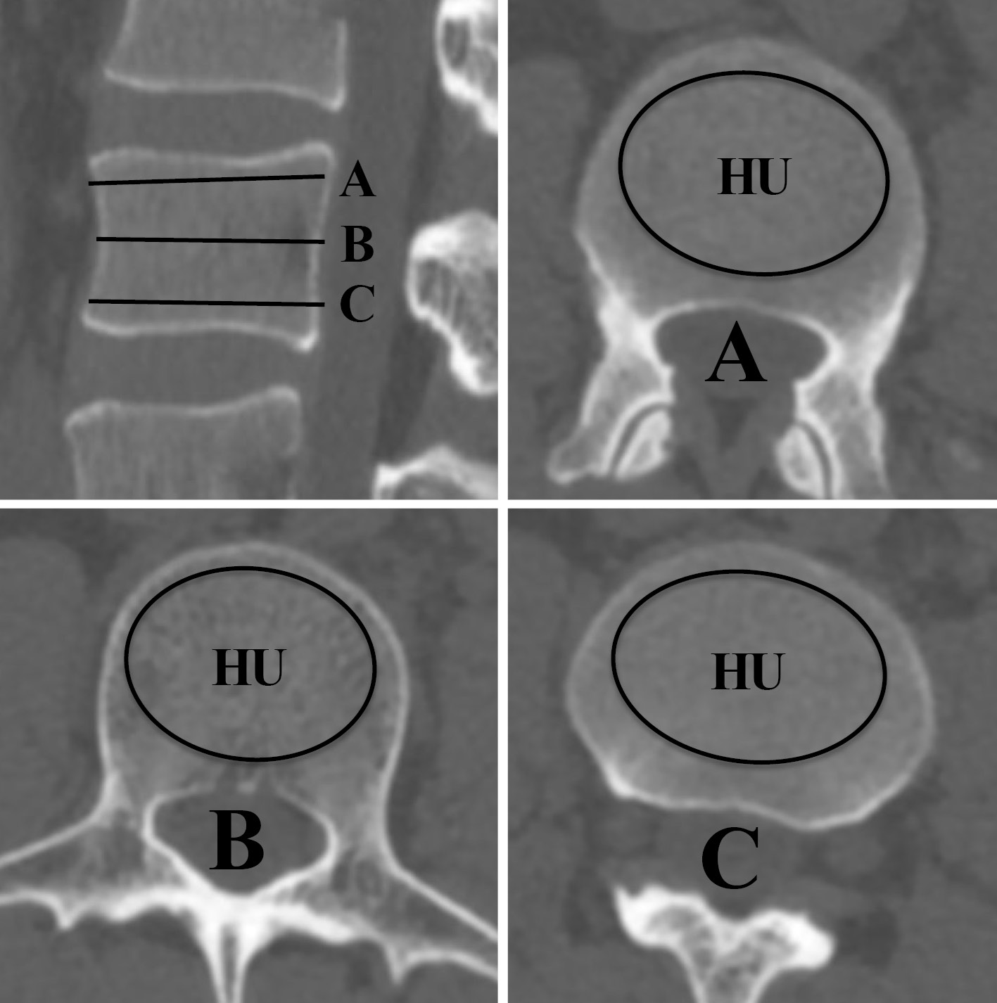
    Introduction: Although bone health assessment prior to spine surgery is imperative, assessment of bone mineral density (BMD) with dual energy X-ray absorptiometry (DXA) is not easily available at all centers before spine surgery. Moreover, it has been recently reported that lumbar Computed Tomography (CT) -based Hounsfield Units (CTHU) correlate with BMD from DXA scans. If CTHU can be utilized for bone health assessment prior to spine surgery, it would be very useful to assess patients' bone health without added expense to the patient. Therefore, we investigated the usefulness of opportunistic assessment of bone health with CTHU.

    Methods: Subjects comprised 138 patients who underwent a lumbar CT and DXA within a 6-month period. Three types of scanners were used for CT. CTHU values between the T12 and L4 vertebral levels were measured, calibrated by asynchronous phantom, compared to T-scores from DXA scans, and compared in patients with and without osteoporotic fractures. Furthermore, we also examined the threshold for DXA-defined osteoporosis (T-score ≤ −2.5) and the presence of osteoporotic fracture using receiver operating characteristic (ROC) curve analysis.

    Results: CTHU values correlated with T-scores with DXA scan of both lumbar spine and hip. CTHU values were significantly lower in patients with osteoporotic fractures than in patients without osteoporotic fractures. Furthermore, the ROC analyses revealed that the thresholds for DXA-defined osteoporosis and the presence of osteoporotic fracture were 74.1 and 69.1, respectively.

    Conclusions: CTHU values from three types of scanners correlates with T-scores with DXA scan of lumbar spine and hip and can be useful to screening of osteoporosis. The thresholds for DXA-defined osteoporosis and the presence of osteoporotic fracture were 74.1 and 69.1, respectively.

    Download PDF (1735K)
  • Yohei Ito, Hisanori Mihara, Yasunori Tatara, Takanori Niimura
    2024 Volume 15 Issue 1 Pages 13-19
    Published: January 20, 2024
    Released on J-STAGE: January 20, 2024
    JOURNAL FREE ACCESS

    Introduction: Postoperative dysphagia is a relatively common complication in anterior cervical decompression and fusion. This study investigated the effect of preoperative ST intervention on prevention of dysphagia following cervical anterior decompression fusion surgery and risk factors for the occurrence of postoperative dysphagia, including preoperative swallowing function.

    Methods: A total of 116 patients who underwent anterior cervical decompression and fusion after preoperative ST evaluation of swallowing function (tongue pressure and repetitive saliva swallowing test [RSST]) on the day before surgery were included. Patients were divided into postoperative dysphagia (+) and (−) groups, and radiological parameters and preoperative swallowing function on the day before surgery were compared. The effectiveness of preoperative swallowing training was also evaluated in 60 patients who underwent ST evaluation at the time of admission for myelography.

    Results: Postoperative dysphagia occurred in 12.9% (15/116). The dysphagia (+) group had higher age, higher level of fixation, and lower preoperative tongue pressure and RSST compared to the dysphagia (−) group. Postoperative C2-7 and ΔC2-7 angles, corresponding to pre- and postoperative change, respectively, were significantly higher in the dysphagia (+) group. Regarding ST intervention at the time of admission for myelography, tongue pressure improved significantly, but there was no significant difference in RSST.

    Conclusions: The results of this study indicate that preoperative swallowing function is related to the occurrence of dysphagia after anterior cervical decompression and fusion. The results also suggest that swallowing training by ST improves swallowing function at the time of surgery.

    Download PDF (1217K)
  • Ryosuke Yanai, Akira Matsumura, Takashi Namikawa, Masatoshi Hoshino, H ...
    2024 Volume 15 Issue 1 Pages 20-27
    Published: January 20, 2024
    Released on J-STAGE: January 20, 2024
    JOURNAL FREE ACCESS

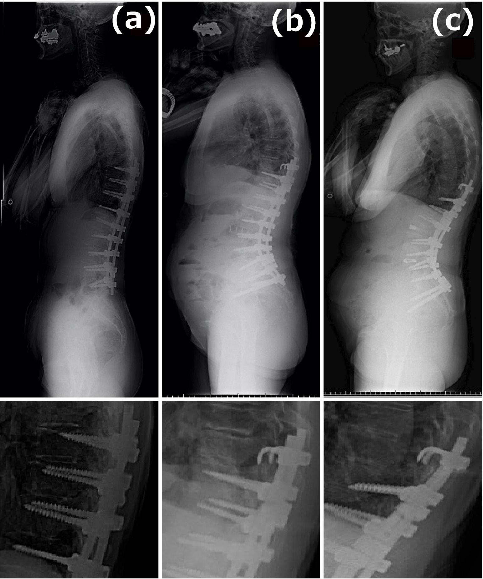
    Introduction: Proximal junctional kyphosis (PJK) is a common mechanical complication which has a potential risk of revision surgery in adult spinal deformity (ASD) surgery. Three different types of upper instrumented vertebra (UIV) anchors were changed to prevent PJK and proximal junctional vertebral fractures (PJVF). The purpose of this study was to compare the incidence of PJVF in ASD surgery among the three types of UIV anchors.

    Methods: Sixty ASD patients with minimum 2-year follow-up were recruited in this study. We divided the patients into three groups according to UIV instruments; pedicle screw (PS) group (n = 15 patients; mean age at surgery, 69.4 years; mean follow-up duration, 78 months), hook group (n = 25, 71.5 years, 59.0 months), and hook-short PS (sPS) group (n = 20, 69.1 years, 13.9 months). We evaluated the radiographic parameters before surgery, one week after surgery, and final follow-up (FFU), and also assessed surgical outcomes using Japanese version SRS-22 questionnaire (SRS-22). We compared the parameters and incidence of PJVF among the three groups. PJVF was defined as the presence of UIV or UIV±1 fracture in this study.

    Results: The incidence of PJVF was 40% (6/15) in the PS grp, 24% (7/25) in the Hook group, and 10% (2/20) in the hook-sPS group. The statistical analysis did not show any significant difference in incidence of PJVF between the three groups (P = 0.1). Comparative analysis of the radiographic parameters among the three groups demonstrated that preoperative pelvic incidence-lumbar lordosis, Pelvic tilt, and thoracolumbar kyphosis (TLK) at FFU differ significantly (P < 0.05); proximal junctional angle (PJA) and TLK in the hook-sPS group at FFU were significantly lower than the hook group (P < 0.05).

    Conclusions: Our study shows that hooks at UIV and short PS at UIV+1 as UIV anchor may prevent PJVF. This UIV construct is considered to match the concept of soft-landing for thoraco-pelvic fixation in the ASD patients. Further biomechanical study is necessary to clarify our results.

    Download PDF (1806K)
  • Takuya Sada, Eiichiro Iwata, Akinori Okuda, Masato Tanaka, Takahiro Mu ...
    2024 Volume 15 Issue 1 Pages 28-33
    Published: January 20, 2024
    Released on J-STAGE: January 20, 2024
    JOURNAL FREE ACCESS

    Introduction: Lateral intervertebral fusion of the lumbar spine is associated with the risk of injury to large blood vessels, their branches, and venous malformations.

    Objective: To evaluate the anatomy that poses a risk of vascular injury in lateral intervertebral fusion using preoperative contrast-enhanced computed tomography (CT).

    Materials: Forty-six patients who underwent contrast-enhanced CT prior to lateral intervertebral fusion at our institution were included, and major arteries and veins were evaluated on horizontal section images at each vertebra from L1/2-L4/5. The presence or absence of lumbar artery branches running longitudinally across the intervertebral disk was evaluated in each vertebral segment. Venous malformations were evaluated for (a) duplicated vena cava, (b) running of the left common iliac vein branch outside the common iliac artery, and (c) dilatation of the ovarian vein or spermatic cord vein.

    Results: Major arteries traveling in Zones 1-4 according to Moro et al.'s evaluation method were found in 5 cases (11%) in L1/2, 3 cases (7%) in L2/3, 2 cases (4%) in L3/4, and 5 cases (11%) in L4/5. Major veins were found in 6 cases (13%) in L1/2, 16 cases (35%) in L2/3, 37 (80%) in L3, 37 (80%) in L1/2, 16 (35%) in L2/3, 39 (85%) in L3/4, and 39 (85%) in L4/5. Longitudinal disk traversal of the lumbar artery branch was present in 1 case (2%) in L3/4 and 15 (33%) in L4/5. Venous malformations were found in (a) 0 (0%), (b) 4 (9%), and (c) 3 (7%) patients, respectively.

    Conclusions: Major arteries and veins may be present within the operative range at any elevation from L1/2 to L4/5, and segmental arteries may traverse near the intervertebral disk at L3/4 and L4/5. Venous malformations should also be assessed preoperatively.

    Download PDF (1352K)
  • Daisuke Kamakura, Keiji Hasegawa, Kazumasa Nakamura, Katsunori Fukutak ...
    2024 Volume 15 Issue 1 Pages 34-39
    Published: January 20, 2024
    Released on J-STAGE: January 20, 2024
    JOURNAL FREE ACCESS

    Introduction: This study examined the risk factors for C5 palsy focusing on the traction and impingement effects of cervical laminoplasty.

    Methods: Ninety three patients who underwent cervical open-door laminoplasty between December 2015 and March 2021 were enrolled in this study.

    We examined the diameter of bilateral C4/5 foramen, anterior protrusion of the superior articular process of C5, gutter position, and pre- and postoperative C2-7 angle in plain radiograph.

    Results: The incidence of C5 palsy was 7.5% (7/93 cases).

    There were substantial differences in the diameter of bilateral C4/5 foramen, anterior protrusion of the superior articular process of C5 and the preoperative C2-7 angle in plain radiograph; however, there were no substantial differences in the gutter position and the postoperative C2-7 angle in plain radiograph.

    Conclusions: The risk factor for C5 palsy after cervical open-door laminoplasty was examined.

    The results suggest that the diameter of bilateral C4/5 foramen, anterior protrusion of the superior articular process of C5, and preoperative C2-7 angle in plain radiograph are major preoperative risk factors.

    Download PDF (1090K)
  • Kiyonori Yo, Eiki Tsushima, Yosuke Oishi, Masaaki Murase, Katsumi Doi, ...
    2024 Volume 15 Issue 1 Pages 40-48
    Published: January 20, 2024
    Released on J-STAGE: January 20, 2024
    JOURNAL FREE ACCESS

    Introduction: This study aimed to clarify the relationships between the increased scores of low back pain and other domains of Japanese Orthopaedic Association Back Pain Evaluation Questionnaire (JOABPEQ) in patients with osteoporotic vertebral fractures (OVFs) treated with balloon kyphoplasty (BKP) during the 1-year follow-up period.

    Methods: Overall, 71 patients with OVFs treated with BKP participated in this study. The scores of each domain of JOABPEQ were compared before surgery and at 1-year follow-up. The patients were divided into lower lumbar pain improved and nonimproved groups based on the increased scores of low back pain of JOABPEQ during the follow-up period. The increased scores of other domains, i.e., lumbar function, walking ability, social life function, and mental health, were compared between the two groups. In addition, correlations were assessed between the increased scores of low back pain and other domains during the follow-up period.

    Results: There were 40 and 31 cases in the lower lumbar pain improved and nonimproved groups, respectively. The lower lumbar pain improved group had significantly higher increased scores of lumbar function, walking ability, and social life function than the nonimproved group. Significant associations were found between the increased scores of low back pain and those of lumbar function, walking ability, and social life function.

    Conclusions: The increased scores of low back pain were associated with those of lumbar function, walking ability, and social life function during the 1-year follow-up period in patients with OVFs treated with BKP.

    Download PDF (1310K)
feedback
Top