Journal of Spine Research
Online ISSN : 2435-1563
Print ISSN : 1884-7137
Volume 12, Issue 7
Displaying 1-19 of 19 articles from this issue
Editorial
Original Article
  • Gakuji Gondo, Tsuyoshi Watanabe, Masahiko Tanaka, Kazuko Hotta, Takahi ...
    2021Volume 12Issue 7 Pages 905-909
    Published: July 20, 2021
    Released on J-STAGE: July 20, 2021
    JOURNAL FREE ACCESS

    Introduction: The number of patients with odontoid fractures has been increasing with the aging of the population. The authors report the clinical characteristics and treatment results of patients with odontoid fractures.

    Method: A retrospective analysis was performed on 53 patients admitted to Shonan-Kamakura General Hospital who were diagnosed with odontoid fracture. They were divided into two groups: type II and type III. Age, cause of injury, combined injuries, neurological findings, treatment methods, and treatment results were compared between the two groups.

    Results: There were 28 patients in the type II group and 28 patients in the type III group. The mean age was 60.4 years in the type II group and 77.2 years in the type III group. The proportion of males was 60.7% and 40% in the type II and type III groups, respectively. Traffic accidents were the main cause of injury in the type II group. Surgery was performed in 17 (61%) and three cases (12%) of the type II and type III groups, respectively. Bony fusion was confirmed in 78% of type II and 81% of type III patients, excluding those lost to follow-up. Four cases of type II and one case of type III were finally diagnosed as nonunion, but no one suffered neurological deterioration.

    Conclusion: Type II odontoid fracture had a higher proportion of patients of younger age, men, traffic accident and surgical treatment compared to type III. Similar results were obtained with respect to treatment, in principle, with surgery for type II and conservative treatment for type III.

    Download PDF (1000K)
  • Isao Kitahara, Hiroaki Shirotori, Hiroshi Yonetani
    2021Volume 12Issue 7 Pages 910-916
    Published: July 20, 2021
    Released on J-STAGE: July 20, 2021
    JOURNAL FREE ACCESS

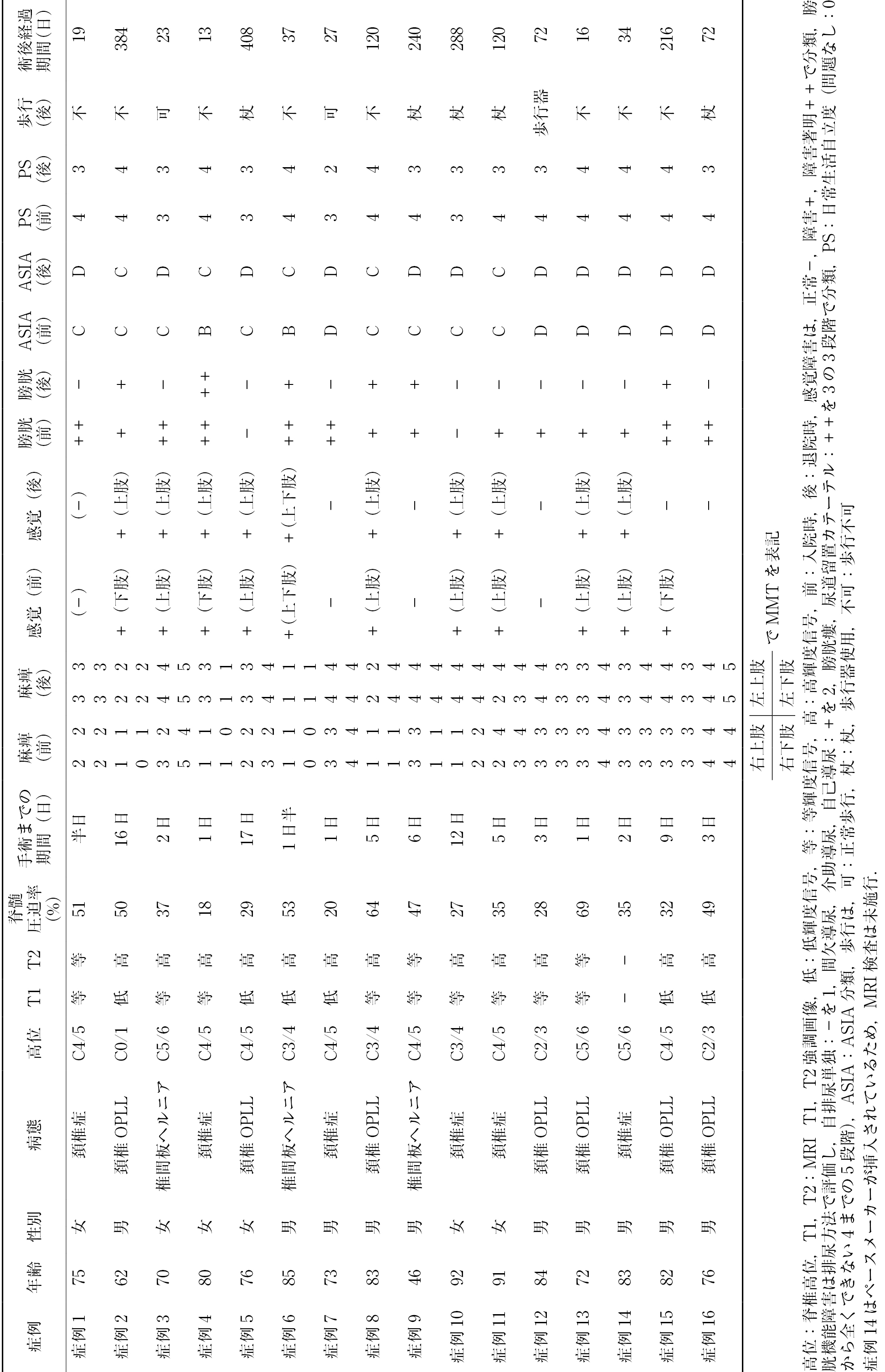
    All 16 patients with cervical cord injuries without bone lesions due to minor hyperextension injuries caused by falls who presented with motor paralysis and underwent acute decompression surgery showed severe symptoms. The results were particularly poor in elderly patients with severe spinal cord compression. Motor paralysis of the lower limbs due to the central cervical spinal cord injury was relatively mild, and there was a trend toward improvement in the bladder functions and gait disturbance. However, the recovery of the upper limb functions was poor and activities of daily living acquisition were insufficient. Patients who underwent acute decompression surgery within 3 days following the cord injury showed a slight improvement even in cases of severe spinal cord compression. However, after 4 days postoperatively, a slow recovery was observed, especially in elderly patients with a high spinal cord compression rate. Regarding perioperative complications, there were no major concerns and no cases that compromised an early discharge from the hospital. The results of this study suggest that early spinal cord decompression, early discharge, and rehabilitation are important for elderly patients with severe spinal cord compression.

    Download PDF (1417K)
  • Tetsuryu Mitsuyama, Kaiji Ota, Takeshi Umebayashi, Ryuta Kono
    2021Volume 12Issue 7 Pages 917-925
    Published: July 20, 2021
    Released on J-STAGE: July 20, 2021
    JOURNAL FREE ACCESS

    Introduction: Effective surgical procedures for dropped head syndrome (DHS) are still not established. The main purpose of this study was to assess the risk factors for postoperative dysphagia and causes of instrumentation failure in surgery for DHS.

    Methods: We retrospectively analysed postoperative dysphagia, instrumentation failure, and surgical procedures in 14 patients who underwent surgery for flexible DHS.

    Results: Two patients presented with postoperative dysphagia. Severe dysphagia soon after posterior fixation surgery alone was resolved by the reoperation of lessening cervical lordosis. Mild dysphagia after three-level anterior fixation surgery spontaneously recovered several months. One patient with the lowest instrumented vertebra (LIV) of C7 required pedicle subtraction osteotomy of C7 and additional posterior fixation of T5 as a revision for caudal instrumentation failure. Another patient with severe osteopenia showed the backing-out of rostral posterior screws several months after surgery. The LIV of posterior fixations were C7 in 1, T1-4 in 10, and T5 or 6 in 3 patients. The upper instrumented vertebrae were C2 in 10 and C3 in 4 patients. The ranges of anterior fixation were five levels in one, four levels in three, and three levels in six patients. All of them included C4/5 and C5/6.

    Conclusions: Excessive surgical correction of cervical lordosis and advanced age after multilevel anterior fixation are risk factors for postoperative dysphagia in surgery for DHS. The LIV of C7 confers a risk of caudal instrumentation failure. One cause of rostral instrumentation failure is an imbalance in the pull-out strength of screws in only posterior fixations. Anterior and posterior fixations are basically recommended as surgery for DHS, after careful assessment of preoperative swallowing function. Posterior fixations range from C2 or C3 to the upper thoracic spine and extend caudally depending on the thoracolumbar alignment. Ranges of anterior fixations extend to more than three levels, including the lower cervical spine.

    Download PDF (1568K)
  • Takeshi Hara, Ryosuke Takahashi, Yukoh Ohara, Hirokazu Iwamuro, Kazuak ...
    2021Volume 12Issue 7 Pages 926-932
    Published: July 20, 2021
    Released on J-STAGE: July 20, 2021
    JOURNAL FREE ACCESS

    Duragen® is an absorptive artificial dura mater formed using a porous collagen matrix. Platelets infiltrate its inside to form a membrane with fibrin clots, which has an early effect of preventing cerebrospinal fluid leakage. We retrospectively examined the effect of Duragen® on preventing cerebrospinal fluid leakage during spinal cord surgery. Of the 34 cases of spinal surgery using Duragen®, 2 had CSF leakage. Duragen® is considered useful in preventing cerebrospinal fluid leakage in spinal cord surgery and a material that can be used in various ways due to its affinity for water.

    Download PDF (1374K)
  • Shinji Kotaka, Yasushi Fujiwara, Naofumi Hashiguchi
    2021Volume 12Issue 7 Pages 933-937
    Published: July 20, 2021
    Released on J-STAGE: July 20, 2021
    JOURNAL FREE ACCESS

    Introduction: The prevalence of osteoporotic vertebral fractures (OVFs) is increasing with the aging of the population. The majority of OVFs heal after 8-12 weeks of conservative treatment. However, some patients with OVF have neurological deficits (NDs). Reconstructive spinal surgery is often performed in patients having OVFs with NDs. However, these patients are usually elderly patients with various major morbidities. Therefore, fewer invasive interventions are required. At our institute, balloon kyphoplasty (BKP), which is a minimally invasive surgery, combined with microscopic posterior decompression was performed in patients having OVFs with NDs. The purpose of this study was to evaluate the clinical outcomes of BKP combined with microscopic posterior decompression for OVFs with NDs.

    Methods: Between November 2012 and March 2020, 31 consecutive patients who underwent BKP combined with microscopic posterior decompression at our institution were included in this study. In addition, 72 patients who underwent BKP for OVF were included in the control group. Lateral views on radiography images were used to measure the local kyphotic angle preoperatively and postoperatively.

    Results: The average local kyphotic angles were 14.3±18.9° preoperatively and 15.4±20.4° postoperatively. The average Japanese Orthopaedic Association (JOA) scores were 9.2±4.3° preoperatively and 13.2±6.3° postoperatively. The improvement rate was 13.0%. The differences were statistically significant in the JOA score but not in the local kyphotic angle. In the control group, the average local kyphotic angles were 15.0±10.6° preoperatively and 8.2±8.9° postoperatively. The differences were statistically significant. Compared to the control group, the correction angle was significantly smaller.

    Conclusions: BKP combined with microscopic posterior decompression may only result in a smaller correction of the local kyphotic angle. However, the JOA score significantly improved following the surgery. The rate of improvement was 13.0% in this study; therefore, this method may be considered a temporary surgery rather than radical surgery. If the indication is limited, BKP combined with microscopic posterior decompression is a useful technique.

    Download PDF (806K)
  • Yoshitaka Suzuki, Tetsuro Hida, Junichi Ukai, Toshihiro Ando, Koji Sat ...
    2021Volume 12Issue 7 Pages 938-946
    Published: July 20, 2021
    Released on J-STAGE: July 20, 2021
    JOURNAL FREE ACCESS

    Surgical intervention for pseudarthrosis after lumbar body fusion surgery in patients with osteoporosis is complicated and difficult. We treated 3 such cases using transpedicular impaction allogenic bone graft to increase the strength of the augmented pedicle screws and processed fibula to reinforce the endplate. All cases achieved bony fusion without loosening or pulling out of the screws and subsidence of cages. Augmentation of pedicle screw fixation with allogenic bone grafts and fibula plates is an alternative option for reconstruction surgery. Furthermore, instrumentation and fusion with this augmentation technique together with the fibula plate offer a possible primary surgery to treat patients with reduced bone quality due to severe osteoporosis and for avoiding pseudarthrosis.

    Download PDF (2953K)
  • Yu-ya Kamizawa, Kenji Kosaka, Sho Fujiwara, Yu-ichiro Ohnishi, Takashi ...
    2021Volume 12Issue 7 Pages 947-951
    Published: July 20, 2021
    Released on J-STAGE: July 20, 2021
    JOURNAL FREE ACCESS

    Introduction: Gait parameters, before and after surgery, were studied in patients undergoing posterior lumbar decompression.

    Methods: The gait of 11 patients who underwent posterior lumbar decompression surgery was analysed preoperatively and 1 month postoperatively using the optical gait analyser OPTOGAIT system. The evaluation items were each gait parameter, JOA score, ODI, and VAS during gait, and the results were compared between the preoperative and postoperative parameters.

    Results: Significant improvements were observed in ODI, JOA score, VAS during gait, and walking rate.

    Conclusions: Posterior lumbar decompression improves walking rate.

    Download PDF (787K)
  • Masayuki Ishihara, Shinichiro Taniguchi, Yoichi Tani, Masaaki Paku, Ta ...
    2021Volume 12Issue 7 Pages 952-957
    Published: July 20, 2021
    Released on J-STAGE: July 20, 2021
    JOURNAL FREE ACCESS

    Purpose: The aim of this study was to examine the short-term results of circumferential minimally invasive surgery (cMIS) using lateral interbody fusion (LIF) and percutaneous pedicle screw (PPS) in the treatment of spinal deformities in patients with adult idiopathic scoliosis (AS).

    Subjects and methods: The study subjects were six patients with AS (five females and one male) who had undergone c-MIS using LIF and PPS between 2018 and 2019, and were followed up for more than 18 months. The parameters examined included the number of fixed vertebral bodies, upper instrumented vertebra (UIV), lower instrumented vertebra (LIV), number of LIF, average volume of blood lost, operation time, various spinopelvic parameters, pre- and postoperative Oswestry Disability Index (ODI), and perioperative complications.

    Results: The UIV was T4, T7, and T9 in two cases. The LIV was the pelvis in all cases. The average numbers of fixed vertebral bodies and LIF were 13.3 and 4.5, respectively. The average volume of blood lost and operation time were 723 ml and 352 min, respectively. The spinal pelvic parameters improved (lumbar lordosis (LL), 11° preoperatively vs. 50° postoperatively; pelvic incidence (PI) -LL, 39° preoperatively vs. 0° postoperatively; pelvic tilt (PT), 32° preoperatively vs. 15° postoperatively; thoracic kyphosis (TK), 19° preoperatively vs. 40° postoperatively; Cobb angle (CA), 66° preoperatively vs. 25° postoperatively; C7 central sacrum vertical line (C7CSVL), 52 mm preoperatively vs. 7 mm postoperatively; and sagittal vertical axis (SVA), 128 mm preoperatively vs. 25 mm finally). The ODI score improved from 51 to 26. Two months after the operation, the S2AI set screw was removed in one patient. The other patients had no complications.

    Conclusion: cMIS is a minimally invasive surgical procedure for the treatment of AS that achieves good correction and suitable short-term results. Even if cMIS is performed without posterior osteotomy, sufficient correction is possible as it corrects the balance of the coronal and sagittal planes without complete correction of scoliosis.

    Download PDF (1489K)
  • Shuntaro Tsuchida, Yoshihiro Kitahama
    2021Volume 12Issue 7 Pages 958-965
    Published: July 20, 2021
    Released on J-STAGE: July 20, 2021
    JOURNAL FREE ACCESS

    Between April 2017 and April 2020, five patients with lumbar disc herniation and posterior ring apophyseal separation were surgically treated using transforaminal full-endoscopic lumbar discectomy (FELD). The surgery was performed under general anaesthesia. The herniated disc, including the bony fragment, was resected in all cases. The results were good, and there were no neurological complications. Transforaminal FELD has the advantages of less soft tissue or bony structure damage. Because it is possible to approach the lesion directly using this method, we can safely resect the herniated disc including the bony fragment without excessive nerve retraction. On the other hand, dural tears should be carefully considered because of dural adhesion to bony fragments.

    Download PDF (1801K)
  • Shu Nakamura, Fujio Ito, Yasushi Miura, Motohide Shibayama, Naoto Hosh ...
    2021Volume 12Issue 7 Pages 966-972
    Published: July 20, 2021
    Released on J-STAGE: July 20, 2021
    JOURNAL FREE ACCESS

    Introduction: We previously reported using an internal retractor for percutaneous full-endoscopic posterior discectomy of the cervical spine. The internal retractor is useful in areas with narrows safety margins, such as the cervical spine. On the other hand, in the lumbar spine, the internal retractor is also useful for placing the outer sheath onto the intervertebral disc.

    Methods: Percutaneous full-endoscopic posterior discectomy using an internal retractor was performed for cases with cervical or lumbar disc herniation.

    Results: A total of 14 cases (7 cases of cervical spine and 7 cases of lumbar spine) were studied. In all cases, the hernia could be resected while maintaining a proper visual field, and there was no postoperative neuropathy.

    Conclusions: Unlike the conventional spatula or dissector, the internal retractor surface can be inserted at the outermost area and safe retraction of the nerve root can be performed by just rotating the instrument and the scope. Moreover, it is possible to operate with forceps or a bipolar coagulator while protecting the nerve root securely with the internal retractor and observing it. A two-step nerve root retraction with an internal retractor and an outer sheath can be performed safely because of a smaller gap with the outer sheath than with a conventional dissector and spatula. Due to these unconventional characteristics, the internal retractor contributes to the safety and facility of percutaneous full-endoscopic posterior discectomy in the whole spine.

    Download PDF (1831K)
  • Yuya Takesue, Akiyoshi Yamazaki, Keiichi Katsumi, Tatsuki Mizouchi, Yu ...
    2021Volume 12Issue 7 Pages 973-978
    Published: July 20, 2021
    Released on J-STAGE: July 20, 2021
    JOURNAL FREE ACCESS

    Introduction: We performed sublaminar taping stabilisation at the proximal adjacent segment to prevent adjacent segmental disease (ASD). The purpose of this study was to examine the effectiveness of sublaminar taping stabilisation.

    Methods: Forty-two patients underwent L4/5/S1 interbody fusion with L3 sublaminar taping stabilisation (SLT group), and 73 patients underwent L4/5/S1 interbody fusion alone (control group). We evaluated the clinical findings, including the operation time, volume of blood loss, recovery rate based on the Japanese Orthopaedic Association score, and reoperation rate. Furthermore, we evaluated the patients radiologically to examine the global alignment, Pfirrmann classification, slip distance, disc height, posterior opening angle, and presence or absence of radiological ASD.

    Results: Clinical examination revealed no significant differences between the two groups. In contrast, the slip distance and posterior opening angle were significantly reduced in the SLT group. In addition, the rate of radiological ASD was significantly decreased in the SLT group.

    Conclusions: These results suggest that sublaminar taping may decrease the risk of radiological ASD. Moreover, sublaminar taping stabilisation may reduce the need for reoperation due to ASD.

    Download PDF (1005K)
  • Kazunari Fushimi, Chizuo Iwai, Satoshi Nozawa, Haruhiko Akiyama
    2021Volume 12Issue 7 Pages 979-983
    Published: July 20, 2021
    Released on J-STAGE: July 20, 2021
    JOURNAL FREE ACCESS

    We recently performed transforaminal lumbar interbody fusion (TLIF) using two kidney-type cages to obtain good bony fusion and prevent cage subsidence. In this comparative study, we investigated the efficacy of TLIF using two kidney-type cages. Fifty-six patients who underwent TLIF were retrospectively evaluated. Twenty-two patients who underwent surgery with two bullet-type cages (2-bullets group), 17 cases with one kidney-type cage (1-kidney group), and 17 cases with two kidney-type cages (2-kidneys group) were compared. Twelve months after the surgery, subsidence of the cage was 1.14 mm in the 2-bullets group, 1.63 mm in the 1-kidney group, and 0.73 mm in the 2-kidneys group. Subsidence of cage was significantly more in 1-kidney group than in the 2-kidneys group. Bony fusion was not achieved in four cases in the 1-kidney group. Improvement of symptoms and recovery of clinical scores did not differ among the groups.

    Download PDF (1054K)
Case Report
  • Yuichi Yoshida, Naohiro Kawamura, Yohei Kakuta, Yuki Onishi, Junichi O ...
    2021Volume 12Issue 7 Pages 984-988
    Published: July 20, 2021
    Released on J-STAGE: July 20, 2021
    JOURNAL FREE ACCESS

    A 45-year-old man with severe hyperuricemia developed back pain and difficulty walking. Magnetic resonance imaging (MRI) revealed a thoracic epidural mass lesion compressing the spinal cord, and CT showed calcification in the mass. Decompression and fusion surgery were performed, and the pathological diagnosis was that of spinal tophaceous gout. Although motor function improved partially, severe spasticity persisted. Retrospectively, the calcified mass in the thoracic spine had been identifiable on CT two months before the onset of back pain. If patients with poor control of hyperuricemia complain of back pain, CT is highly recommended to rule out spinal tophaceous gout.

    Download PDF (1064K)
  • Sawako Ogiso, Yusuke Nakao, Kenichi Hirabayashi, Shigeo Sano
    2021Volume 12Issue 7 Pages 989-995
    Published: July 20, 2021
    Released on J-STAGE: July 20, 2021
    JOURNAL FREE ACCESS

    Introduction: Dropped head syndrome is broadly classified into two as the neck-oriented and the back-oriented groups. Patients with Parkinson's disease have a low ability to compensate for sagittal imbalance and therefore present with dropped head syndrome, which is frequently of the back-oriented type.

    Case Report: We report three cases of dropped head syndrome in Parkinson's disease. They had three different kinds of spinal deformity, namely, kyphoscoliosis, iatrogenic flat back, and post-traumatic kyphosis; all of them also had sagittal imbalance. Surgical correction of the thoracolumbar spine improved not only the sagittal imbalance but also the dropped head syndrome.

    Conclusion: Back-oriented dropped head syndrome can be improved by surgical correction of the thoracolumbar spine.

    Download PDF (3114K)
  • Katsuji Shimizu, Kei Miyamoto, Hideo Hosoe
    2021Volume 12Issue 7 Pages 996-1001
    Published: July 20, 2021
    Released on J-STAGE: July 20, 2021
    JOURNAL FREE ACCESS

    Giant cell tumor (GCT) of the bone is a benign lesion which can be cured by surgical resection. However, when this lesion affects a vertebra, it can produce severe neurological deficits due to compression of the spinal cord or cauda equina, especially if the lesion recurs after incomplete excision. In the present case, a 13-year-old female with GCT of the L6 was treated with total spondylectomy and spinal shortening, followed by simple internal fixation with no strut bone graft. After two years of follow-up, we reported her case (Lancet 1996), because it was a novel concept of surgical reconstruction of lumbar spine after total spondylectomy. This patient's case has been revisited 20 years later.

    Download PDF (1257K)
  • Daiki Saito, Koichiro Ono, Naoto Kotani, Toshihiko Itou, Tokifumi Maji ...
    2021Volume 12Issue 7 Pages 1002-1006
    Published: July 20, 2021
    Released on J-STAGE: July 20, 2021
    JOURNAL FREE ACCESS

    Introduction: A combination of full-endoscopic debridement and drainage (FEDD) with minimally invasive spine stabilisation (MISt) using percutaneous pedicle screwing (PPS) was applied to a case of pyogenic thoracic spondylitis.

    Case: A seventy-year-old man visited the hospital because of back pain. He was diagnosed with pyogenic thoracic spondylitis at the T8/9 level. Methicillin-resistant Staphylococcus aureus (MRSA) was detected in the blood culture, and vancomycin was administered. Conservative treatment failed, and surgery was performed. FEDD was performed on the T8/9 disc using the right posterolateral approach, followed by MISt between T5 and T12, excluding the infected vertebrae. MRSA was detected in the disc material sampled during surgery, and antibiotic administration was continued. Antibiotics were continued for 6 weeks after surgery. The patient's general condition and laboratory data gradually improved, and he was discharged on the 55th hospital day. No recurrence of spondylitis was observed after six months.

    Conclusions: The combination of FEDD and MISt ca n be an option for patients with pyogenic thoracic spondylitis. As the skin incisions for PPS used in MISt and FEDD are different, it is possible to perform both at the same time.

    Download PDF (1031K)
  • Atsushi Shimizu, Tetsuryu Mitsuyama, Takeru Umebayashi, Takashi Komori ...
    2021Volume 12Issue 7 Pages 1007-1011
    Published: July 20, 2021
    Released on J-STAGE: July 20, 2021
    JOURNAL FREE ACCESS

    Introduction: Most intradural extramedullary tumours of the lumbar spine are schwannomas or meningiomas. Rarely, ependymomas or paragangliomas originating from the filum terminale can occur in the lumbar spine. Using case presentations of spinal tumours originating from the filum terminale, we discuss their imaging features and intraoperative monitoring method.

    Case Report: Here, we present two cases of spinal tumours from the filum terminale. Axial MRI images showed that the tumours were located in the central part of the lumbar spinal canal and that the remaining cauda equina nerves were symmetrically displaced. For intraoperative monitoring, the bulbocavernous reflex (BCR) was performed together with somatosensory evoked potentials (SEPs) and motor evoked potentials (MEPs). Both tumours originated from the filum terminale, and the craniocaudal sides of the filum terminale had to be cut to remove the tumour completely. The pathological diagnoses were ependymoma and paraganglioma, respectively.

    Conclusion: In cases of cauda equina nerves displaced symmetrically by centrally located tumours, as seen on axial MRI images, the tumour might originate from the filum terminale, such as in ependymomas or paragangliomas. During surgery for tumours in the filum terminale, intraoperative BCR may be useful to preserve the adjacent pundendal nerves.

    Download PDF (983K)
  • Takashi Hashimoto, Yasuo Ohori, Makoto Yoshida, Yasumitsu Ajiro, Ikuho ...
    2021Volume 12Issue 7 Pages 1012-1017
    Published: July 20, 2021
    Released on J-STAGE: July 20, 2021
    JOURNAL FREE ACCESS

    Introduction: We report a case of tardive cauda equina incarceration after dural tear during discectomy.

    Case Report: The patient was a 26-year-old woman who had a dural tear during lumbar discectomy. More than a month later, headache and sciatica recurred, but various examinations did not show any remarkable or abnormal findings. Therefore, although diagnosis was difficult, the characteristic feature of leg pain was obtained on compressing the wound. After surgical reduction of incarceration, the symptoms disappeared, with no recurrence.

    Conclusion: If a patient has neurological symptoms after dural tear but no examination findings, cauda equina incarceration should be suspected.

    Download PDF (1517K)
feedback
Top