Journal of Spine Research
Online ISSN : 2435-1563
Print ISSN : 1884-7137
Volume 11, Issue 8
Displaying 1-13 of 13 articles from this issue
Original Article
  • Koichi Yoshikane, Katsuhiko Kikuchi
    2020Volume 11Issue 8 Pages 997-1003
    Published: August 20, 2020
    Released on J-STAGE: August 20, 2020
    JOURNAL FREE ACCESS

    Introduction: Full-endoscopic lumbar laminoplasty (FEL) is thought to be the least minimal invasive spine surgery for lumbar spinal canal stenosis (LSS), using an 8 mm diameter cannula and full-endoscopic instruments. On the other hand, several complications have been reported. In this study, a retrospective study was made to reveal the incidence of perioperative complications as well as to figure out the appropriate management of complications.

    Methods: 564 patients who underwent FEL for LSS in our institute were included. The involved levels were L1/2 : 1, L2/3 : 3, L3/4 : 107, L4/5 : 440, L5/S1 : 13. Intraoperative videos, medical records including surgical records, X-ray, CT, and MRI imaging findings were retrospectively examined.

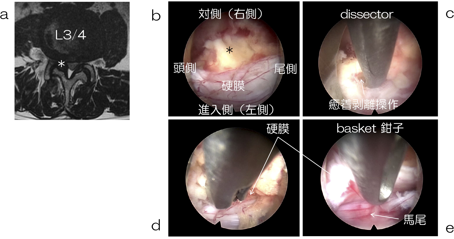
    Results: Complications occurred in 53 out of 564 cases, leading to 9.4% of incidences. Dural tears occurred in 23 cases. Leg pain due to a nerve stimulation was caused by the inappropriate position of the drain tube in 9 cases. Postoperative epidural hematoma was found in 7 cases. The causes of 14 cases of postoperative cauda equina injury, including 3 with neurogenic bladder, were not identified.

    Conclusions: These complications occurred at any given time during this investigation period and not just during a specific time period. Unfortunately, complications cannot be eliminated, but must be dealt with appropriately to overcome the difficulty of problem. Dural tears were caused using basket forceps; therefore, the use of basket forceps should be avoided especially during excision of the deep layers of ligamentum flavum. After the operation, the tip of the drain tube should be placed on the dorsal surface of the lamina in the lateral fluoroscopic view, and which can minimize the occurrence of pain due to a nerve root irritation. We should keep in mind the possibility of delayed onset postoperative epidural hematoma for at least several days after operation. In 14 of the cases found, the causes of postoperative injury to the cauda equina were not identified. Unintentional injury to the cauda equina may have occurred during the decompression procedure in the narrow spinal canal. So, we must pay meticulous attention to the location of the tip of the cannula in the spinal canal throughout the entire operation.

    Download PDF (1604K)
  • Shu Nakamura, Fujio Ito, Yasushi Miura, Motohide Shibayama, Naoto Hosh ...
    2020Volume 11Issue 8 Pages 1004-1009
    Published: August 20, 2020
    Released on J-STAGE: August 20, 2020
    JOURNAL FREE ACCESS

    Introduction: We have already reported percutaneous lumbar interbody fusion (PELIF) that can be performed percutaneously and safely by using an L-shaped retractor etc. We report clinical results over one year after surgery.

    Methods: PELIF with hybrid facet screwing or conventional percutaneous pedicle screwing was used for severe intervertebral disc degeneration and spondylolisthesis, and also at L5-S1 level.

    Results: The subjects consisted of 16 cases; hybrid facet screw fixation was performed in 9, L5/S1 bilateral transfacet pedicle screw fixation in 2, and pedicle screw fixation in 5 cases. Severe intervertebral disc degeneration was performed in 14, spinal stenosis in 5, foraminal stenosis in 5, spondylolisthesis in 4 cases (with duplication). The mean VAS score (0-10) for low back pain was decreased from preoperative 6.4 to 1.9, and for leg symptoms was decreased from preoperative 4.9 to 1.1. No cage-insertion-related complications were detected.

    Conclusion: PELIF is a possible therapeutic option that should be considered for not only spondylolisthesis at various intervertebral levels but also for severe disk degeneration because of its minimal invasiveness.

    Download PDF (1664K)
  • Katsuhiko Kikuchi, Koichi Yoshikane
    2020Volume 11Issue 8 Pages 1010-1015
    Published: August 20, 2020
    Released on J-STAGE: August 20, 2020
    JOURNAL FREE ACCESS

    Objective: The purpose of this study was to evaluate the outcome of FELD with FEL (FEL) and transforaminal FELD (FELD TF) for lumbar disc herniation with canal stenosis (LDH with LCS).

    Method: From July 2015 to June 2018, we analyzed the severity of canal stenosis in MRI, type of neurological symptoms, operation time, visual analog scale (VAS) of pre and post operation and Macnab criteria for LDH with LCS patients. FEL was performed under general anesthesia. On the other hand, FELD TF was done by transforaminal approach under local anesthesia.

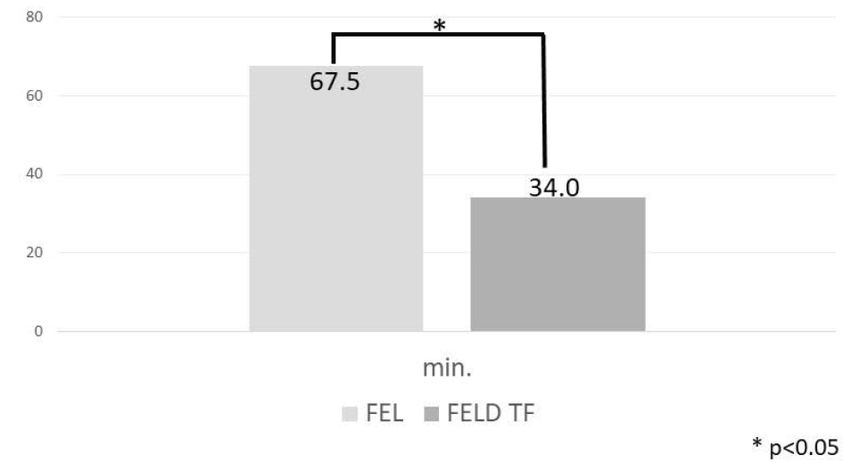
    Results: The results were mentioned in order of FEL:FELD TF. 27 (16 male and 11 female):59 (36 male and 23 female) patients met the inclusion criteria. Mean age was 73.2:64.3 years. Mean follow up periods was 13.4:11.8 months. In MRI, severe stenosis was 14:6, moderate was 12:39, mild was 1:14 patients. 17:41 patients had unilateral and 10:18 patients had bilateral neurological symptoms. Mean operation time was 67.5:34.0 minutes (p < 0.05). Preoperative VAS scores of low back pain were 63.4:58.6, of leg pain were 80.2:75.4 and of numbness were 76.3:68.5. Postoperative VAS scores of low back pain were 37.0:35.7, of leg pain were 39.4:36.9 and of numbness were 47.6:36.3. Mean improvement rates of VAS of low back pain were 41.6:39.1, of leg pain were 50.8:51.0 and of numbness were 37.6:46.9%. In MacNab criteria, 19 (70.4%):38 (64.4%) were satisfactory and 2 (0.7%):11 (18.6%) were poor. Postoperative VAS scores of both were significantly lower than preoperative one. Operative time of FELD TF was significantly shorter than that of FEL. In MacNab criteria, poor rate of FELD TF was higher than that of FEL. In 10 patients with poor results in FELD TF, 5 patients had stenosis and 4 had herniation.

    Conclusion: FELD TF was less invasive surgery, but functional outcomes were inferior, so we should choose it carefully.

    Download PDF (1023K)
  • Shuntaro Tsuchida, Yoshihiro Kitahama
    2020Volume 11Issue 8 Pages 1016-1025
    Published: August 20, 2020
    Released on J-STAGE: August 20, 2020
    JOURNAL FREE ACCESS

    We developed a novel full-endoscopic lumbar foraminoplasty (FELF) for lumbar isthmic spondylolisthesis. To decompress the exiting nerve root safely, we resected upper posterior edge of the lower vertebral body before root decompression. This procedure was performed on 7 patients. The results were good in 6 cases. At the first case, reoperation (FELF) was required because of sustained leg pain for insufficient decompression. The result of reoperation was good. FELF is an effective procedure for lumbar foraminal stenosis not only with spondylosis but with lumbar isthmic spondylolisthesis.

    Download PDF (1872K)
  • Keisuke Ishii, Yoshinori Kyoh
    2020Volume 11Issue 8 Pages 1026-1031
    Published: August 20, 2020
    Released on J-STAGE: August 20, 2020
    JOURNAL FREE ACCESS

    To treat lumbar spondylolysis in athletes, conservative treatment is often preferred over fusion surgery, where performance degradation is a concern. This paper reports the case we experienced, in which a national-level high school rubber-ball baseball player had to stop playing baseball due to pain caused by lumbar spondylolysis. The athlete received a full endoscopic direct repair resulting in successful bone fusion and showed high performance after returning. It is believed that full endoscopic direct repair can be an option in some cases involving a desire to return to high-level sports.

    Download PDF (1414K)
  • Tadatoshi Inoue, Takeshi Kikuchi, Takuya Morita, Hisakazu Shitozawa, Y ...
    2020Volume 11Issue 8 Pages 1032-1037
    Published: August 20, 2020
    Released on J-STAGE: August 20, 2020
    JOURNAL FREE ACCESS

    Introduction: The C1 lateral mass screws (LMS) are useful tool because they have firm stability that can allow us to perform reduction. Generally, when inserting the screws, there are risk to damage the vertebral artery and the venous plexus because the width of the C1 vertebral arch is narrow. We have performed posterior C1/2 fixation via Tan method using Stealth-Midas (Medtronic plc). The purpose of this study is to report the advantage of surgery using Stealth-Midas.

    Methods: A total of 14 patients (male: 6, female: 8, mean age: 74.2) who underwent C1/2 fixation using Stealth-Midas from September 2017 to June 2019 were included. We investigated the accuracy of inserting screws, operation time, blood loss, and complications. We used Vertex Select (Medtronic plc) in all cases, and the diameter and the length of the screws were determined by the navigation system.

    Results: In this series, 5 patients had odontoid fracture, 5 had atlantoaxial fracture, 3 had atlantoaxial subluxation and 1 had posterior odontoid pseudotumor. Mean width of the narrowest part of C1 vertebral arch was 4.06 mm (3.27-5.24 mm). Diameter of screws were φ3.5 mm used in 22, φ4.0 mm in 4, and φ4.5 mm in 2. The mean operation time was 115 minutes (90-157 min), mean blood loss was 36 ml (small quantity-185 ml). There were no screw deviation and complications such as neurovascular injury.

    Conclusions: It is hard to attach the reference arc on the atlas, therefore it tends to misguide us when inserting screws. Furthermore, in the fracture or the subluxation case, the atlas usually moves and rotates because of lack of stability. In our experience, by using Stealth-Midas, the C1 movement was restricted because making the burr hole reduced need to push the vertebral arch hard. It could allow us to perform surgery safely and accurately. Navigation assisted surgery with Stealth-Midas can be useful for C1/2 posterior fixation.

    Download PDF (1349K)
  • Ryo Sasaki, Takafumi Maeno, Hiroshi Kono, Masayuki Umano, Hiroaki Naka ...
    2020Volume 11Issue 8 Pages 1038-1043
    Published: August 20, 2020
    Released on J-STAGE: August 20, 2020
    JOURNAL FREE ACCESS

    Introduction: Osteoporotic lower lumbar vertebral fracture causes loss of lumbar lordosis and abnormal spinal alignment, and to maintain the correction with osteoporotic bone, we need long fusion containing iliac bone. And, several clinical studies have reported poor surgical results of short fusion avoiding long fusion for elderly patient.

    Purpose: To investigate surgical outcomes of short fusion preserving posterior element with X-core which could produce sound anterior column support for delayed paralysis after osteoporotic L4 vertebral fracture in our institution.

    Materials and Methods: We retrospectively reviewed the patient records of 3 women, who were diagnosed with delayed paralysis after L4 osteoporotic vertebral fracture and performed anterior reconstruction and posterior fixation (1 above 1 below) with X-core. Their average age at the operation was 74.3 years old and average follow-up period was 17.3 months. Clinical results were evaluated by JOA score at the preoperative and final follow-up. Radiological results were evaluated by local lordosis (L3-L5), height of the inter-vertebral body (between caudal endplate of L3 to cranial endplate of L5), implant subsidence (more than 2 mm), pedicle screw loosening, and bone union.

    Results: Average JOA score was 7.3 at the preoperation and 23.7 at final follow-up. Average local lordosis was -3.2 degrees at the preoperation, 15.9 after surgery, and 16.0 at final follow-up. Average height of the inter-vertebral body was 28.5 mm at the preoperation, 35.9 after surgery, and 35.2 at final follow-up. 1 case showed implant subsidence and pedicle screw loosening, however, all cases showed bone union finally.

    Discussion: It is difficult to maintain tolerable spinal alignment after fixation for osteoporotic vertebral fracture. Anterior column reconstruction with X-core could correct kyphosis potently, and more, preserving posterior element reduce stress of posterior implants, as a result, correction was preserved.

    Clinical results of short fusion preserving posterior element with X-core for delayed paralysis after osteoporotic L4 vertebral fracture were satisfactory, local lordosis was acquired and maintained.

    Conclusions: Short fusion preserving posterior element with X-core may be one of surgery for delayed paralysis after osteoporotic L4 vertebral fracture.

    Download PDF (1267K)
  • Shigeyuki Kitanaka, Ryota Takatori, Hitoshi Tonomura, Kan Oyabu, Tomon ...
    2020Volume 11Issue 8 Pages 1044-1048
    Published: August 20, 2020
    Released on J-STAGE: August 20, 2020
    JOURNAL FREE ACCESS

    We investigated the clinical outcomes of the lateral lumbar interbody fusion (LLIF) with percutaneous pedicle screw (PPS) procedure for dialysis-related spondyloarthropathy and lumbar degenerative disease. We retrospectively compared the clinical parameters of 9 patients (6 males, 3 females) with dialysis-related spondyloarthropathy (group D) and 9 patients (5 males, 4 females) with lumbar degenerative disease (group L) who were treated in our institution between April 2014 and April 2018. There were no differences between the two groups for operative time or total blood loss. However, there was a significant between-group difference in the peri- and postoperative complications. Three cases of endplate injury and one case of postoperative infection occurred in group D. There was no difference between the two groups in JOA score. One case in group D required reoperation because of postoperative cage subsidence. The clinical outcome of the procedure was acceptable for cases of dialysis-related spondyloarthropathy, making LLIF with PPS an effective treatment option for this patient group. However, further study is necessary regarding the indications for indirect decompression and the optimal postoperative therapy and adjuvant therapy for this procedure.

    Download PDF (919K)
  • Kai Fujita, Masaki Tatsumura, Fumihiko Eto, Kohei Murakami, Masashi Ya ...
    2020Volume 11Issue 8 Pages 1049-1055
    Published: August 20, 2020
    Released on J-STAGE: August 20, 2020
    JOURNAL FREE ACCESS

    Background: Lateral Interbody Fusion (LIF), which is minimally invasive, is rapidly spreading in spinal fusion for adult spinal deformities. We investigated which wedged-shape had an effect on the intervertebral coronal correction in lateral interbody fusion. Also we investigated the incidence of endplate and pedicle fractures.

    Method: 14 cases (2 males, 12 females), 46 intervertebral space, 120 pedicles who performed lumbar anterior fusion using LIF. The intervertebral coronal angle between each vertebra was measured by preoperative and post operative CT. <Investigation 1> We divided each intervertebral space for two groups (Convex group: cage inserted from open wedge or Concave group: cage inserted from closed wedge) <Investigation 2> We divided each intervertebral space for two groups (HW group: a group with intervertebral angle of greator or equal 10° or LW group: a group with the angle less than 10°). We examine the correcting angle and the incidence of endplate and pedicle fractures. Statistical examination was performed using Fisher exact probability test and chi-square test.

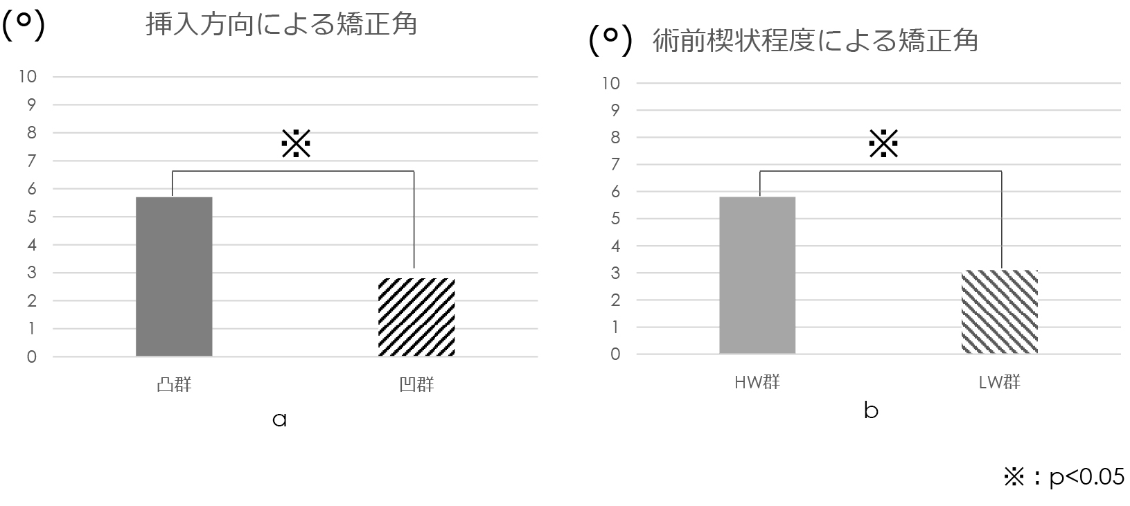
    Result: The endplate fractures occurred in 10 out of 46 intervertebral discs (22%) and the pedicle fracture occurred in 4 cases (3.3%) of 120 pedicles. The average correction angle of the convex group was 5.7 °, and the average correction angle of the concave group was 2.7 ° (p <0.001). The incidence of endplate fractures was 6 in 19 (32%) in the convex group and 3 out of 22 (14%) in the concave group (p >0.05). The incidence of pedicle fractures was 3 in 58 pedicles (5.2%) in the convex group and 1 in 62 pedicles (1.6%) in the concave group (p >0.05). The average correction angle for the HW group was 5.8 °, and the average correction angle for the LW group was 3.0 ° (p <0.01). The endplate fractures were 6 of 14 (43%) in the HW group and 4 of 32 (13%) in the LW group (p=0.046). Pedicle fractures occurred in 1 of 54 pedicles (1.9%) in the HW group and 3 of 66 pedicles in the LW group (4.5%) (p >0.05).

    Discussion: A larger correction can be obtained by cage insertion from convex side with lumbar degenerative kyphoscoliosis. Also a larger correction can be obtained by cage insertion into large wedge but endplate fracture is more frequent. It is necessary to pay attention to postoperative correction loss due to endplate fractures in intervertebral vertebrae with strong wedge-shaped deformation.

    Download PDF (1248K)
  • Tomowaki Nakagawa, Masako Tokunaga, Eiji Takahashi, Hironori Hyoudo, T ...
    2020Volume 11Issue 8 Pages 1056-1060
    Published: August 20, 2020
    Released on J-STAGE: August 20, 2020
    JOURNAL FREE ACCESS

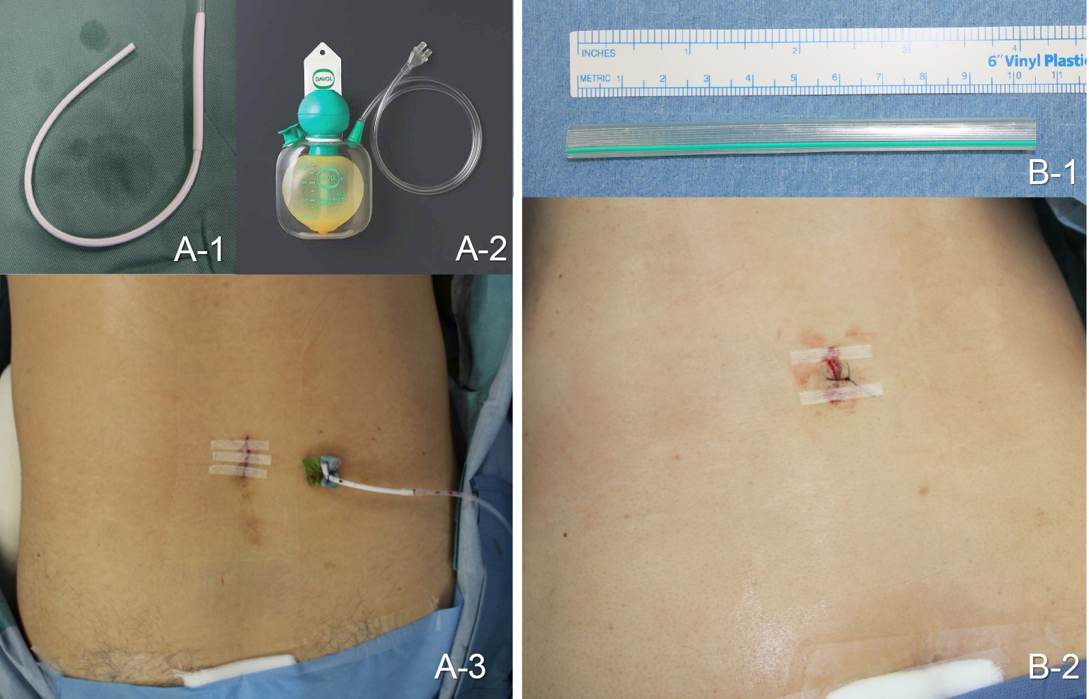
    The efficacy and safety of open drain in Microendoscopic laminotmy (MEL) for lumbar spinal canal stenosis were examined. The subjects: 176 patients who underwent MEL. The closed drainage group (closed group: 76 cases), the open drainage group (open group: 100 cases). There was no infection, and one emergency operation with acute epidural hematoma was seen in the closed group. The average postoperative bleeding was significantly higher in the open group than in the closed group. The degree of compression of the dural sac was lower in the open group than in the closed group. Lower limb pain occurred more frequently in the open group. In this study, open drainage was suitable for MEL.

    Download PDF (963K)
Case Report
  • Masaki Tatsumura, Shun Okuwaki, Hisanori Gamada, Kai Fujita, Kohei Mur ...
    2020Volume 11Issue 8 Pages 1061-1067
    Published: August 20, 2020
    Released on J-STAGE: August 20, 2020
    JOURNAL FREE ACCESS

    Introduction: Pars defects repair is effective for end-stage lumbar spondylolysis in which pain remains even after conservative therapy. We avoided interbody fusion which causes adjacent intervertebral disorders and selected pars defects repair because of preservation lumbar mobile segments. Modified Smiley Face Rod method was applied to two judo players with lumbar spondylolysis with pseudoarthrosis. In both cased, bony fusion was achieved, and it was possible to return to the competition without compromising performance.

    Cases: Case 1 was 14-year-old male Judo player with black belt. At the first consultation, a lumbar spondylolysis with slip of grade 1 in the Meyerding classification was found in the 5th lumbar. Since conservative therapy did not improve pain and was unable to return to the competition, a modified Smiley Face Rod repair procedure was performed 7 months after the start of the treatment. The reduction of the anterior slip of the 5th lumbar vertebra was obtained with the operation. Wearing brace was continued for 3 months after the operation, and judo was resumed 6 months after the operation. He returned to the game 8 months after the operation and participated in the prefectural tournament and achieved a prize. Case 2 was a 14-year-old female with five years of judo history. At the first consultation, a lumbar spondylolysis with slip of grade 1 in the Meyerding classification was found in the 5th lumbar. As conservative treatment with brace and physiotherapy were performed, and back pain improved in 2 months and judo was resumed. However, the pain became severe again, and it became difficult to continue the competition. Therefore, a pars defect repair using a modified Smiley Face Rod method was performed, and reduction of the slip of the 5th lumbar was obtained. She finished wearing the brace 3 months after the operation, resumed judo 4 months after the operation, and returned to the game 11 months after the operation. Bone union was performed partially 12 months after the operation and confirmed absolutely 36 months after the operation.

    Conclusion: The modified Smiley Face Rod method was possible to correct slips of grade 1 in the Meyerding classification, and was able to return to Judo without removing implants.

    Download PDF (1841K)
  • Fumihiro Miyaguchi, Naoya Kawabata, Yoshiharu Horikawa, Masahide Nakaj ...
    2020Volume 11Issue 8 Pages 1068-1074
    Published: August 20, 2020
    Released on J-STAGE: August 20, 2020
    JOURNAL FREE ACCESS

    Fragility fractures of the pelvic ring (FFPs) are increasing with the aging of the population.

    ORIF with ilioinguinal approach or pararectus approach for FFPs is maximally invasive (long time of operation and massive bleeding). We performed percutaneous posterior fusion by spine system for FFPs. The iliac screw and ilio-sacral screw (IS screw) are inserted by percutaneous pedicle screw (PPS). Left and right I-S screw are connected by the rod add tow connectors. The rod of iliac screw is through between the spinous processes S1 and S2. The rod of IS screw is through between the spinous processes L5 and S1. The rod of iliac screw and the rod of IS screw are connected with two cross links. Skin incision are only 40 mm incision on the posterior iliac processes and 15 mm incision of buttocks.

    This method is applicable in the case of narrow space between the posterior iliac processes and the sacral posterior wall. We performed this method for forth FFPs (mean 77.6 years). The mean operating time was 162 minutes and mean blood loss was 375 g. The clinical results of this method were excellent. This method is useful for FFPs.

    Download PDF (1807K)
  • Masayuki Umano, Hiroshi Kono, Takafumi Maeno, Ryo Sasaki
    2020Volume 11Issue 8 Pages 1075-1080
    Published: August 20, 2020
    Released on J-STAGE: August 20, 2020
    JOURNAL FREE ACCESS

    Introduction: We report a case of an intradural arachnoid cyst in the thoracic spine with ossification of the yellow ligament (OYL) that was successfully treated via a unilateral microsurgical approach.

    Case report: A 65-year-old man was referred to our clinic for gait disturbance and numbness of the left upper limb. His medical history was unremarkable except for diabetes mellitus and lumbar canal stenosis. CT myelography showed cervical ossification of the posterior longitudinal ligament, thoracic OYL, and a thoracic arachnoid cyst. We performed laminoplasty for the cervical ossification of the posterior longitudinal ligament and removed the OYL at T10/11. The symptoms improved immediately postoperatively. Therefore, we decided to monitor the cyst without further intervention. At 1.5 years after the initial surgery, the gait disturbance worsened. Although the size of the intradural cyst from the level of T6 to T8 and the OYL at T8/9 had not significantly changed, we decided to remove the cyst and the OYL. After the performance of right-sided hemilaminectomy from T5/6 to T8/9 and excision of the OYL at T8/9 via a microsurgical unilateral approach, we incised the dura, carefully explored the arachnoid cyst, and performed an en bloc excision. The symptoms were improved after surgery.

    Conclusions: The causes of arachnoid cyst development remain unclarified. As the lower end of the cyst coincided with the canal stenosis due to OYL, the development of the arachnoid cyst may be related to the preexistence of the OYL. We usually treat thoracic OYL with microsurgical decompression via a unilateral approach. This minimally invasive technique is very useful because it spares the spinal process ligament complex, paravertebral muscles, and facet joint of the contralateral side, and does not require any instrumentation.

    Download PDF (1497K)
feedback
Top