Journal of Spine Research
Online ISSN : 2435-1563
Print ISSN : 1884-7137
Volume 15, Issue 2
Displaying 1-7 of 7 articles from this issue
Editorial
Original Article
  • Keigo Nagasawa, Kengo Fujii, Toru Funayama, Shota Nakagawa, Sayaka Tan ...
    2024 Volume 15 Issue 2 Pages 50-56
    Published: February 20, 2024
    Released on J-STAGE: February 20, 2024
    JOURNAL FREE ACCESS

    Introduction: This study aimed to examine factors associated with poor outcomes in osteoporotic vertebral fracture (OVF) treatment; medical complications and decreased activities of daily living (ADL) at discharge.

    Methods: We retrospectively studied 144 consecutive inpatients with thoracolumbar vertebral fractures in our hospital and statistically investigated factors associated with medical complications and decreased ADL at discharge for treating OVFs. The clinical course of OVF, presence of medical complications during bed rest, ADL evaluation, bone mineral density, and lumbar spine X-ray and MRI images were investigated in 129 patients with 151 vertebral bodies after excluding 15 patients with diffuse idiopathic skeletal hyperostosis-related fractures. Lumbar spine MRI images were used to measure the cross-sectional area of the paraspinal and psoas major muscles and evaluate fatty degeneration according to the Goutallier classification.

    Results: Of the 129 patients, 4 underwent surgery. Medical complications were observed in 27 cases, and multivariate analysis showed that emergency transport and low body mass index were independently associated factors. Of the 95 patients with preinjury ADLs of walking with a cane or better, 38 had decreased ADLs at discharge, and multivariate analysis showed that paraspinal fatty degeneration was an independent associated factor.

    Conclusions: Evaluation of fatty degeneration of paraspinal muscles might be useful for predicting ADL in patients with OVF, and medical or social intervention for high-risk cases of ADL decline should be considered.

    Download PDF (1498K)
  • Hirokazu Shoji, Yuya Ishikawa, Jin Watanabe, Kimihiko Sawakami, Kei Wa ...
    2024 Volume 15 Issue 2 Pages 57-63
    Published: February 20, 2024
    Released on J-STAGE: February 20, 2024
    JOURNAL FREE ACCESS

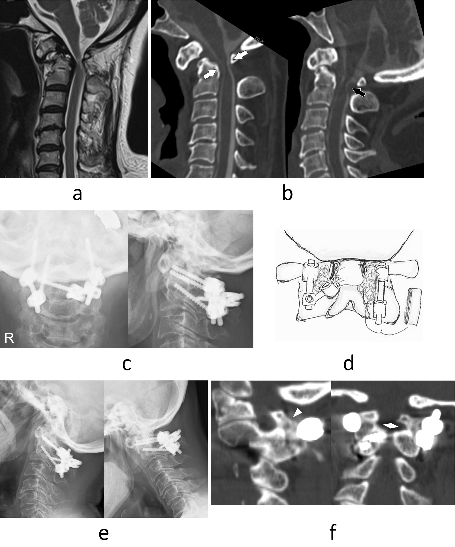
    Introduction: Posterior atlantoaxial fixation is a common procedure for upper cervical spine diseases, including retro-odontoid pseudotumor and atlantoaxial instability. Resection of C1 posterior arch is additionally performed for patients with severe stenosis or irreducible atlantoaxial dislocation. Resection of the C1 posterior arch has a potential risk of nonunion because of a decreased posterior area for bone grafting, although a few reports on posterior atlantoaxial fusion combined with C1 posterior arch resection (C1/2 DF) exist. The purpose of this study was to clarify clinical outcomes of the C1/2 DF, including fusion rate.

    Materials: We included 52 patients who had undergone atlantoaxial fixation for upper cervical spine diseases between 2012 and 2021 and were followed over 1 year after surgery. Bony union rates were compared between 10 patients in DF group and 42 patients in only fusion (F) group. Bony union, fibrous union, and nonunion were determined by CT and flexion-extension X-ray at 1 year postoperatively.

    Results: No significant difference in the methods of spinal instrumentation, transarticular screws, or screw-rod constructs between the two groups was observed. The status of fusion in the DF group was bony union in seven patients, fibrous union in two, and nonunion in one. No significant difference in either the rate of union (DF/F; 70/ 86%, p = 0.35) or nonunion (10/2.4%, p = 0.32) between the two groups was observed. No patients required revision surgery for nonunion.

    Conclusions: The addition of resection of the C1 posterior arch does not affect the rate of bony union after posterior atlantoaxial fixation.

    Download PDF (1208K)
  • Arihiko Tsukamoto, Mitsunori Yoshimoto, Ken Miyashita, Akimitsu Oyama, ...
    2024 Volume 15 Issue 2 Pages 64-70
    Published: February 20, 2024
    Released on J-STAGE: February 20, 2024
    JOURNAL FREE ACCESS

    Introduction: There are several reports that quantitatively evaluated the effects of intradiscal injection therapy of condoliase on intervertebral discs and vertebral body endplates.

    Methods: Eleven patients who underwent intradiscal injection therapy with condoliase and could undergo MRI UTE method for 1 year were enrolled. The intervertebral disc and vertebral endplate were evaluated by Pfirrmann classification, Endplate Classification and Score, respectively, and the T2* value of the intervertebral disc nucleus pulposus and vertebral cartilage endplate was measured. Intervertebral disc height measurements and the presence or absence of Modic changes were also evaluated.

    Results: Intervertebral disc nucleus pulposus T2* values decreased after treatment with condoliase injections in eight cases and increased again in 3 weeks to 3 months after treatment. The intervertebral disc height decreased after treatment, and eight patients showed mild recovery from 3 months to 1 year after treatment. A negative correlation was observed between the rate of disc height reduction 1 year after treatment and the pretreatment disc nucleus pulposus T2* value. Vertebral cartilage endplate T2* values changed from 6 months to 1 year after treatment.

    Conclusions: All patients who experienced a decrease in the intervertebral disc water content after condoliase treatment recovered to a mild degree. After the water content of the intervertebral disc recovered, the intervertebral disc height also slightly recovered. The changes in the vertebral cartilage endplates were considered as the result of intervertebral disc degeneration rather than the direct effect of condoliase.

    Download PDF (1157K)
Case Report
  • Shimei Tanida
    2024 Volume 15 Issue 2 Pages 71-77
    Published: February 20, 2024
    Released on J-STAGE: February 20, 2024
    JOURNAL FREE ACCESS

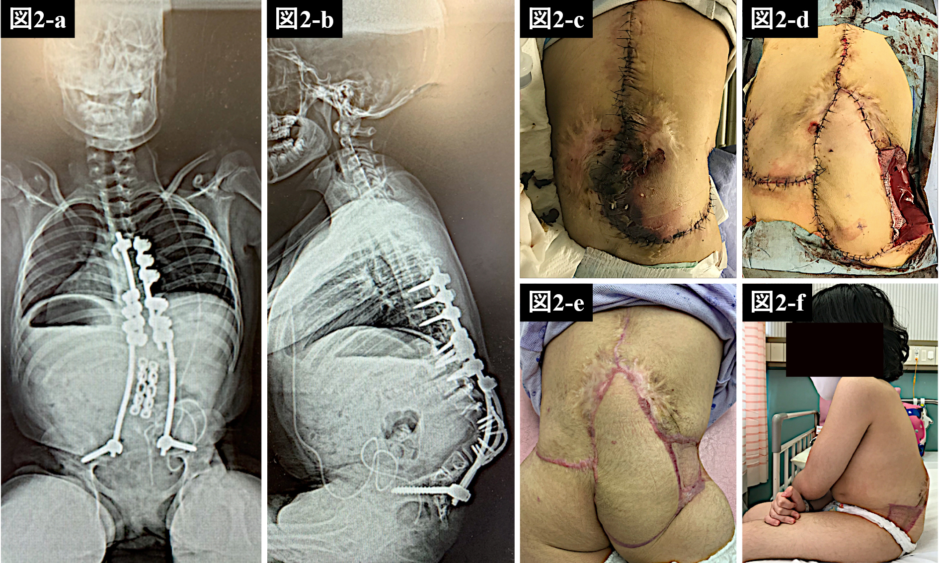
    Introduction: Spinal deformities associated with meningomyelocele, including thinning of the soft tissues in the lumbar region and scar contractures, can cause difficulty in posterior fusion wound closure, resulting in postoperative wound healing failure. In particular, with kyphosis, the skin of the bony prominence is in poor condition and may also be highly strained during closure, leading to postoperative wound necrosis. The lack of soft tissue coverage may result in deep infection due to poor wound healing, and further deep infection may lead to bone fusion failure, orthodontic loss, and implant failure.

    Case Report: Two cases of skin necrosis and wound dehiscence after posterior corrective fusion for spinal deformity associated with meningomyelocele were treated with the gluteal artery perforator flap during reoperation: a 10-year-old female with kyphosis and a 35-year-old female with scoliosis. The latter was undergoing dialysis. In both cases, the wounds healed without developing deep infection.

    Conclusions: Our results indicate that the gluteal artery perforator flap is an effective salvage technique to compensate for inadequate soft tissue at the lower lumbar level and to control wound healing and infection even in spinal deformities associated with meningomyelocele.

    Download PDF (1894K)
  • Yusuke Takahashi, Takashi Shimauchi, Issei Tanaka, Midori Suyama, Ryus ...
    2024 Volume 15 Issue 2 Pages 78-83
    Published: February 20, 2024
    Released on J-STAGE: February 20, 2024
    JOURNAL FREE ACCESS

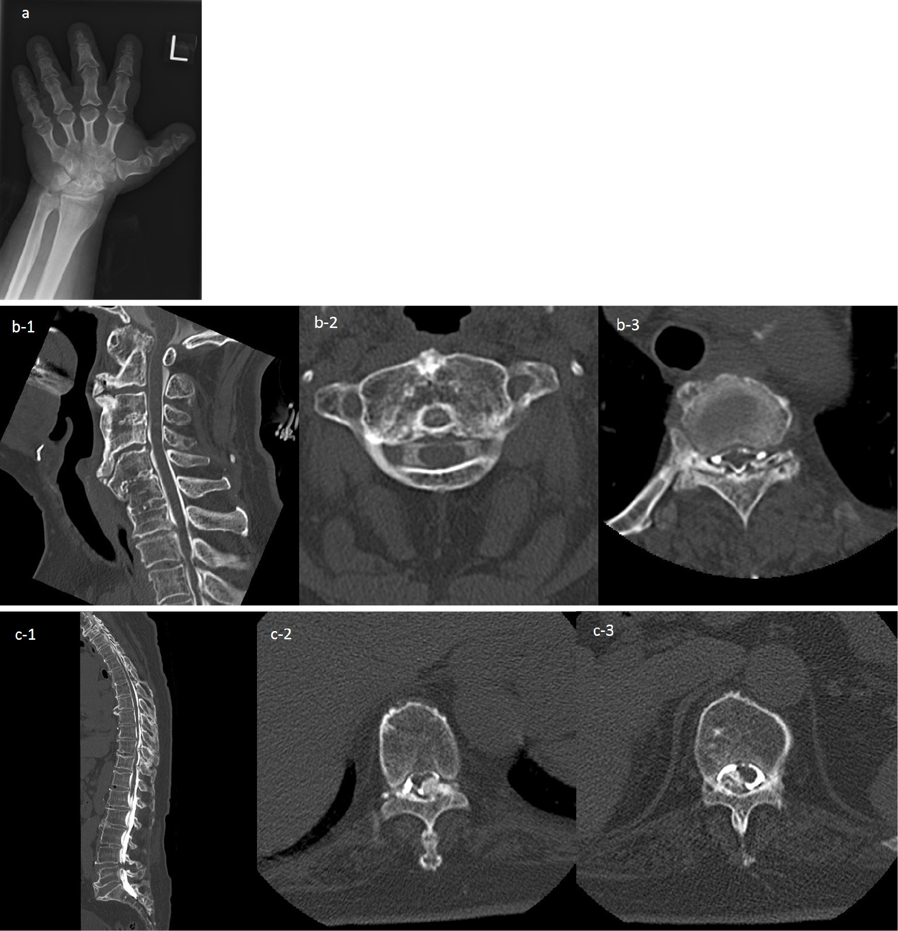
    Introduction: Pseudopseudohypoparathyroidism (PPHP) is a condition of physical features called Albright's hereditary osteodystrophy (AHO) accompanied by mental retardation. PPHP shows various bone lesions with osteophytes and ligamentous ossification of the vertebral body that causes spinal canal stenosis.

    Case Report: A 72-year-old woman visited our hospital because of neurological symptoms in the extremities and gait disturbance due to spinal nerve compression. Imaging findings showed spinal canal stenosis due to osteophytes at the atlas level, and narrowing of the spinal canal due to osteophytes, and ligamentum flavum ossification at the thoracic level. The ataxic gait disturbance worsened and required a wheelchair. Surgery was performed after admission. It included decompression of lesions at the atlas level and decompression with implant fixation of thoracic lesions. The postoperative course was good, and the patient was able to walk with a cane at the time of discharge.

    Conclusions: We report a clinical case of PPHP with the narrowing of multiple spinal canals owing to the ossification of the ligamentum flavum.

    Download PDF (1280K)
feedback
Top