Journal of Spine Research
Online ISSN : 2435-1563
Print ISSN : 1884-7137
Current issue
Displaying 1-7 of 7 articles from this issue
Editorial
Original Article
  • Hiroki Ito, Taro Inoue, Hisatake Yoshihara
    2024 Volume 15 Issue 4 Pages 695-699
    Published: April 20, 2024
    Released on J-STAGE: April 20, 2024
    JOURNAL FREE ACCESS

    A 7-year-old woman presented with severe neck pain without any specific triggers. She was diagnosed with cervical disc calcification at another hospital. Cervical Computed Tomography showed calcification within the intervertebral disc space at C3/4 and protrusion toward the spinal canal. Cervical Magnetic Resonance Imaging revealed severe compression of the spinal cord with intramedullary intensity change. She was treated conservatively with a cervical collar. Severe neck pain was gradually decreased and finally disappeared. Similarly, calcification protruding toward the spinal canal was gradually disappeared. Conservative treatment should be considered for pediatric cervical disc calcification even if there was a compressive spinal cord lesion.

    Download PDF (1194K)
  • Taro Inoue, Hisatake Yoshihara
    2024 Volume 15 Issue 4 Pages 700-706
    Published: April 20, 2024
    Released on J-STAGE: April 20, 2024
    JOURNAL FREE ACCESS

    Introduction: This study aimed to investigate the incidence and clinical characteristics of neurological deterioration after posterior fixation surgery for cervical spine injuries.

    Methods: We retrospectively reviewed 51 patients (12 women and 39 men) with traumatic cervical fractures or dislocations who underwent posterior fixation surgery at our hospital between April 2015 and March 2022. Of these, 37 patients had cervical fractures and 14 had cervical dislocations or subluxations without fractures.

    Results: Of the 51 patients, 9 (18%) showed neurological deterioration after cervical posterior fixation surgery. Six patients had C5 palsy, 2 experienced other nerve root palsies, and 1 experienced severe tetraplegia. Additional surgeries were performed on 4 patients (8%), with 3 patients undergoing foraminotomy and 1 undergoing laminectomy. Two of the procedures were performed on the same day as the posterior fixation surgery.

    Conclusion: Surgeons should be aware that patients with cervical spine injuries who undergo posterior fixation surgery have a relatively high frequency of neurological deterioration.

    Download PDF (2208K)
  • Yuya Ito, Shunsuke Kanbara, Tomohiro Matsumoto, Keigo Ito
    2024 Volume 15 Issue 4 Pages 707-712
    Published: April 20, 2024
    Released on J-STAGE: April 20, 2024
    JOURNAL FREE ACCESS

    Introduction: The lumbar juxta-facet cyst (LJFC) reportedly affects 1.7% of patients who undergo magnetic resonance imaging (MRI) examination for lower back pain. Owing to the high risk of LJFC recurrence, the intervertebral arthrodesis was selected for surgery in the absence of conservative treatment. In our hospital, patients with LJFC were treated with facet block (FB) and selective nerve root block before surgery. We retrospectively reported the outcomes of these nonsurgical treatments and investigated their effectiveness factors.

    Methods: A total of 59 patients with LJFC diagnosed by MRI or myelo-computed tomography and treated conservatively at our hospital since 2014 were included in the study. The patients who did not undergo surgery were considered to be effectively treated by conservative treatment. Conversely, conservative treatment was considered ineffective on patients who underwent surgery. The effectiveness factors of conservative treatment were also examined.

    Results: The conservative treatment was effective in 34 cases (58%). Additionally, it was significantly effective in patients with high signal inside the cyst on T2-weighted images and mild symptoms after FB alone.

    Conclusions: A prior minimally invasive conservative treatment for LJFC before surgery is important.

    Download PDF (982K)
  • Hirofumi Sakaeda, Yuji Matsuoka, Tatsuki Matsuoka, Akinori Hattori, Hi ...
    2024 Volume 15 Issue 4 Pages 713-720
    Published: April 20, 2024
    Released on J-STAGE: April 20, 2024
    JOURNAL FREE ACCESS

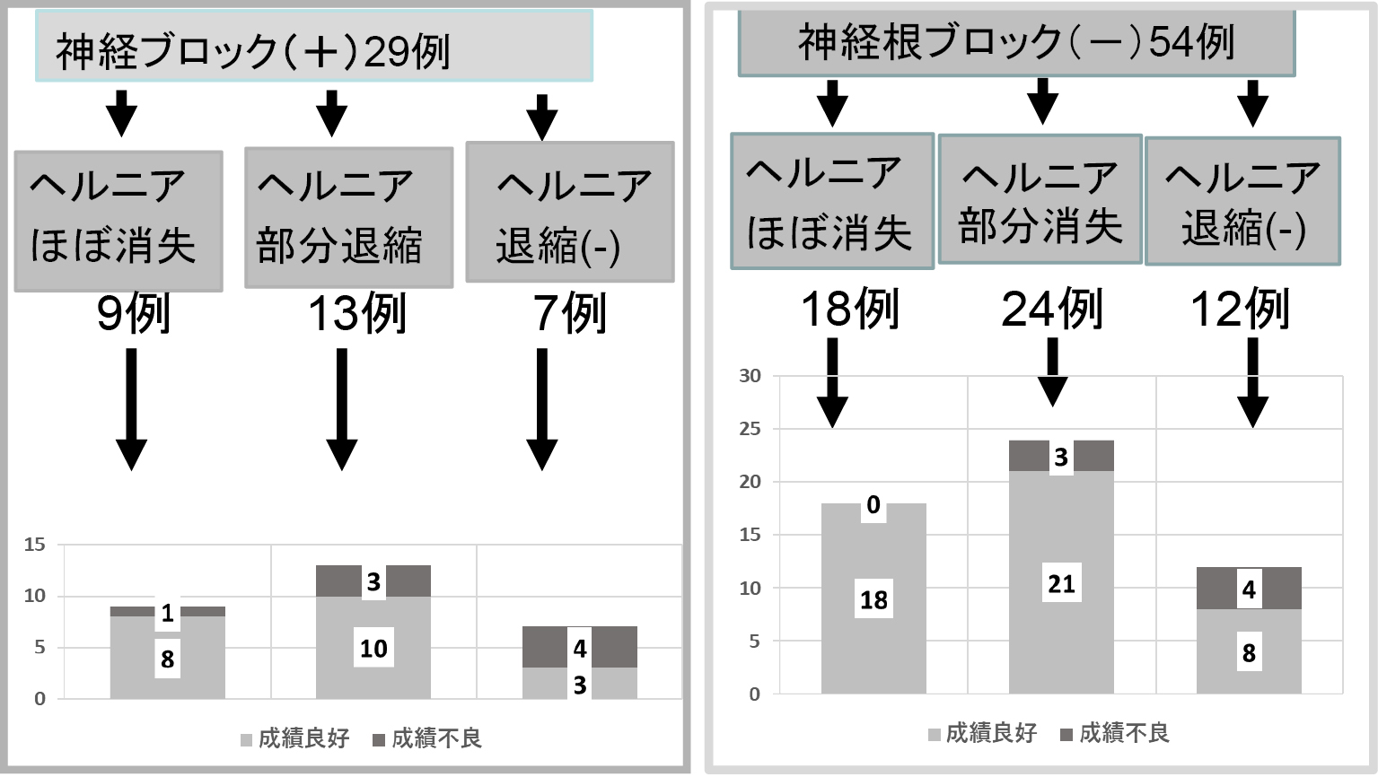
    OBJECTIVE: We investigated whether condoliase-induced chemonucleolysis could be a conservative treatment to avoid surgery in the treatment of lumbar disc herniation.

    Methods: 83 patients with herniated discs with Pfirrmann grade III or higher who were treated with intradiscal condoliase injection therapies and observed for more than 6 months were analyzed. Nerve root blocks were preferentially selected for painful patients who resisted conservative treatments, and condoliase injection therapies were recommended for patients who still did not improve. History at before the procedure and 2 weeks, 4 months, 6 months, 12 months after the procedure were analyzed. MRI at before the procedure and 4 months, 6 months, 12 months after the procedure were analyzed.

    Results: (1) Condoliase injection therapies showed that the onset of effect were as early as 2 weeks, and 92% of improvements of final arriable points were reached at 6 months, and symptoms improved until 1 year. (2) Dropout operative cases in other hospitals were added as poor performers. Our results showed an efficacy rate of 79%, with 3 cases requiring surgery. (3) MRI showed hernia reductions in 77% of patients. In cases where even partial hernia reductions were observed, good results were expected in 89% of the cases. (4) Spinal canal stenosis, multiple vertebral disc disorders, instability, past surgery, and heavy labor were associated with poor results. (5) Intervertebral disc height decreased to 85% after enzyme injection and recovered to 93% after 1 year, compared with the initial height.

    Conclusion: Condoliase injection therapy may be the next non-surgical treatment of choice for patients even with moderate disc degeneration if risk factors such as spinal canal stenosis, multiple intervertebral disc disorders, instability, past surgery, and heavy labor are avoided.

    Download PDF (1546K)
Case Report
  • Ron Fujii, Masayoshi Morozumi, Akio Muramoto, Yuji Matsubara
    2024 Volume 15 Issue 4 Pages 721-725
    Published: April 20, 2024
    Released on J-STAGE: April 20, 2024
    JOURNAL FREE ACCESS

    Introduction: Metastatic intramedullary spinal cord tumors have a poor prognosis, and chemotherapy is considered ineffective. We report a case of a patient who had a good outcome with an immune checkpoint inhibitor after resection of a metastatic intramedullary tumor.

    Case Report: 74-year-old male underwent upper lobectomy for lung cancer. Seven months after surgery, he developed urinary retention and difficulty in walking. Contrast-enhanced MRI revealed an intradural extramedullary tumor with homogeneous internal contrast at C7~Th1 level, and an emergent tumor resection was performed. Intraoperative findings revealed that part of the tumor was firmly adhered to the spinal cord, so the adhered area was cauterized. Immediately after surgery, the patient's paraparesis improved and he was able to walk with a cane. The histopathological diagnosis was lung cancer metastasis, and postoperative MRI showed residual intramedullary lesions, which led to the diagnosis of metastatic intramedullary tumor. Since the tumor cells highly expressed PD-L1, treatment with an immune checkpoint inhibitor was started, and the spinal cord edema improved and the patient was able to maintain his ambulatory function.

    Conclusion: Recently, the efficacy of immune checkpoint inhibitors for brain metastases of lung cancer with high PD-L1 expression was demonstrated. In this case, spinal cord edema improved and good neurological function was maintained, suggesting that immune checkpoint inhibitors may also be effective in metastatic intramedullary spinal cord tumors.

    Download PDF (1307K)
  • Shingo Kainuma, Muneyoshi Fukuoka, Nobuyuki Watanabe, Hiroki Yamada, K ...
    2024 Volume 15 Issue 4 Pages 726-731
    Published: April 20, 2024
    Released on J-STAGE: April 20, 2024
    JOURNAL FREE ACCESS

    Introduction: Tight filum terminale (TFT) is a stretch-induced functional disorder of the spinal cord due to the fact that its caudal portion is anchored by an inelastic structure, and it has been reported as a potential cause of persistent low back pain and leg pain. TFT is often overlooked and untreated because standard imaging studies such as MRI do not reveal any abnormalities.

    Case Report 1: 13-year-old boy. He presented with the onset of low back pain during exercise in the past two months. He had difficulty sitting due to the low back pain. A provocation test for spinal cord extension (TFT provocation test) was positive. CT myelography confirmed a tense filum terminale in both prone and supine positions. After surgical transection of the filum terminale, the low back pain improved, and subsequent TFT provocation test became negative.

    Case Report 2: 47-year-old man. He developed low back pain after driving two months ago. The low back pain worsened with forward bending of the lumbar spine, and TFT provocation test was positive. Gait was unstable due to spasticity in the lower extremities. Although there were no abnormal findings in the cervical and thoracic spine, CT myelography revealed a tense filum terminale in both prone and supine positions. After surgical transection of the filum terminale, the low back pain improved, and there was a slight improvement in gait. TFT provocation test also became negative.

    Conclusions: TFT should be considered as a potential diagnosis for low back pain, especially when standard imaging studies fail to identify any abnormalities.

    Download PDF (1957K)
feedback
Top