Journal of Spine Research
Online ISSN : 2435-1563
Print ISSN : 1884-7137
Volume 14, Issue 9
Displaying 1-13 of 13 articles from this issue
Editorial
Original Article
  • Yoshihito Sakai, Norimitsu Wakao, Hiroki Matsui, Naoaki Osada, Tsuyosh ...
    2023 Volume 14 Issue 9 Pages 1184-1191
    Published: September 20, 2023
    Released on J-STAGE: September 20, 2023
    JOURNAL FREE ACCESS

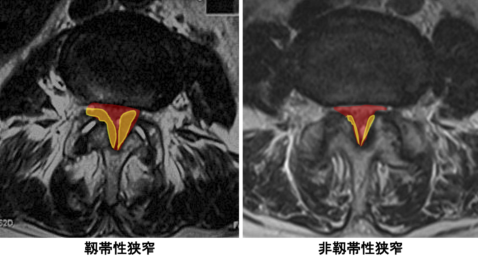
    Introduction: The mechanism of the thickness of the ligament flavum in lumbar spinal stenosis has not been elucidated. We clinically defined the thickness using MRI and demonstrated the involvement of insulin receptor signaling at the genetic level using transcriptome analysis. Present study was conducted to investigate the involvement of endogenous insulin as a risk factor in flavum hypertrophy.

    Methods: In 1,119 patients (average age: 76.1 years, 65-96 years, male: 584 cases, female: 535 cases) who underwent treatment for lumbar spinal stenosis, flavum thickness was defined according to the Sakai's criteria, and the homeostasis model assessment of insulin resistance (HOMA-IR) was calculated via fasting insulin and glucose levels. Logistic regression analysis was performed to examine risk factors for flavum hypertrophy.

    Results: Flavum thickness was observed in 51% of the patients, who were significantly older, male, diabetic, and had higher BMI, HOMA-IR, and creatinine level. Body composition and imaging findings showed significantly higher extremity and trunk muscle mass, lumbar lordosis, sacral tilt, and sagittal vertical axis. HOMA-IR was significantly higher in flavum thickness in the analysis of covariance adjusted for age and sex, and was also recognized as a significant factor in logistic regression analysis.

    Conclusions: Insulin resistance was high in elderly patients with lumbar spinal stenosis with ligamentum flavum thickness, suggesting the possibility that factors other than mechanical stress, such as inflammation and senescence for flavum hypertrophy.

    Download PDF (1383K)
  • Minoru Yamada, Fujio Ito, Zenya Ito, Motohide Shibayama, Shu Nakamura, ...
    2023 Volume 14 Issue 9 Pages 1192-1196
    Published: September 20, 2023
    Released on J-STAGE: September 20, 2023
    JOURNAL FREE ACCESS

    Introduction: Full endoscopic lumbar discectomy transforaminal approach (FELD TF) was performed for revision surgery of recurrent lumbar disc herniation with surgery performed by the same spine surgeon, and the clinical results were evaluated retrospectively.

    Methods: Thirty-four cases had a history of surgery at the same level. Of these, twenty-one cases had recurrent hernias, five cases of new disc herniation after posterior decompression, each of FELD and microendoscopic discectomy (MED) insufficient removal, and six cases after percutaneous laser disc decompression. All patients had lower extremity pain as the main issue.

    Results: All patients had herniated discs removed and considerable improvement in postoperative symptoms was observed (JOABPEQ: JOA Back Pain Evaluation Questionnaire, VAS: Visual Analog Scale, JOA score: Japanese Orthopedic Association score, Macnab). There were no intraoperative complications. The FELD TF approach through the intervertebral foramen allows direct access to the herniated disc and removal of the herniated disc without osteotomy, avoiding the epidural space adhesions.

    Conclusions: The FELD TF technique is a useful surgical treatment option for herniated discs with previous surgery.

    Download PDF (920K)
  • Kimihiko Sawakami, Kei Watanabe, Kazuhiro Hasegawa, Noriaki Yamamoto, ...
    2023 Volume 14 Issue 9 Pages 1197-1203
    Published: September 20, 2023
    Released on J-STAGE: September 20, 2023
    JOURNAL FREE ACCESS

    Introduction: Teriparatide (TPTD) is expected to be an adjuvant therapy, targeting the patients with osteoporotic spine because of its strong bone formation promoting action from an early stage. From the viewpoint of cancellous bone histomorphometry, we reported that at least 3 months of preoperative administration is desirable to provide a more substantial anabolic effect from the early postoperative stage. Alternatively, cortical bone makes a significant biomechanical contribution to vertebral body strength in the osteoporotic spine. The aim of this study was to examine the effect of TPTD administration on cortical bone with respect to bone histomorphometry and to elucidate the significance of the administration period in patients with osteoporosis.

    Methods: Thirty-nine patients with spinal fusion and osteoporosis, who consented to undergo iliac biopsy, were allocated to the following treatment groups: TPTD neoadjuvant therapy, TPTD group (n = 32) and no neoadjuvant therapy, NTC group (n = 7). Patients in the TPTD group were categorized into subgroups based on preoperative TPTD administration periods as follows: 1 month (n = 6), 2 months (n = 7), 3 months (n = 7), 4 months (n = 6), and 6 months (n = 6). All patients were double labeled with tetracycline preoperatively. Iliac biopsy was performed during spinal fusion surgery. Histomorphometric analyses were performed on nondecalcified, thinsliced specimens. Specimens were classified based on the TPTD administration period and subsequently compared with those in the NTC group.

    Results: P1NP and TRACP5b were significantly increased in the TPTD group compared to the NTC group at the time of biopsy (144.7±78.3 vs. 67.42±25.6; p = 0.0017 and 586.3±292.0 vs. 368.1±147.3; p = 0.0233, respectively). There was a difference between endocortical and periosteal surfaces. In the endocortical, mineralizing surface (MS/BS) reached a peak at 3 months of the administration, with a 2.3-fold increase relative to that in the NTC group. In periosteal, MS/BS finally reached a significant level at 6 months (p = 0.0446). The value 4.4% was nearly equal to the result of 24 months administration study reported in a previous study.

    Conclusions: Anabolic bone response in the endocortical was similar to cancellous bone. However, in periosteal, anabolic response became to a plateau at 6 months of the administration. In other words, 6 months or more is desirable to rebuild cortical bone. To maximize the effect of TPTD on biomechanical strength, we suggest continuing the administration for 24 months.

    Download PDF (1225K)
  • Atsushi Yoshioka, Mikihiro Kondo, Kenichiro Tanaka, Yudo Hachiya
    2023 Volume 14 Issue 9 Pages 1204-1212
    Published: September 20, 2023
    Released on J-STAGE: September 20, 2023
    JOURNAL FREE ACCESS

    Introduction: This study aimed to analyze the clinical results of chemonucleolysis with condoliase for lumbar disc herniation (LDH).

    Methods: We retrospectively evaluated the first 100 patients who underwent condoliase injection for LDH from August 2018 to September 2021. In total, 76 males and 24 females (mean age 36.3 years (15-62) ) were included. Visual analog scale (VAS) scores for back pain, leg pain and leg numbness, complications, surgical conversion, recurrence, changes in disc height and herniation size were evaluated.

    Results: The mean VAS score was considerably improved from baseline to 3 month after the injection. Skin rashes were observed in three patients, one patient complained low back and leg pain immediately after the injection. No severe adverse complications were reported. Eight patients underwent surgery. There were two cases of recurrence of herniation at the follow-up period. The mean disc height decreased at 3 weeks after the injection; however, no considerable decrease at 3 months and thereafter was observed. On Magnetic Resonance Imaging (MRI), 58 patients showed a reduction in disc herniations size.

    Conclusions: We reported clinical results of condoliase for LDH in first 100 cases. Chemonucleolysis with condoliase is a safe and significantly improved symptoms.

    Download PDF (2607K)
  • Eiichiro Iwata, Yusuke Yamamoto, Takuya Sada, Sachiko Kawasaki, Akinor ...
    2023 Volume 14 Issue 9 Pages 1213-1218
    Published: September 20, 2023
    Released on J-STAGE: September 20, 2023
    JOURNAL FREE ACCESS

    Introduction: Needle biopsy is frequently used to confirm the diagnosis of infectious spondylitis and is submitted for bacterial culture to identify the causative organism. However, in many cases, culture cannot be performed, and diagnostic performance may be improved by submitting the biopsy for histopathological examination. This study aimed to compare the diagnostic performance of needle biopsy for infectious spondylitis with bacterial culture test, histopathological test, or a combination of both tests.

    Methods: We retrospectively reviewed 41 patients who underwent needle biopsy for suspected infectious spondylitis. We calculated the sensitivity, specificity, positive predictive value, negative predictive value, and accuracy rate of bacterial culture test, histopathological test, and a combination of both tests.

    Results: The sensitivity, specificity, positive predictive value, negative predictive value, accuracy rate, and P value of the bacterial culture test, histopathological test, and the combination of both tests were (58%, 90%, 95%, 41%, 66%, 0.008), (74%, 100%, 100%, 56%, 81%, <0.001), and (90%, 90%, 97%, 75%, 90%, <0.001), respectively. Of the 13 cases in the infected group with negative culture results, 10 (77%) cases were positive in histopathological examination.

    Conclusions: In needle biopsy for infectious spondylitis, not only bacterial culture but also histopathological examination must be performed to prevent delay in diagnosis.

    Download PDF (1048K)
  • Masaru Hatano, Keishi Maruo, Shoji Nishio, Yoshiteru Nakamura, Yoshihi ...
    2023 Volume 14 Issue 9 Pages 1219-1224
    Published: September 20, 2023
    Released on J-STAGE: September 20, 2023
    JOURNAL FREE ACCESS

    Introduction: Our previous study has demonstrated that polyetheretherketone (PEEK) cages and titanium-coated PEEK (TCP) cages exhibit similar fusion rates at 3 and 12 months. Thus, a three-dimensional (3D)-printed porous titanium alloy (PTA) cage has been introduced as a new interbody cage material. The aim of this study was to compare radiographic outcomes between the TCP and PTA cages.

    Methods: A total of 97 patients, who underwent 1- or 2- level transforaminal lumbar interbody fusion between October 2015 and July 2020, were enrolled. A total of 97 cases (TCP: 37 cases and PTA: 60 cases) and 121 levels (TCP: 50 levels and PTA: 71 levels) were studied. Patient and surgical factors included age, sex, number of levels, operative time, and estimated blood loss. Moreover, radiographic outcomes including endplate cyst, pedicle screw (PS) loosening, cage subsidence (>2 mm), and trabecular bone remodeling (TBR) were assessed by computed tomography (CT) at 3 months after surgery. Bony union was assessed by CT at 12 months after surgery.

    Results: There was no significant difference observed in patients and surgical factors between the two groups. Furthermore, positive cyst sign was significantly less in the PTA group than in the TCP group (30% vs. 54%, p = 0.01). The TBR was significantly more in the PTA group than in the TCP group (52% vs. 72%, p = 0.03). The fusion rate was equivalent between the two groups at 12-months postoperatively (TCP: 86% vs. PTA: 87.3%, p = 0.83). The incidence of PS loosening was also similar between the two groups.

    Conclusions: This study revealed that the fusion rate was similar between the two groups at 12 months. However, endplate cyst signs were significantly less in the PTA group and TBR was significantly more in the PTA group at 3 months after surgery. These results suggest that the PTA cages may have an advantage due to the initial fixation strength.

    Download PDF (1669K)
  • Hiroshi Nomura, Shigeharu Nomura
    2023 Volume 14 Issue 9 Pages 1225-1233
    Published: September 20, 2023
    Released on J-STAGE: September 20, 2023
    JOURNAL FREE ACCESS

    Introduction: Herein, we investigated the clinical outcomes of patients with osteoporosis at high risk of fractures who were treated with romosozumab following fresh osteoporotic vertebral fractures (OVFs).

    Methods: This study included 60 patients with osteoporosis who underwent romosozumab treatment at our clinic and whose follow-up period was >12 months. The bony union of the fractured vertebrae was diagnosed with a vertebral segmental angle (VSA) of the fractured vertebrae in the lateral views of the spine on regular radiographs, both in lateral and sitting positions, and the presence of an intervertebral cleft sign (ICS) in the lateral position 12 months after the treatment. To measure the correct bone mineral density (BMD) of the lumbar spine, all patients were divided into 2 groups: the "L1-4" group, comprising patients with L1, 2, 3, or 4 OVFs, and the "non-L1-4" group, comprising patients without L1-4 OVFs. The average BMD of the nonfractured vertebrae of L1, 2, 3, or 4, excluding the fractured vertebrae, was calculated in the "L1-4" group. The corrected BMD was then calculated using the values in the "non-L1-4" group and those of the nonfractured vertebrae in the "L1-4" group. Clinical assessment was measured using a visual analog scale (VAS) and the Roland-Morris Disability Questionnaire (RDQ).

    Results: The difference between the lateral and sitting positions in VSA was within 5° in all cases and revealed excellent vertebral stability of the fractured vertebrae within 2-3 months of treatment. The ICS examination revealed that the bony union rate was 96.3% 12 months after the treatment. The BMD of the lumbar spine at 6 and 12 months and the proximal femur at 12 months were considerably higher than those before treatment. The BMD of the lumbar spine increased by 11.7% and 17.4%, and that of the proximal femur increased by 1% and 2.1% at 6 and 12 months, respectively. The VAS and RDQ of all patients considerably improved within 1 month of treatment.

    Conclusions: Treatment with romosozumab considerably increased the BMD of the lumbar spine and proximal femur.

    Download PDF (1278K)
  • Yasuyuki Tamaki, Kazuhiro Murotani
    2023 Volume 14 Issue 9 Pages 1234-1238
    Published: September 20, 2023
    Released on J-STAGE: September 20, 2023
    JOURNAL FREE ACCESS

    Introduction: This study aims to find the predictive factors of acute pyogenic spondylitis that resist conservative treatment.

    Methods: We studied 76 cases (men: 49 and women: 27, average age: 74 years, average follow-up period: 198 days) of acute pyogenic spondylitis where conservative treatment was followed. Subjects were compared by dividing into groups R of 30 and NR of 46 cases with and without resist of conservative treatment, respectively.

    Results: Average of age, men, periods from medical institution consultation to a diagnosis, the number of the infected vertebral bodies, compromised host, infection progress case of the MRI and resistant bacteria in groups R and NR were 76 and 72 years old, 20 and 29 cases, 7.4 and 8.3 days, 2 and 1.7 vertebral bodies, 20 and 23 cases, 9 and 14 cases, 7 and 8 cases, respectively. Notable differences were observed in the number of infected vertebral bodies. The average of body temperature at the first examination, 1 week, 2 weeks, 3 weeks, and 4 weeks after treatment, were 38.5°C and 38.6°C, 37.3°C and 37.2°C, 36.9°C and 37.0°C, and 37.0°C and 36.9°C, and 36.9°C and 36.8°C, respectively. The average of WBC at the first examination, 1 week, 2 weeks, 3 weeks, and 4 weeks after treatment were 10,847 and 13,776, 8,253 and 9,254, 8,011 and 7,533, 6,370 and 6,460, and 6,642 and 6,639 μL, respectively. Notable difference was observed in the WBC of the first examination. Average of CRP at the first examination, 1 week, 2 weeks, 3 weeks, and 4 weeks after treatment were 15.0 and 15.7, 8.6 and 6.4, 5.4 and 3.0, 5.4 and 2.9, and 4.8 and 1.9 mg/dl, respectively. Notable differences were observed among the CRP of 1 week, 2 weeks, 3 weeks, and 4 weeks after treatment. Using the multiplex logistic-regression analysis, only CRP of 4 weeks was notably different and the cutoff level was CRP = 2.0 mg/dl.

    Conclusions: Acute pyogenic spondylitis that resists conservative treatment was predictable using CRP of 4 weeks after treatment and the cutoff level was CRP = 2.0 mg/dl.

    Download PDF (801K)
  • Fuyuki Tominaga, Eiji Mori, Hiroya Ikari, Takaaki Yoshimoto
    2023 Volume 14 Issue 9 Pages 1239-1245
    Published: September 20, 2023
    Released on J-STAGE: September 20, 2023
    JOURNAL FREE ACCESS

    Introduction: Intradiscal injection therapy using condoliase is a new minimally invasive treatment for lumbar disc herniation. This study aimed to examine the short-term outcomes of intradiscal condoliase injection for lumbar disc herniation and reveal the factors influencing the effectiveness of this therapy.

    Methods: Sixty-nine patients (48 men and 21 women), who were treated with condoliase injections and followed-up for >3 months, were enrolled in this prospective study. Their mean age was 45.0 years (range, 17-89 years). We investigated the Visual Analog Scale scores for low back and leg pain as clinical assessments, and the herniation size on magnetic resonance imaging as radiological assessments, preinjection, and 1 month and 3 months postinjection. An improvement of ≥50% in the Visual Analog Scale (VAS) score for leg pain was considered to indicate effectiveness.

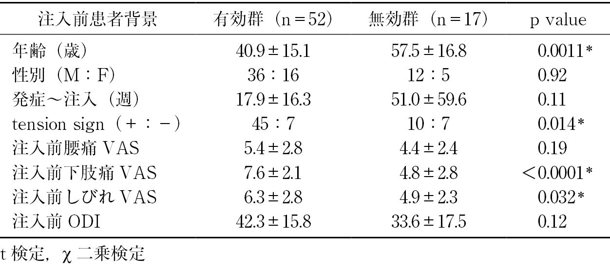
    Results: The mean VAS score for low back pain notably improved from 5.1 preinjection to 2.8 postinjection, and that of leg pain notably improved from 6.9 to 2.4. The herniation size decreased from 53.2 mm2 preinjection to 24.4 mm2 postinjection. Fifty-two patients (75%) at 3 months postinjection showed improvement, and compared with those who did not show effectiveness, these patients were significantly younger (40.9±15.1 vs. 57.5±16.8 years, t-test, p = 0.0011) and positive tension sign was seen in effective cases (86.5% vs. 58.8%, p = 0.014). Furthermore, the effective group improved the VAS score for the lower leg at 1 month postinjection (−4.5±2.7 vs. −0.2±2.1, t-test, p < 0.0001).

    Conclusions: Condoliase injection significantly improved the VAS scores for low back pain and leg pain. Young age, positive tension sign, and improvement in the VAS score for the lower leg after 1month were associated with effectiveness at 3 months postinjection.

    Download PDF (1413K)
  • Ryosuke Nishi, Nodoka Manabe, Daisuke Tsunoda, Kazuro Tsuge, Kunio Kam ...
    2023 Volume 14 Issue 9 Pages 1246-1251
    Published: September 20, 2023
    Released on J-STAGE: September 20, 2023
    JOURNAL FREE ACCESS

    Introduction: Previous studies have reported various factors related to the prognosis after lumbar spine surgery. However, it is unclear to what extent each factor affects the postoperative outcome. This study aimed to clarify the factors affecting postoperative hospitalization using preoperative physical function, a questionnaire, intraoperative information, and patient information as independent variables.

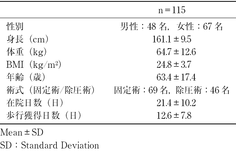
    Methods: This study comprised 115 patients with lumbar spinal canal stenosis and herniation of lumbar discs who underwent posterior lumbar spinal decompression (n = 46) and fusion surgery (n = 69) at our hospital. At the time of the surgery, the average age of the 48 men and 67 females was 63.4±17.4 years. Physical functions, including 6-min walk test (6MWT) and Manual Muscle Test (MMT), were performed before the surgery. The questionnaire included the RDQ, which was provided before the surgery. Additionally, we investigated operative information (the presence or absence of fusion surgery) and patient information (BMI, gender, and the number of days to acquire ambulation). Multiple regression analysis was performed with these factors as the independent variables and the duration of hospitalization as the dependent variables.

    Results: The number of days to acquire ambulation, the presence or absence of fusion surgery, age, and the muscle strength of the hip extension were found to be significant related factors (p < 0.01).

    Conclusions: The factors influencing the duration of hospitalization following lumbar spine surgery were identified in this study. The duration of hospitalization depend on the prolonged walking acquisition period, the fusion surgery, old age, and the weakness of hip extension muscles.

    Download PDF (973K)
  • Daisuke Inoue, Hideki Shigematsu, Hiroaki Matsumori, Yurito Ueda, Tosh ...
    2023 Volume 14 Issue 9 Pages 1252-1259
    Published: September 20, 2023
    Released on J-STAGE: September 20, 2023
    JOURNAL FREE ACCESS

    Introduction: We have combined high-angle cage and posterior column osteotomy (HAP) in posterior lumbar interbody fusion (PLIF) surgery (PLIF with HAP), focusing on local lumbar alignment correction and bone fusion. We report the postoperative outcomes of PLIF with HAP at our hospital.

    Methods: PLIF with HAP was performed in 70 patients at our institution between April 2020 and November 2021. In surgery, total facetectomy was performed, and a 12° titanium cage was used. Percutaneous pedicle screws were inserted, and all cases underwent compression with the screws. The following items were examined: operative time, blood loss, preoperative lumbar BMD, at pre-/post-/postoperative 6-month slip length (%slip), segmental lumbar lordosis (SLL), ratios of mean disc height to vertebral body height and posterior disc height to vertebral body height on lumbar lateral X-ray, at pre-/postoperative 6-month JOA score, VAS for lumbar pain, bone fusion rate on computed tomography (CT) at 6 months postoperatively, and postoperative complications.

    Results: There were considerable differences in %slip and SLL at pre-/postoperatively and pre-/6 months postoperatively. And there was considerable differences in JOA and lumbar pain VAS at pre-/6 months postoperatively. The bone fusion rate was 92.9% at 6 months postoperatively. Complications included one case of transient neuralgia.

    Conclusions: Corrected local alignment and bone fusion for PLIF with HAP were good.

    Download PDF (1444K)
Case Report
  • Yutaro Okamura, Kazuyuki Segami, Shu Takahashi, Kazumichi Yagura, Koji ...
    2023 Volume 14 Issue 9 Pages 1260-1265
    Published: September 20, 2023
    Released on J-STAGE: September 20, 2023
    JOURNAL FREE ACCESS

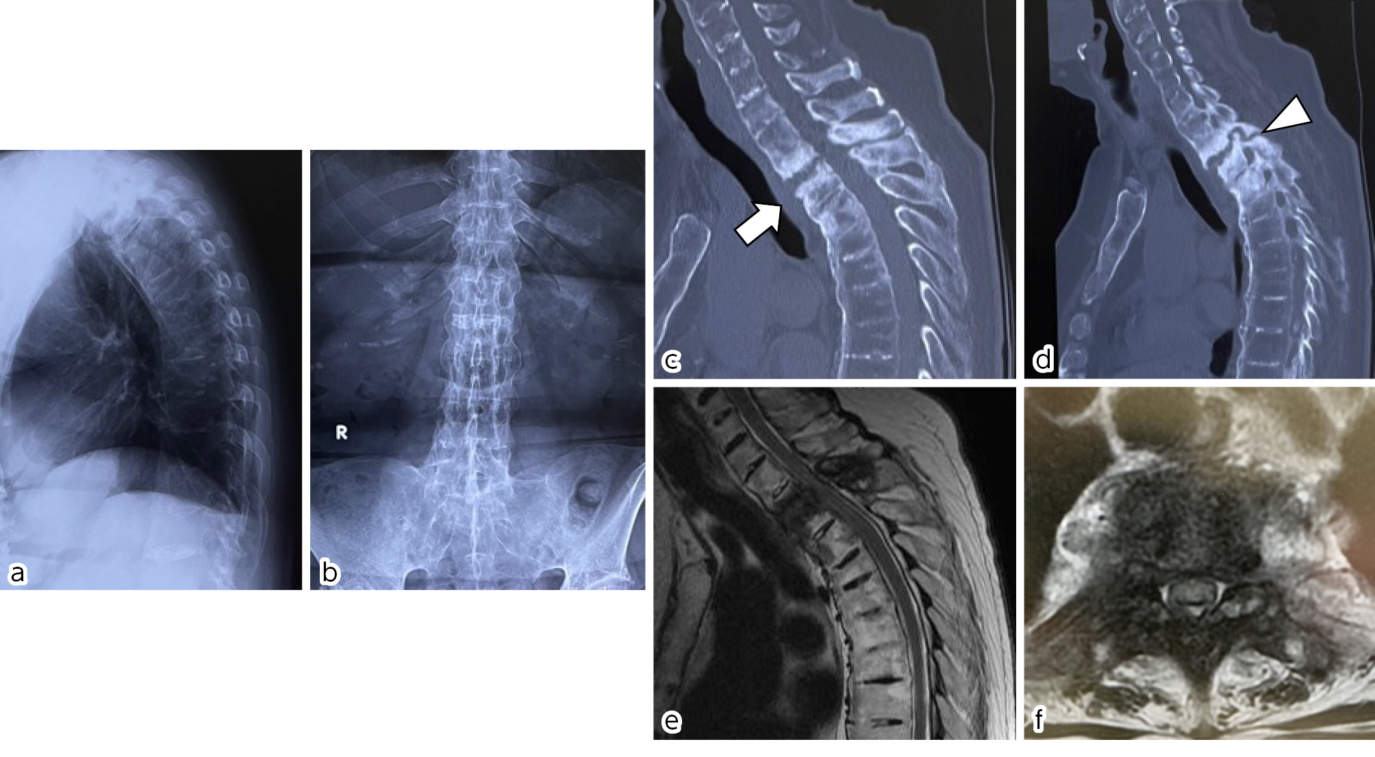
    Introduction: Extensive discovertebral destruction is a little-known complication associated with advanced ankylosing spondylitis (AS). These lesions are either inflammatory or traumatic in nature, the latter often associated with deformity, severe back pain, and neurologic sequelae. Therefore, surgical treatment is often recommended in patients with traumatic lesions.

    Case Report: A 68-year-old female patient being treated for osteoporosis associated with AS was admitted to our hospital for progressive leg paresis. Computed tomography and magnetic resonance imaging scans revealed widespread destruction of T2-T3 vertebral bodies and intervertebral disc with a resulting posterior column like-pseudarthrosis. Surgery was performed using a posterior approach, combining posterior fixation with screws and rods encompassing C6-T6, and decompression without anterior fusion. Subsequently, solid fusion was achieved, and the patient had a favorable recovery.

    Conclusions: Posterior fixation without anterior fusion is a viable operative strategy in patients with extensive discovertebral destruction associated with AS.

    Download PDF (1452K)
feedback
Top