Journal of Spine Research
Online ISSN : 2435-1563
Print ISSN : 1884-7137
Volume 11, Issue 5
Displaying 1-13 of 13 articles from this issue
Editorial
Review Article
  • Kanji Mori, Toshitaka Yoshii, Takashi Hirai, Narihito Nagoshi, Soraya ...
    2020Volume 11Issue 5 Pages 805-810
    Published: May 20, 2020
    Released on J-STAGE: May 20, 2020
    JOURNAL FREE ACCESS

    Ossification of the posterior longitudinal ligament of the spine (OPLL) is characterized by heterotopic bone formation in the posterior longitudinal ligament of the spine. We collected the whole-spine computed tomography (CT) sagittal images of symptomatic patients who were diagnosed with cervical OPLL using standard X-rays of the cervical spine from 20 institutions belonging to the Japanese Multicenter Research Organization for Ossification of the Spinal Ligament (JOSL). Retrospective studies of these cases were conducted using basic data. These studies elucidated some epidemiological data; however, the characteristics of patients with radiologically severe cervical OPLL remained unknown. Therefore, we conducted additional analyses using this cohort to elucidate the characteristics of patients with radiologically severe cervical OPLL.

    We have advocated a grading system according to the number of levels affected by OPLL (OS-index) in our previous study; using this system, patients with cervical OPLL were divided into three subgroups (grades 1-3) based on OS-index. We compared patients in the severe group (S-group, grade 3) and non-severe group (NS-group, grades 1 and 2) to delineate the characteristics of patients with radiologically severe cervical OPLL.

    Consequently, a total of 234 patients with a mean age of 65 years were recruited. The S-group comprised 48 patients (21%, 12 women and 36 men) and the NS-group comprised 92 patients (79%, 22 women and 70 men). The mean age of men in the S-group (68 years) was significantly higher than that of those in the NS-group (64 years); however there was no significant difference in the mean age of women between the S-group (69 years) and the NS-group (66 years). There were no significant differences in body mass index, ossification of the nuchal ligament-positivity, and the presence of diabetes mellitus between the S- and the NS-groups.

    The results demonstrated that the cases where OPLL spreads over almost the entire cervical spine (severe cases) have a different background from non-severe cases. In addition, it is likely that the background of OPLL is different between male and female patients. We need to keep these findings in mind for further investigation.

    Download PDF (829K)
  • Shinji Takahashi, Masatoshi Hoshino, Hiroaki Nakamura
    2020Volume 11Issue 5 Pages 811-819
    Published: May 20, 2020
    Released on J-STAGE: May 20, 2020
    JOURNAL FREE ACCESS

    Adjacent vertebral fractures (AVF) are well-known complications following balloon kyphoplasty. However, their clinical and radiological impact has not been formally elucidated. This review summarizes the findings of previous studies on AVF and introduces our previous report. We conducted a prospective cohort study to investigate in detail the clinical and radiological impact of AVF following balloon kyphoplasty for osteoporotic vertebral fractures and to establish a scoring system for predicting their incidence. In the study, 109 patients who underwent balloon kyphoplasty were analyzed and followed up for at least 6 months. Overall, 32 patients (29%) had AVFs. No significant difference was observed in each clinical outcome at the 6-month follow-up, although a higher visual analog scale score for back pain was observed at the 1-month follow-up in the AVF group than in the nonAVF group (p < 0.001). Multiple logistic regression model showed increased odds ratio of thoracic or thoracolumbar spine, presence of prior AVF, >25° kyphosis before surgery, and >10° correction for AVF. This study developed a simple scoring system for predicting AVF incidence. The total AVF score varied from 0 to 6. All the patients with total AVF scores of 5-6 showed AVF.

    Download PDF (1466K)
Original Article
  • Akira Matsumura, Takashi Namikawa, Minori Kato, Yusuke Hori, Masayoshi ...
    2020Volume 11Issue 5 Pages 820-826
    Published: May 20, 2020
    Released on J-STAGE: May 20, 2020
    JOURNAL FREE ACCESS

    Introduction: A previous study has shown that the incidence of postoperative distal adding-on (DA) in Lenke 1 or 2 adolescent idiopathic scoliosis (AIS) patients is higher in those with AR lumbar modifier than in those with AL lumbar modifier. However, the risk factors associated with DA in Lenke 1 or 2 AIS patients with AR lumbar modifier remain unclear. The purpose of this study was to determine the risk factors associated with DA in these patients.

    Methods: Thirty-six Lenke 1 or 2 AIS patients with AR lumbar modifier with a minimum of 2-year follow-up were recruited in this study (Lenke type 1: 13 patients and type 2: 21 patients; mean age at surgery: 15.2 years; average follow-up period: 44.1 months). We evaluated radiographic parameters before surgery (PreO), 1 week after surgery (PO), and at final follow-up (FFU). DA was defined as (1) an increase in Cobb angle of at least 5° and distalization of the end vertebra or (2) a change in disc angulation of 5° or greater below the lowest instrumented vertebra (LIV) from PO to FFU. LIV levels were also determined as the distance from neutral vertebra, stable vertebra, and last substantially touched vertebra (LSTV).

    Results: DA occurred in 10 patients (29.4%), and additional surgery was required in 1 patient. The mean age at surgery was 14.0 years in the DA (+) group and 15.7 years in the DA (−) group (P = 0.14). Radiographic parameters (PreO/PO/FFU) in the DA (+) group were as follows: PT: 39.2/18.8/19.5°, MT: 66.4/13.3/19.1°, TL/L: 31.1/3.9/9.2°, RSH: −10.5/10/8.9 mm, TAVT: 55.5/10.0/17.1 mm, LIV-tilt: 25.3/0.1/8.6°, L4-tilt: 25.3/0.1/8.6°, CVA: 11.9/−8.8/−2.9 mm, and TK: 11.0/18.5/18.5°. The parameters in the DA (−) group were as follows: PT: 33.6/14.1/14.6°, MT: 60.0/12.3/13.4°, TL/L: 29.8/4.7/5.2°, RSH: −14.0/10.1/8.4 mm, TAVT: 48.1/9.6/8.3 mm, LIV-tilt: 20.5/0.2/1.4°, L4-tilt: 25.3/0.1/8.6°, CVA: 10.2/−2.3/−1.0 mm, and TK: 11.0/17.0/17.5°. Regarding preoperative and postoperative parameters, there was no significant difference between the two groups. However, the statistical analysis of the LIV level indicated that LIV more proximal than LSTV was a significant risk factor for DA (P = 0.02).

    Conclusions: The incidence of DA was 29.4% in this series. An LIV level proximal to LSTV is a possible risk factor for postoperative DA in Lenke type 1 or 2 AIS patients with AR lumbar modifier.

    Download PDF (1931K)
  • Masayuki Ishihara, Shinichiro Taniguchi, Yoichi Tani, Masaaki Pak, Tak ...
    2020Volume 11Issue 5 Pages 827-834
    Published: May 20, 2020
    Released on J-STAGE: May 20, 2020
    JOURNAL FREE ACCESS

    Introduction: A common complication after corrective surgery for adult spinal deformity (ASD) is PJK (proximal junctional kyphosis), that is supposed to be caused by multifactorial etiology. As a strategy for reducing the incidence of PJK after corrective surgery for ASD, we have been carrying out the following 4 points; (1) Inserting pedicle screw at thoracic level using the PPS (percutaneous pedicle screw) technique to preserve the posterior soft tissues such as the back muscle and ligaments, (2) Inserting longer pedicle screws in the caudal direction obliquely at the upper instrumental vertebra (UIV), (3) Making the bending direction of the proximal end of rod slightly kyphotic along the surface of thoracic spine, (4) Setting the target lordosis between PI-10 and PI to avoid overcorrection. In this study, we compared the incidence of PJK before and after introducing this strategy in corrective surgery for ASD.

    Methods: A total of fifty-three ASD patients who underwent corrective surgery at a single institution from 2016 to 2017 and had minimum 2-year follow-up were included. In the corrective surgery for ASD, lateral lumbar interbody fusion followed by posterior thoraco-lumbar fixation with percutaneous pedicle-screw technique was performed. Posterior fixation with PPS technique was performed from the lower thoracic spine to the pelvis in all cases. Out of 53 ASD patients, 28 patients underwent corrective surgery before introducing the strategy (Group C) and the rests (25 patients) underwent this surgery after introducing the strategies (Group X). Radiological assessment, including various spinopelvic parameters and proximal junctional angle (PJA), and the incidence of PJK were evaluated. PJA was defined as the angle between the lower endplate of UIV and upper endplate of 2-supra adjacent vertebra and PJK was defined as post-operative PJA greater than 10°.

    Results: There was no significant difference between the two groups in various spinopelvic parameters. The incidence of PJK (Group X/Group C) was 0%/25% at post-op 6 months, 8%/32% at post-op 12 months and 12%/32% at post-op 18 months, showing statistically significant lower incidence at any follow-up periods in Group X (p< 0.05). The reoperation rate was 0% in Group X and 17% in group C, indicating significant lower rate in Group X (p< 0.05).

    Conclusions: The result of this study is suggesting that our strategy for reducing the incidence of PJK after corrective surgery for ASD would be effective.

    Download PDF (2307K)
  • Kanichiro Wada, Gentaro Kumagai, Hitoishi Kudo, Sunao Tanaka, Toru Asa ...
    2020Volume 11Issue 5 Pages 835-841
    Published: May 20, 2020
    Released on J-STAGE: May 20, 2020
    JOURNAL FREE ACCESS

    Introduction: This study aimed to elucidate the characteristics of cervical spine function in the ossification of the posterior longitudinal ligament (OPLL) of the cervical spine using a patient-oriented scoring system in a longitudinal survey of the general population.

    Methods: The subjects were 538 people who participated in the Iwaki Health Promotion Project in 2008 or 2010 at the time of the first survey and revisited 7 years later (at the time of the second survey). We evaluated plain radiographs of the lateral cervical spine, cervical spine function, upper extremity function, and lower extremity function using the Japanese Orthopaedic Association Cervical Myelopathy Evaluation Questionnaire (JOACMEQ). These scores were compared between the OPLL and the non-OPLL groups.

    Results: The prevalence of cervical OPLL was 4.8%. During the 7 years between evaluations, the cervical spine function score of JOACMEQ decreased by 3.5 points, the upper extremity function decreased by 0.2 points, and the lower extremity function decreased by 3.1 points in the OPLL group, The change of cervical spine function was significantly correlated with the changes of upper and lower extremity function and cervical function score at the first survey in the OPLL group.

    Conclusions: Cervical spine function in cervical OPLL may decrease in relation to functional decline of the extremities.

    Download PDF (905K)
  • Hiroki Ushirozako, Go Yoshida, Tomohiko Hasegawa, Yu Yamato, Tatsuya Y ...
    2020Volume 11Issue 5 Pages 842-847
    Published: May 20, 2020
    Released on J-STAGE: May 20, 2020
    JOURNAL FREE ACCESS

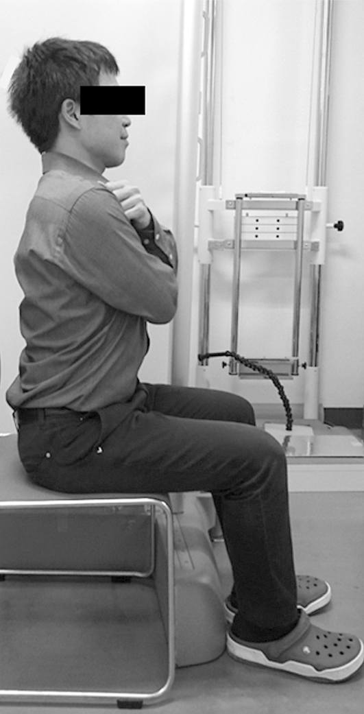
    Introduction: Spinopelvic alignment in the sitting position remains unclear. We investigated the differences in spinopelvic alignment in the standing and sitting positions in outpatients.

    Methods: We enrolled a total of 84 outpatients with low back pain (43 men, 41 women; average age: 64.4 years). Patients with a history of spinal surgery were excluded. Whole-spine lateral radiographs were obtained for all patients in the standing and sitting positions without using a backrest. The sagittal vertical axis (SVA), L1-S1 lumbar lordosis (LL), sacral slope (SS), and pelvic incidence (PI) were measured. Patients with ΔSS of <5° (change in SS between the standing and sitting positions) were categorized as the group showing lesser pelvic retroversion. Statistical analysis was performed using unpaired t-test and chi-square test. The SPSS software (version 23.0; IBM, Armonk, NY, USA) was used for statistical analysis. P-values of < 0.05 were considered statistically significant.

    Results: The group with lesser pelvic retroversion included 28 patients (33%) and that with pelvic retroversion included 56 patients (77%). In the group with lesser pelvic retroversion, there were primarily women, PI was significantly lower, SVA significantly increased from the standing to sitting positions (P < 0.05), and change in LL was significantly smaller (P < 0.01).

    Conclusions: An anterior trunk tilt compensates for the lower reduction in LL in patients with lesser pelvic retroversion from the standing to sitting positions.

    Download PDF (1686K)
  • Chihiro Maeda, Takahiro Hozumi, Hiroyuki Oka, Kiyofumi Yamakawa, Shure ...
    2020Volume 11Issue 5 Pages 848-852
    Published: May 20, 2020
    Released on J-STAGE: May 20, 2020
    JOURNAL FREE ACCESS

    Introduction: Diagnostic tumor markers for identifying spinal metastases of unknown origin (SMUO) at initial presentation are unknown. Therefore, we aimed to determine the predictive ability of 17 tumor markers for identifying primary sites of spinal metastases.

    Methods: We recruited 240 patients with SMUO at the Komagome Hospital between January 2007 and August 2018. The patients had primary pathologies of the lung (n = 60), prostate (n = 34), breast (n = 15), thyroid (n = 11), pancreatic and bile duct (n = 11), stomach and colon (n = 13), and other digestive organs (n = 33) and all named cancers (n = 184), as well as multiple myeloma (n = 28), malignant lymphoma (n = 18), and others (n = 10). Diagnostic values were statistically determined as areas under the receiver operating characteristics curve (AUCs).

    Results: Significant tumor markers determined using AUCs comprised thyroglobulin (Tg) for thyroid cancer (AUC = 1.0), prostate-specific antigen (PSA) for prostate cancer (AUC = 0.94), carbohydrate antigen 19-9 (CA19-9) and pancreatic cancer-associated antigen for pancreatic and bile duct cancer (AUC = 0.89 and 0.83, respectively), sialyl SSEA-1 antigen (SLX) for lung cancer (AUC = 0.81), CA19-9 for digestive cancer (AUC = 0.74), carbohydrate antigen 153 (CA153) for breast cancer (AUC = 0.73), carcinoembryonic antigen (CEA) for stomach and colon cancer (AUC = 0.73), soluble interleukin 2 receptor (sIL-2R) for malignant lymphoma (AUC = 0.73), and tissue polypeptide antigen/CEA for all named cancers (AUC = 0.72). Serum and urinary values determined using immunoelectrophoresis (IEP) to diagnose multiple myeloma were statistically significant (P < 0.0001, two-tailed test). On the other hand, analysis using sIL-2R is expensive, and CA153 can be substituted by inspection, palpation, and contrast-enhanced computed tomography for detecting breast cancer.

    Conclusions: We recommend analyses using Tg, PSA, CA19-9, SLX, and CEA and IEP to cost-effectively identify primary sites of spinal metastases.

    Download PDF (773K)
  • Yasuyuki Tamaki, Katsufumi Hyakuna, Akira Uchikoshi
    2020Volume 11Issue 5 Pages 853-857
    Published: May 20, 2020
    Released on J-STAGE: May 20, 2020
    JOURNAL FREE ACCESS

    Introduction: It is often difficult to determine the treatment option for thoracolumbar burst fractures with a Thoracolumbar Injury Classification and Severity Score of 4 (TLICS 4).

    Methods: We retrospectively reviewed and compared six patients who underwent conservative treatment and nine patients who underwent surgical treatment for thoracolumbar burst fracture with TLICS 4.

    Results: Vertebral body height loss was significantly less and bed rest duration was significantly shorter in the operation group. There were no significant between-group differences in local kyphosis, Denis pain scale score, and neurological symptoms.

    Conclusions: We found that conservative treatment in TLICS 4 provides favorable outcomes, but surgical treatment provides the advantages of early ambulation and the prevention of deformity in the damaged vertebral body.

    Download PDF (885K)
  • Sachiko Kawasaki, Hideki Shigematsu, Masato Tanaka, Yuma Suga, Yusuke ...
    2020Volume 11Issue 5 Pages 858-865
    Published: May 20, 2020
    Released on J-STAGE: May 20, 2020
    JOURNAL FREE ACCESS

    Introduction: We assessed which factors affected postoperative cervical kyphosis after laminoplasty for cervical spondylotic myelopathy (CSM).

    Methods: We retrospectively recruited 57 cases that underwent laminoplasty for CSM and were followed-up for at least 2 years after operation. Regarding clinical outcomes, we assessed the Japanese Orthopaedic Association (JOA) recovery rate of these individuals.

    Results: In these 57 cases, 11 cases (19.3%) was found to be kyphotic (ΔC2-7 Cobb angle > 5°) at neutral position in the 2 years after operation (Group K, the others were Group L). Their statistical analysis showed that Group K had smaller preoperative C7 slope than Group L (23.2°±10.3°, 31.6°±10.1°, respectively) (p < 0.05). From the receiver operator characteristic curve, the cut-off value was 26.5°, the sensitivity was 67.4%, and the specificity was 72.7%. Furthermore, the odds ratio was 9.3 (95%CI: 1.78-48.50, p < 0.01). No other preoperative factors affected postoperative kyphosis in our study: age, C2-7 Cobb angle, C2-7 sagittal vertical axis (SVA), center of gravity of the head-C7 SVA, and C7 short cervical length. Furthermore, the progression degree of kyphosis did not have correlation with each JOA recovery rate.

    Conclusions: The risk factor of postoperative cervical kyphosis after laminoplasty for CSM was only a small preoperative C7 slope. The cut-off value of preoperative C7 slope was 26.5°.

    Download PDF (1470K)
Case Report
  • Michita Noma, Masato Anno, Masayoshi Fukushima
    2020Volume 11Issue 5 Pages 866-871
    Published: May 20, 2020
    Released on J-STAGE: May 20, 2020
    JOURNAL FREE ACCESS

    Introduction: Postoperative disc herniation, epidural hematoma, and implant malposition are major complications following cervical spine surgery for trauma and can lead to neurological dysfunction. Neurological deterioration due to a posterior pathology is relatively rare. This study reports a rare case of postoperative neurological deficit due to buckling of a ligamentum flavum.

    Case report: A 70-year-old man presented at our hospital with a C4/5 cervical spine injury without neurological deficit. Posterior fixation was performed using lateral mass screws. The patient developed paraplegia after the surgery. Magnetic resonance imaging revealed spinal cord compression at C4/5 due to buckling of a ligamentum flavum. Therefore, laminectomy was performed 6 hours after the primary fixation, which resulted in rapid neurological recovery.

    Conclusions: Postoperative neurological deficit can occur due to a collapsed ligamentum flavum following posterior cervical fusion surgery. Therefore, an accurate diagnosis is essential for determining the most appropriate surgical intervention.

    Download PDF (1573K)
  • Yasuyuki Tamaki, Katsufumi Hyakuna, Akira Uchikoshi, Masashi Neo
    2020Volume 11Issue 5 Pages 872-876
    Published: May 20, 2020
    Released on J-STAGE: May 20, 2020
    JOURNAL FREE ACCESS

    Introduction: We report a case of minimally invasive posterior lumbar interbody fusion for high-grade spondylolisthesis.

    Case Report: The patient was an 80-year-old woman with L5/S high-grade lumbar spondylolisthesis, and her Japanese orthopedic association (JOA) score was 11 points. L5/S posterior interbody fusion by in situ fixation was performed because the patient's pelvic incidence-lumbar lordosis mismatch was small. The operation was performed using a transdiscal screw (hereafter referred as TDS), and a bone graft of L5/S was used for holes in the bone of S1 TDS. The patient's JOA score at 2 years postoperatively was improved to 26 points, and bone union was confirmed.

    Conclusions: This operation was a minimally invasive and useful method.

    Download PDF (1381K)
feedback
Top