Journal of Spine Research
Online ISSN : 2435-1563
Print ISSN : 1884-7137
Volume 12, Issue 8
Displaying 1-11 of 11 articles from this issue
Original Article
  • Katsuhiko Kikuchi, Koichi Yoshikane
    2021 Volume 12 Issue 8 Pages 1018-1024
    Published: August 20, 2021
    Released on J-STAGE: August 20, 2021
    JOURNAL FREE ACCESS

    Objective: The purpose of this study was to evaluate the usefulness of Full-endoscopic spine surgery (FESS) under local anesthesia for elderly people.

    Method: From September 2015 to September 2018, we had 46 elderly patients for whom we performed FESS under local anesthesia. We analyzed operation time, visual analog scale (VAS) of pre and post operation, systematic complications during perioperative period and Macnab criteria.

    Results: 18 male and 31 female were included in this study. Mean age was 79.5 years. Mean follow up periods was 11.5 months. Preoperative VAS scores of low back pain was 66.3, of leg pain was 79.5 and of numbness was 69.4. Postoperative VAS scores of low back pain was 53, of leg pain was 49.7 and of numbness was 32.3. In Macnab criteria, 10 patients (20%) were poor and 7 patients had reoperation.

    Conclusion: Although FESS under local anesthesia for elderly people was less invasive and usefull, functional outcomes in 20% of patients were inferior.

    Download PDF (1636K)
  • Keishi Maruo, Fumihiro Arizumi, Kazuki Kusuyama, Norichika Yoshie, Tom ...
    2021 Volume 12 Issue 8 Pages 1025-1029
    Published: August 20, 2021
    Released on J-STAGE: August 20, 2021
    JOURNAL FREE ACCESS

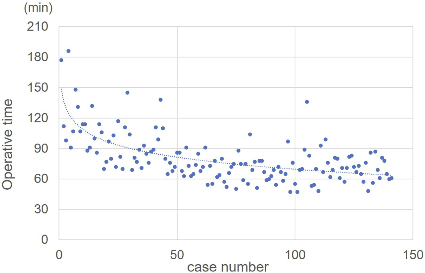
    Introduction: The learning curve of microendoscopic discectomy (MED) was investigated by an inexperienced surgeon in spinal endoscopy.

    Methods: A total of 191 consecutive patients who underwent lumbar endoscopic surgeries between November 2014 and February were retrospectively reviewed, 141 consecutive patients were treated with MED. The mean age was 48.9±17.5 years, 84 were males and 57 were females. Operative time, blood loss, and complications were investigated. The learning curve was calculated by dividing the operation time by 30 cases.

    Results: The mean operative time was 80.4±23.9 min, and the mean blood loss was 2.3 ml±10.5 ml. The mean operative time for the first 30 patients was 107.8±28.9 minutes, 83.1±16.8 minutes for patients 31 to 60, and 68.8±12.8 minutes for patients 61 to 90. Complications included dural tear in 3 patients (2.1%), inferior articular process fracture in 1 patient (0.7%), hematoma in 1 patient (0.7%), and recurrent herniation in 1 patient (0.7%). There were no cases of infection, wrong site surgery, or open conversion.

    Conclusions: MED is a minimally invasive and safe technique even for an inexperienced surgeon with a domestic spinal endoscopic educational system.

    Download PDF (947K)
  • Reiko Yoneyama, Kazuo Ohmori, Osamu Matsushige
    2021 Volume 12 Issue 8 Pages 1030-1034
    Published: August 20, 2021
    Released on J-STAGE: August 20, 2021
    JOURNAL FREE ACCESS

    Introduction: The learning curve of full-endoscopic discectomy (FED) is well known. However, the learning curve changes due to the contribution of several training courses. The purpose of this study is to compare the learning curve between a surgeon who started the FED-IL procedure in 2012 and a surgeon in 2019.

    Methods: We included case from the 1st case of FED to the cases of 6 months. Surgeon A performed FED-IL procedure on 14 patients (10 males and 4 females with a mean age of 48.8±13.5 years) between November 2019 and May 2020. Surgeon B performed FED-IL procedure on 30 patients (14 males and 16 females, mean age 49.9±14.6 years) between October 2012 and April 2013.

    Results: The mean operative time for surgeons A and B was 95.8±37.8 minutes and 114.7±37.1 minutes, respectively. The tendency to reduce the operative time was similar for both surgeons A and B (P for interaction=0.319). Using multivariate linear regression analysis, the mean operative time was 23.4 minutes shorter for surgeon A than for surgeon B adjusted for patient age, gender, and surgical level (P<0.05).

    Conclusion: Compared to EFD beginner surgeon in 2012, beginner surgeon in 2019 had a shorter EFD-IL surgery time.

    Compared to the early years of 2012, EFD-IL learning curve in 2019 shortened. This may be due to the widespread availability of specialized EFD-IL training and other educational programs.

    Download PDF (769K)
  • Osamu Matsushige, Kazuo Ohmori, Reiko Yoneyama
    2021 Volume 12 Issue 8 Pages 1035-1039
    Published: August 20, 2021
    Released on J-STAGE: August 20, 2021
    JOURNAL FREE ACCESS

    The inside-out transforaminal (TF) method often results in exiting nerve damage, and the outside-in method has been reported as a countermeasure. Therefore, operation time and presence or absence of foraminoplasty were compared between 12 patients who underwent full-endoscopic disectomy via the inside-out TF method (Group I) and 12 patients who underwent it via the outside-in TF method (Group O) for lumbar foraminal herniation. The endoscopic operation time and time to identify the exiting nerve were longer in Group O; this Group also showed higher foraminoplasty rate. Therfore, this study did not confirm the superiority of the outside-in method over the inside-out method.

    Download PDF (1006K)
  • Fumihiko Eto, Masaki Tatsumura, Katsuya Nagashima, Toru Funayama, Masa ...
    2021 Volume 12 Issue 8 Pages 1040-1046
    Published: August 20, 2021
    Released on J-STAGE: August 20, 2021
    JOURNAL FREE ACCESS

    Introduction: The spinous process splitting approach, which is less invasive to the paraspinal muscles, is often used for posterior decompression of the lumbar spine. However, if the spinous process is separated from the lamina after surgery, the strength of the paraspinal muscles attached to the spinous process cannot be efficiently transmitted to the vertebral body. We believe that it is important to fuse the spinous process and lamina after surgery. Therefore, we repositioned the spinous process to the lamina with absorbable sutures after decompression. In the present study, we investigated the bone union rate with this method 2 years after surgery.

    Methods: We evaluated 74 patients who underwent multi-level posterior decompression with the spinous process splitting approach for lumbar spinal canal stenosis. Spinous processes were repositioned with absorbable sutures before wound closure in 203 laminas. A computed tomography (CT) scan obtained 2 years post-surgery was used to evaluate the bone union rate. Bone union was confirmed if the spinous process was found to be in contact with the lamina. In addition, complete fusion was confirmed if bone union was seen in all laminas.

    Results: Two years post-surgery, bone union was achieved in 181 laminas (89.2%). Furthermore, there were 56 cases of complete fusion (75.7%).

    Conclusions: Posterior columns, such as the spinous process, supraspinous ligaments, and interspinous ligaments, are involved in spinal stabilization. Therefore, a fusion between the spinous process and lamina should be maintained after posterior decompression surgery. We found that the bone union rate between the spinous process and lamina was 64.4% at 6 months after surgery. Even in patients who used a brace for only 3 months after surgery and followed no exercise restrictions, the bone union between the spinous process and lamina continued to progress even 6 months after surgery. In addition, the absorbable sutures used in this study are easily available and inexpensive for normal wound closure. Our technique of repositioning the spinous process to the lamina using absorbable sutures has proven to be useful for bone union, and may subsequently prevent spinal instability.

    Download PDF (1452K)
  • Kuniyoshi Tsuchiya
    2021 Volume 12 Issue 8 Pages 1047-1052
    Published: August 20, 2021
    Released on J-STAGE: August 20, 2021
    JOURNAL FREE ACCESS

    Introduction: Cases of posterior surgery for cervical radiculopathy using FED (Full Endoscopic Discectomy) were retrospectively reviewed.

    Methods: Seven patients that underwent posterior decompression surgery using FED systems have completed 12 months follow up. All of the patients were suffered from radiculopathy due to cervical disc herniation.

    Results: Mean surgical time was 97 minutes and bleeding was minimum in all cases. Average radiculopathy scores of the patients before surgery was 10.8, which was improved to 17.5 at three months and 18.0 at one year.

    Discussion: Surgery using FED systems has several advantages compared to other methods. Closer view point to the neural tissue makes it possible to observe neural structures more clearly, thus makes the procedure safer and more comfortable. To appreciate this advantage, precise orientation and strict bleeding control are mandatory. Perfusion used in FED surgery enables releasing vessels from other surrounding tissues by hydrostatic pressure, making it easier to control bleeding from epidural vessels. Although small diameter of the device gives benefit on FED surgery, this feature can result in higher possibility of wrong level surgery or migration of the scope into spinal canal. Thus this technique should be carefully performed to prevent neurologic complications and enough training will be required.

    Conclusions: Deep and closer eye point is a substantial advantage in FED surgery.

    Download PDF (1476K)
  • Tomohiro Hikata, Tsuyoshi Iida, Morito Takano
    2021 Volume 12 Issue 8 Pages 1053-1059
    Published: August 20, 2021
    Released on J-STAGE: August 20, 2021
    JOURNAL FREE ACCESS

    Introduction: The purpose of this study was to clarify the risk factors for anterior longitudinal ligament injury (ALL injury) in posterior corrective surgery for adult spinal deformity (ASD) using lateral interbody fusion (LIF) procedure.

    Methods: We analyzed 38 consecutive patients (7 male and 31 female patients) who underwent posterior corrective surgery for ASD from March 2017 to May 2020. Patients were divided into two cohorts (patients with intraoperative ALL injury were R-group, and no ALL injury were N-group). We assessed patient' characteristics (age, gender, BMI, prior spinal surgery, and preexisting vertebral fracture), operative information (operative time, estimated blood loss, number of LIF segments, number of fusion segments, and three column osteotomy), cage height, cage angle, cage position, and radiological parameter (SVA, C7-CSVL, LL [L1-S1 angle], LLL [L4-S1 angle]), LL on FBB [fulcrum backward bending], LLL on FBB, PI, PT, TK, and PI-LL).

    Result: ALL injury occurred in 8 patients (21.1%). Preoperative SVA (R; 197.9 mm, N; 118.5 mm, p=0.004), LL (R; -9.3°, N; 10.9°, p=0.017), LLL (R; 7.3°, N; 18.5°, p=0.008), PI (R; 42.9°, N; 50.5°, p=0.008), LL on FBB (R; 12.8°, N; 37.1°, p=0.002), and LLL on FBB (R; 11.6°, N; 27.7°, P< 0.001) were statistically significant difference between two groups. Multivariate logistic regression analysis revealed that LLL on FBB was statistically significant risk factor for ALL injury (p=0.005, odds ratio; 0.750, 95% CI; 0.612-0.918).

    Conclusion: In patients with severe rigid sagittal imbalance with decreased lower lumbar lordosis (L4-S1), ALL injury was expected to occur during posterior correction surgery.

    Download PDF (1196K)
  • Masayuki Ishihara, Shinichiro Taniguchi, Yoichi Tani, Takashi Adachi, ...
    2021 Volume 12 Issue 8 Pages 1060-1066
    Published: August 20, 2021
    Released on J-STAGE: August 20, 2021
    JOURNAL FREE ACCESS

    Purpose: We investigated factors causing poor postoperative local lordosis acquisition-aside from cage placement position-in minimally invasive spine stabilization (MISt) using lateral interbody fusion (LIF) and percutaneous pedicle screws (PPS) in patients with lumbar spinal canal stenosis (LSCS).

    Methods: Forty-six LSCS patients (106 levels) underwent level 2 or 3 MISt with LIF and PPS between 2015 and 2017 and were followed up for more than 12 months. We examined pathology, surgical level, preoperative disc condition, segmental lordosis (SL), anterior (ADH) and posterior disc height (PDH), and other spinopelvic parameters. The patients were divided into two groups for comparison. The first group consisted of postoperative SL < 7° disc levels (Group P). The other group consisted of postoperative SL ≥to 7° disc levels (Group G). We classified disc levels based on condition: the less-degenerative type with lordosis (type N), less-degenerative type with kyphosis (type K), unilateral degeneration type (type U), disc-cavity disappearance type (type D), anterior-degeneration type (type A), and posterior-degeneration type (type P). Pathologic results were classified as single-slip, double-slip, LSCS with degenerative kyphosis (PI-LL > 20°), and LSCS/degenerative scoliosis.

    Results: At the surgical level, L2/3 was significantly lower than L3/4 and L4/5. There were no significant differences in the preoperative disc condition and pathology. After surgery, significant improvement was noted in various parameters, except for pelvic tilt (PT). The average SL was 8.7°. Postoperative SL was significantly lower for types K and A. Disc types N and D were significantly higher in the G group. Meanwhile, types K and A were significantly higher in the P group. LSCS with kyphosis was significantly higher in the P group. Preoperative SL, postoperative ADH, and the change in ADH and PDH were significantly smaller in group P. Meanwhile, the pre- and postoperative PDH was significantly larger in group P. Logistic regression analysis was performed with a postoperative SL < 7° as the dependent variable. Preoperative SL, preoperative PDH, and disc properties (K and A) were detected as risk factors. ROC analysis revealed that the preoperative SL cutoff value was 4° (AUC 0.75), and the preoperative PDH cutoff value was 4.5 mm (AUC 0.63).

    Conclusions: Factors causing poor SL acquisition, aside from cage placement position, were investigated in patients who underwent MISt using LIF and PPS. The residual posterior disc component and residual posterior element contributed to the poor acquisition of local lordosis.

    Download PDF (1934K)
  • Yoshikazu Yanagisawa, Masayoshi Ohga
    2021 Volume 12 Issue 8 Pages 1067-1073
    Published: August 20, 2021
    Released on J-STAGE: August 20, 2021
    JOURNAL FREE ACCESS

    Introduction: In our department, intraoperative nerve monitoring (INM) is used in combination, and microscopic minimally invasive posterior cervical herniation / foraminotomy (P-PMCF) is performed in combination with an ultrasonic surgical device. This time, we report whether INM is useful for preventing postoperative neurological complications.

    Methods: Since April 2018,8 patients (male:female = 6:2, average age: 47.0±3.2 years) who underwent P-PMCF under INM were included. INM recorded somatosensory evoked potential (SSEP), transcranial evoked muscle recording evoked potential (Br-MsEP), spontaneous electromyography (fEMG), and nerve root stimulating peripheral muscle recording (tEMG). The survey items included the presence or absence of INM abnormalities, the presence or absence of tEMG records, postoperative complications, changes in the JOA score, which is the criterion for cervical myelopathy before and after surgery, and the presence or absence of additional surgery.

    Results: The average operation time was 145±30.4 minutes and the average bleeding volume was 16.3±20.8 ml. There were 2 cases of fEMG as abnormal INM, but 1 case of increased intraoperative blood pressure and BIS value without abnormal INM. In addition, tEMG could be recorded in 6 of 8 cases. No postoperative complications were observed, and the JOA score, which is the criterion for cervical myelopathy before and after surgery, showed a significant improvement from a preoperative average of 14.19±1.28 to an average of 16.44±0.18 at the final stage (P = 0.0010269). Laminoplasty was performed in one case as additional surgery.

    Conclusions: Although P-PMCF is minimally invasive, there is a risk of intraoperative nerve damage due to small cutaneous incision. In this operation, fEMG and tEMG can prevent radiculopathy and are considered to be useful.

    Download PDF (1247K)
  • Takaki Yoshimizu, Tetsutaro Mizuno, Ushio Nosaka, Takuma Hayashi, Keis ...
    2021 Volume 12 Issue 8 Pages 1074-1080
    Published: August 20, 2021
    Released on J-STAGE: August 20, 2021
    JOURNAL FREE ACCESS

    Introduction: This study aimed to analyze the usefulness of endoscopic lumbar herniotomy by Biportal Endoscopic Spine Surgery (BESS).

    Methods: An observational retrospective study was performed. 89 cases (59 males/30 females; average 42 years old) were investigated. 49 cases of lumbar disc herniation (LDH) were treated by BESS. For comparative cases, 40 cases undergone Microscopic surgery (Micro). Examination items are below. Operative time, amount of bleeding, hospitalization period, postoperative pain of postoperative first day and discharge day measured by Numerical rating scale (NRS). We also checked postoperative improvement from preoperative leg pain, numbness and recurrence rate. For comparison between two groups, the Wilcoxon test was used for statistical analysis.

    Results: Surgical time of BESS was significantly longer than Micro (77 minutes vs 60 minutes). The mean amount of bleeding during BESS was tend to be less than Micro (7.9 g vs 9.9 g). There was no significant difference in postoperative wound pain at the first day (BESS: 3.7, Micro: 3.6), but tend to less at discharge day (BESS: 1.2, Micro: 1.7). Postoperative improvements of leg pain (BESS: 84%, Micro: 80%) and numbness (BESS: 82%, Micro: 75%) are tend to better in BESS. Recurrence cases are six in each groups.

    Conclusions: BESS is two portal surgery which take on a role of camera and working and don't use any cannula and retractor. Normal saline is perfused from camera to working portal to make vision. Thus we can perform surgery with very small incision (5 mm*2). This technic has advantages that we can move camera and surgical instruments more freely. The results of BESS is same as conventional and take it to good option.

    Download PDF (1407K)
Case Report
  • Hideaki Iba, Kazuo Nakanishi, Kazuya Uchino, Seiya Watanabe, Kosuke Mi ...
    2021 Volume 12 Issue 8 Pages 1081-1085
    Published: August 20, 2021
    Released on J-STAGE: August 20, 2021
    JOURNAL FREE ACCESS

    Introduction: Intradural disc herniation is rare pathological condition. This case report was an experience of an intradural disc hernia encountered during revision MED (Re-MED) for recurrent of disc herniation.

    Case report: A 48-year-old woman with a chief complaint of right leg pain. L5/S right disk hernia was recognized and MED was performed. Two years after the operation, right lower extremity pain reappeared. An MRI scan showed a recurrent disk herniation at the same site. Re-MED was performed. However, the expected disk herniation mass was not confirmed. The herniation mass was confirmed intradural. An incision was made in dura mater and the herniation mass was removed. The JOA score after Re-MED improved.

    Conclusion: Re-MED is a useful technique for recurrent of disc herniation. However, considering the possibility of intradural disc herniation during Re-MED, appropriate preoperative preparation is necessary.

    Download PDF (1125K)
feedback
Top