Journal of Spine Research
Online ISSN : 2435-1563
Print ISSN : 1884-7137
Volume 12, Issue 10
Displaying 1-9 of 9 articles from this issue
Editorial
Original Article
  • Hiroaki Kimura, Juichi Miura, Bunichiro Wadayama
    2021Volume 12Issue 10 Pages 1228-1234
    Published: October 20, 2021
    Released on J-STAGE: October 20, 2021
    JOURNAL FREE ACCESS

    Introduction: A previous study showed that pear-shaped discs are a risk factor of cage retropulsion in posterior lumbar interbody fusion (PLIF) because a pear-shaped disc does not tend to make contact with the four cage corners in sagittal plane, and that expandable cages should be used in cases of the unstable contact between the endplates and the cage. Therefore we hypothesized that a pear-shaped disc may be a risk factor of nonunion after PLIF, and I evaluated surgical results of PLIF using expandable cages for pear-shaped discs.

    Methods: We analyzed 68 patients who underwent single- or 2-level PLIF for degenerative lumbar diseases between 2015 and 2018. Ninety-four discs of 68 patients were categorized to 2-groups, the group P with pear-shaped discs (20 discs) and the group C without it (74 discs).

    Results: Fusion rates on CT 6 months after surgery were 50% in the group P and 79.7% in the group C, that made a significant difference (P = 0.01), and fusion rates evaluating flexion-extension X-ray images were 85% in the group P and 94.6% in the group C, that showed no significant difference (P = 0.16).

    Conclusions: Although fusion rates on CT 6 months after surgery were higher in the group C than those in the group P, fusion rates on X-ray images a year after surgery were similar in both groups. Therefore, expandable cages were useful in undergoing PLIF for pear-shaped discs.

    Download PDF (1293K)
  • Motoshi Gomi, Hiroshi Sakuramoto, Koju Hayashi, Ryosuke Takahashi, Dai ...
    2021Volume 12Issue 10 Pages 1235-1239
    Published: October 20, 2021
    Released on J-STAGE: October 20, 2021
    JOURNAL FREE ACCESS

    Purpose: There are various materials for filling the cage of the lateral lumbar interbody fusion (LLIF), and each material has different ability of bone fusion. Although the autogenous bone grafts have a better bone fusion ability, it has a problem of postoperative donor site pain such as a fracture. Since 2019, the demineralized bone matrices (DBM) have become available in Japan. This time, we investigated the early postoperative interbody fusion using both DBM and bone marrow aspirates in the LLIF cage.

    Subjects: The subjects were 7 patients and 14 intervertebral discs that underwent LLIF at our hospital from April 2019 to December 2019. In all cases, DBM and bone marrow aspirates from the iliac bone were mixed, and fixation from the posterior was performed percutaneously. Bone fusion was evaluated by CT images 6 months after the operation.

    Results: Bone fusion with the upper and lower endplates appeared on 3/14 intervertebral space (21.4%).

    Discussion: LLIF often causes postoperative donor site pain. Although it has been reported that the long-term fusion rate of artificial bone and autologous bone graft or allografts is not significantly different from that of autologous bone graft, the facilities where allografts can be used were limited in Japan. DBM is expected to be a new option as allografts that can be used in any hospital. In this study, we were able to avoid donor site pain by using both DBM and bone marrow aspirates, but the fusion was not as good as that of artificial bone graft or autogenous bone grafts in the short term.

    Download PDF (1242K)
  • Masakazu Toi, Keishi Maruo, Fumihiro Arizumi, Kazuki Kusuyama, Kazuya ...
    2021Volume 12Issue 10 Pages 1240-1245
    Published: October 20, 2021
    Released on J-STAGE: October 20, 2021
    JOURNAL FREE ACCESS

    Introduction: CT values of vertebral cancellous bone (Hounsfield unit values; HU values) show a high correlation with bone density. It has also been reported to be a risk factor for pedicle screw loosening after PLIF and TLIF. However, whether HU values affect bone fusion after PLIF and TLIF is not known.

    Methods: The subject were 106 consecutive patients who underwent mini-open TLIF/PLIF in our hospital between 2016 and 2019 with minimum one-year follow-up. The mean age was 69.9±11.6 years and 65 female and 41 males. The HU values were measured at the center of vertebrae (L1-5) on preoperative CT. Existence of pseudoarthrosis on 12 months follow-up X-ray and CT was investigated. PS loosening, cage subsidence (CS), positive endplate cyst sign (PCS) was also assessed. The factors considered were patient factors, surgery-related factors. The HU values are the mean of L1-4 (L1-4 HU), L1 (L1 HU), and the mean HU values of the fixed vertebrae (F-HU) was measured. Clinical outcomes were investigated for low back pain VAS, ODI and JOABPEQ. We compared the above results between the bone fusion group (F group) and the bone fusion failure group (NF group).

    Results: The fusion rate at 12 months after surgery was 84%. Mean age was significantly higher in the F group compare to NF groups. There were no significant difference in sex, BMI, bone density, smoking, steroid user, and PTH user. There were no significant difference in L1-4 HU, L1HU and F HU between the two groups. All surgery-related factors, including the number of fixed vertebrae and the number of cages, did not differ significantly between the two groups. Incidence of PS loosening, CS and PCS were significantly higher in the NF group than in the F group. HU values were significantly lower in the presence of PS loosening, but significantly no different between CS and PCS. ODI at 12 months postoperatively was significantly higher in the NF group. Pain-related disorders, lumbar spine dysfunction, gait dysfunction and social impairment of JOABPEQ were significantly lower in the NF group.

    Conclusion: The HU value may be a predictor of PS loosening, but it was not associated with fusion rate after PLIF or TLIF. Incomplete fusion after TLIF of PLIF associated with poor clinical outcomes. Therefore, the achievement of complete fusion is important to obtain better clinical outcomes.

    Download PDF (945K)
  • Tomonori Ozaki, Kentaro Yamada, Hiroaki Nakamura
    2021Volume 12Issue 10 Pages 1246-1250
    Published: October 20, 2021
    Released on J-STAGE: October 20, 2021
    JOURNAL FREE ACCESS

    Introduction: In spinal fusion surgery, deviation of the pedicle screw (PS) causes complications such as nerve and vascular injury and pedicle fracture. In this study, we investigated the accuracy of PS placement in thoracolumbar fusion using 3D preoperative planning software.

    Methods: Thirty-three patients (263 screws) who underwent a thoracolumbar fusion with PS from April 2019 to March 2020 were included in the study. The accuracy of PS insertion was evaluated by postoperative CT. The difference in the deviation rate according to the implantation method (percutaneous or open) and the surgeon (A; more than 10 years of experience in spine surgery, B; less than 5 years of experience in spine surgery, C; general orthopedic surgeon) was also examined.

    Results: A total of 12 PS deviations (4.6%) were observed, and no nerve or vascular injuries were observed. There was no significant difference in the deviation rate between percutaneous and open techniques when grade 1 or higher was defined as the deviation group (percutaneous 5.0% (6/119), open 4.2% (6/144). p=0.73). There was also no difference between surgeons (A 4.5% (7/157), B 4.3% (3/69), C 5.4% (2/37), p=0.96).

    Conclusions: Preoperative planning with 3D software can help to reduce the deviation rate in PS insertion.

    Download PDF (1025K)
Case Report
  • Taiji Iwasawa, Tetsuya Ohara, Toshiki Saito, Ryoji Tauchi, Ippei Yamau ...
    2021Volume 12Issue 10 Pages 1251-1256
    Published: October 20, 2021
    Released on J-STAGE: October 20, 2021
    JOURNAL FREE ACCESS

    Introduction: Surgical treatment for adult spinal deformity (ASD) improves the patient's quality of life; however, ASD surgery is associated with a high complication rate. The most frequent complication of ASD surgery is rod fracture after long-range fixation as an implant-related complication. Rod fracture often occurs due to pseudarthrosis or non-union and is most commonly observed after 5 years of the surgery with a lower incidence of late onset. Here, we report the cases of two patients with late-onset rod fracture who were treated at our hospital. Case 1 was that of a 64-year-old woman with paralytic scoliosis. Thirteen years after her surgery, she had a fracture of the vertebral body and a fracture of the rod at the L2 level, which had achieved bone union within the range of fixation. She had persistent low back pain; therefore, she underwent revision surgery to replace the rod and add an anterior fixation.

    Case 2 was that of a 39-year-old man with congenital scoliosis. Six years after his surgery, he had rod fracture at the L3-4 level without interbody fusion. The pain persisted; therefore, he underwent revision surgery to replace the rod and add an anterior fixation.

    Conclusions: In both cases, rod fracture occurred after >5 years of the initial surgery. In case 1, the fracture and rod fracture occurred at the same time at the L2 level where the anchor was weak owing to osteoporosis of the long-term fixation vertebral body. In case 2, the rod fracture occurred at the L2 level where interbody fusion was not performed. In cases where mechanically vulnerable areas persist after the surgery, long-term follow-up is necessary owing to the possibility of late-onset rod fracture even after bone union.

    Download PDF (1802K)
  • Kentaro Yamada, Tomonori Ozaki, Hiroaki Nakamura
    2021Volume 12Issue 10 Pages 1257-1263
    Published: October 20, 2021
    Released on J-STAGE: October 20, 2021
    JOURNAL FREE ACCESS

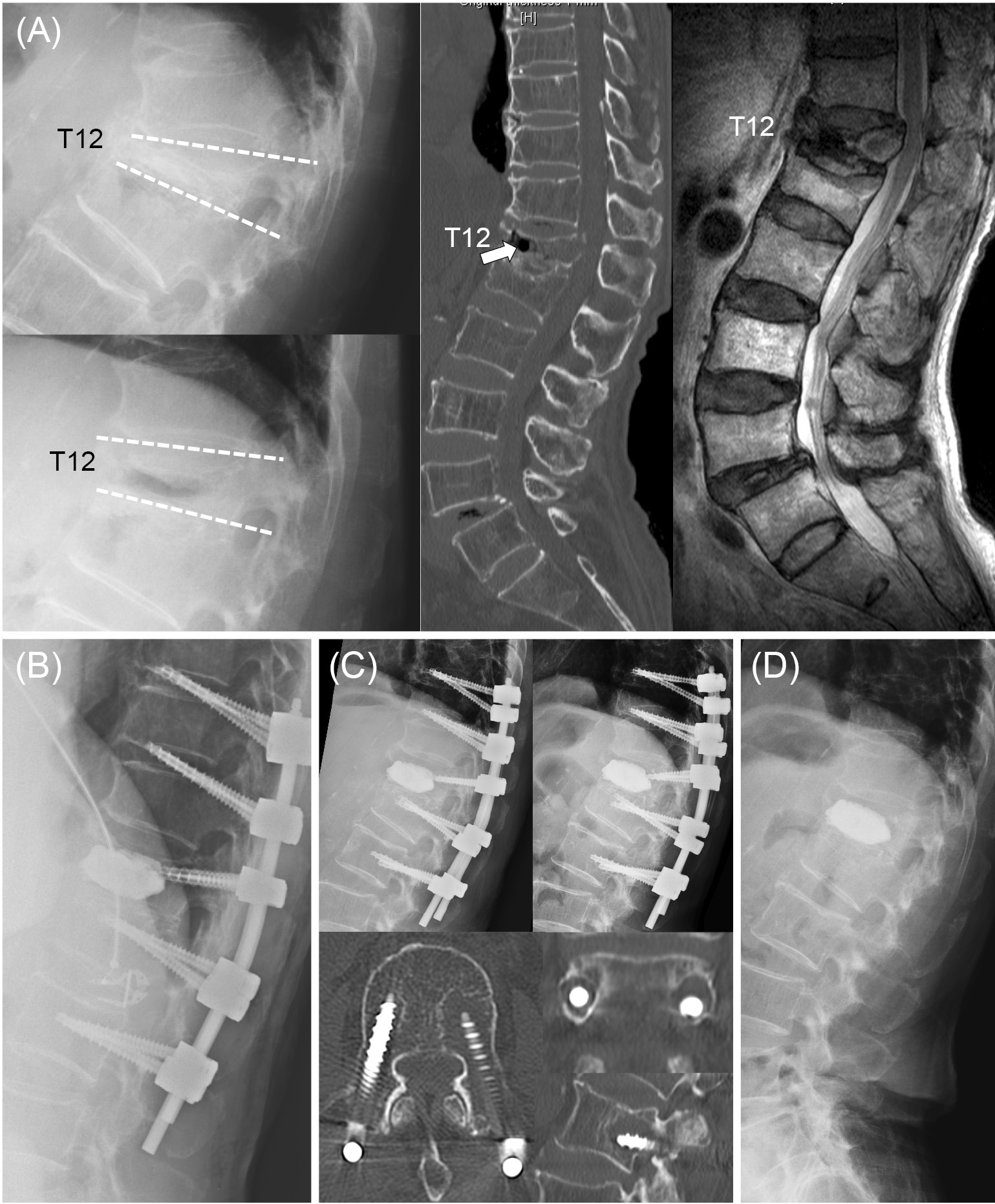
    Introduction: Surgical treatment of thoracolumbar vertebral fractures with diffuse idiopathic skeletal hyperostosis (DISH) needs longer fixation than fractures without DISH, because the segments without ossification by DISH are exposed to higher mechanical stress due to a longer lever arm in the spinal column. Recently, the usefulness of penetrating endplate screw (PES) for patients with DISH have been reported in several reports. We have performed 3 cases using the PES technique for thoracolumbar vertebral fracture with DISH. The purpose of this report was to report the result of the surgery and discuss the indication of the PES technique.

    Case report: One case was fracture with acute phase and two cases were fractures with sub-acute phase. PES technique was used at the ossified segment by DISH and conventional pedicle screw technique was used non-ossified segment. Balloon kyphoplasty was used for fractures with sub-acute phase. The two cases showed good outcomes both in the clinical and radiological assessment. However, one case (T12 vertebral fracture with T8-12 DISH) showed screw loosening at the most caudal screw 1week after the surgery. Screw loosening was getting worse, and revision surgery of implant removal was performed 6 months after the index surgery.

    Conclusions: Screws with the PES technique have reportedly 1.4-fold pull-out strength compared with conventional pedicle screws. The poor outcome case in this study was set screws with PES technique in cranial 2 vertebrae and conventional pedicle screw in caudal two vertebrae, therefore, the reason of poor outcomes might arise from the difference of the screw fixation strength between cranial and caudal. PES technique is useful for fixation to the DISH segment. However, additional maneuver considering fixation strength of screws with the PES technique is necessary for fixation at non-ossified vertebra in case of the end fracture of ossification by DISH.

    Download PDF (2438K)
  • Shintaro Obata, Masaaki Chazono
    2021Volume 12Issue 10 Pages 1264-1268
    Published: October 20, 2021
    Released on J-STAGE: October 20, 2021
    JOURNAL FREE ACCESS

    Introduction: Antiphospholipid syndrome (APS) has been considered to be one of the highest risk of venous thromboembolism (VTE) which is a severe perioperative complication. We herein present a case of lumbar burst fracture concomitant APS, which was performed by posterior spinal fixation surgery.

    Case Report: A 18-year-old male has been undergoing anti-coagulant treatment for APS for 6 years at another hospital. He was involved in a traffic accident while driving on December 2019 and immediately sustained intolerable low back pain. The cause of persisting pain could not be identified by a primary physician, and then the patient was referred to our hospital. On physical examination, he had severe low back pain around L1 spinous process, but he had no neurological deficits. Plain radiography demonstrated a suspected burst fracture of widening of interpedicular distance at L1 in AP view and protrusion of the posterior wall of the vertebral body at L1 in lateral view. CT revealed 40 % of a bone fragment occupancy rate in the spinal canal, but MRI demonstrated no direct compression of the spinal cord. Prior to surgery, unfractionated heparin was administered intravenously for 7 days. With the patient in the prone position, posterior fixation from T11 to L3 with autologous iliac bone graft supplemented with the synthetic bone graft (b-TCP) was performed. On day 1 post surgery, anticoagulant therapy was immediately resumed, following physical rehabilitation on day 2. The postoperative course was uneventful, and the patient was discharged 2 weeks after surgery.

    Conclusions: APS has a one of the possible disease of causing a catastrophic damage and a poor prognosis due to surgical invasiveness. If abnormal values of platelet and coagulant factors on presurgical testing are detected, it is important to preoperatively investigate aPL in potential APS patients and perform meticulous perioperative anticoagulant therapy to prevent such complications.

    Download PDF (1418K)
Secondary Publication
  • Katsuhisa Yamada, Hideki Sudo, Norimasa Iwasaki
    2021Volume 12Issue 10 Pages 1269-1276
    Published: October 20, 2021
    Released on J-STAGE: October 20, 2021
    JOURNAL FREE ACCESS

    This study aimed to analyze the mechanical properties of notch-free curved rods compared with conventional notched rods. Cobalt chromium alloy (φ5.5 mm) spinal rods were assessed by four-point bending tests in accordance with the ASTM F2193. Static four-point bending tests for the curved spinal rods showed that notch-free cobalt chromium alloy rods had a significantly higher ultimate load than the conventional notched cobalt chromium alloy rods. The dynamic four-point bending test showed that force/displacement at a minimum force at 2,500,000 cycles was larger in the notch-free cobalt chromium alloy rod than in the notched cobalt chromium alloy rod. The notch-free curved cobalt chromium alloy rod is likely to maintain its curvature after spinal deformity surgery with a decreased risk of breakage and could overcome the problems of the conventional notched rod such as breakage and spring-back.

    Download PDF (1414K)
feedback
Top