
- |<
- <
- 1
- >
- >|
-
 2021 Volume 44 Issue 1 Pages 1-6
Published: January 01, 2021
Released on J-STAGE: January 01, 2021
 Editor's pick
Synthetic polymers have the potential to work as protein affinity reagents by mimicking protein–protein interactions. The authors report the recent research in the design of synthetic polymer nanoparticles (NPs) that capture and neutralize target molecules for intractable disease therapy. The authors found that lightly crosslinked (2%) N-isopropylacrylamide (pNIPAm)-based NPs bind to target small molecules, peptides, and proteins in vivo by the inclusion of several functional monomers, such as charged and hydrophobic monomers. In addition, modification of linear polymer onto lipid nanoparticles improved polymer circulation time in vivo and binding affinity for the target. These results will provide information for the in vivo application of synthetic polymers.
Download PDF (1951K) Full view HTML
-
2021 Volume 44 Issue 1 Pages 7-17
Published: January 01, 2021
Released on J-STAGE: January 01, 2021
 Download PDF (2838K) Full view HTML
 -
2021 Volume 44 Issue 1 Pages 18-24
Published: January 01, 2021
Released on J-STAGE: January 01, 2021
 Download PDF (1070K) Full view HTML
 -
2021 Volume 44 Issue 1 Pages 25-31
Published: January 01, 2021
Released on J-STAGE: January 01, 2021
 Download PDF (4851K) Full view HTML
 -
2021 Volume 44 Issue 1 Pages 32-38
Published: January 01, 2021
Released on J-STAGE: January 01, 2021
 Download PDF (2155K) Full view HTML
 -
2021 Volume 44 Issue 1 Pages 39-45
Published: January 01, 2021
Released on J-STAGE: January 01, 2021
 Download PDF (1614K) Full view HTML
 -
2021 Volume 44 Issue 1 Pages 46-50
Published: January 01, 2021
Released on J-STAGE: January 01, 2021
 Download PDF (1112K) Full view HTML
 -
2021 Volume 44 Issue 1 Pages 51-56
Published: January 01, 2021
Released on J-STAGE: January 01, 2021
Advance online publication: November 06, 2020 Download PDF (661K) Full view HTML
 -
2021 Volume 44 Issue 1 Pages 57-62
Published: January 01, 2021
Released on J-STAGE: January 01, 2021
 Download PDF (951K) Full view HTML
 -
2021 Volume 44 Issue 1 Pages 63-68
Published: January 01, 2021
Released on J-STAGE: January 01, 2021
 Download PDF (873K) Full view HTML
 -
2021 Volume 44 Issue 1 Pages 69-74
Published: January 01, 2021
Released on J-STAGE: January 01, 2021
Advance online publication: October 28, 2020 Download PDF (747K) Full view HTML
 -
2021 Volume 44 Issue 1 Pages 75-81
Published: January 01, 2021
Released on J-STAGE: January 01, 2021
 Download PDF (3234K) Full view HTML
 -
2021 Volume 44 Issue 1 Pages 82-87
Published: January 01, 2021
Released on J-STAGE: January 01, 2021
 Editor's pick
Acetylcholine (ACh) dilates retinal blood vessels through nitric oxide (NO)-dependent and NO-independent mechanisms. In the rat retinal arteriole, NO stimulates the cyclooxygenase-1 (COX-1)/prostaglandin I2 (PGI2)/prostanoid IP receptor/cAMP signaling pathway and activates 4-aminopyridine-sensitive KV channels (KV). The article by Mori et al. provides evidence suggesting that cytochrome P450 (CYP) epoxygenase-derived epoxyeicosatrienoic acids (EETs) activate large-conductance Ca2+-activated K+ channels (BKCa) in rat retinal arterioles. The NO-independent component of ACh-induced retinal vasodilator response is mediated partly by an endothelium-dependent hyperpolarization mechanism, through CYP epoxygenase-derived EETs. EETs may function as an endothelium-derived hyperpolarizing factor in rat retinal arterioles.
Download PDF (1417K) Full view HTML -
2021 Volume 44 Issue 1 Pages 88-95
Published: January 01, 2021
Released on J-STAGE: January 01, 2021
Advance online publication: November 05, 2020 Download PDF (2942K) Full view HTML
 -
2021 Volume 44 Issue 1 Pages 96-102
Published: January 01, 2021
Released on J-STAGE: January 01, 2021
 Download PDF (652K) Full view HTML
 -
2021 Volume 44 Issue 1 Pages 103-111
Published: January 01, 2021
Released on J-STAGE: January 01, 2021
 Editor's pick
Snail-induced epithelial-mesenchymal transition (EMT) enhanced P-glycoprotein (P-gp) function and drug resistance to P-gp substrate anticancer drug in a human NSCLC cell line, HCC827. This study indicates that multidrug resistance-associated proteins (MRPs) and breast cancer resistance protein (BCRP) are regulated differently in HCC827 cells with Snail-induced EMT. Specifically, the function of MRP5 appears to be enhanced via an increase in membrane localization, whereas the function of BCRP is reduced via a decrease in the expression level. Therefore, it was suggested that MRPs and BCRP are regulated differently in HCC827 cells with Snail-induced EMT.
Download PDF (1265K) Full view HTML -
2021 Volume 44 Issue 1 Pages 112-117
Published: January 01, 2021
Released on J-STAGE: January 01, 2021
 Editor's pick
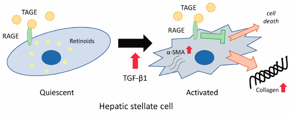
Advanced glycation end-products (AGEs) are produced by the non-enzymatic reaction of sugars with proteins. It has been revealed that glyceraldehyde-derived toxic AGEs (TAGE) are elevated with the non-alcoholic steatohepatitis (NASH) that causes such as liver fibrosis. Liver fibrosis is caused by activation of hepatic stellate cells (HSCs). Herein, it was found that the suppression of apoptosis in activated LX-2 cells which are HSCs by TGF-β1 and TAGE co-treatment is related to an increase in the production of the extracellular matrix such as collagen Ⅰ. This result suggests that TAGE might aggravate the liver fibrosis of chronic hepatitis, such as NASH.
Download PDF (856K) Full view HTML -
2021 Volume 44 Issue 1 Pages 118-124
Published: January 01, 2021
Released on J-STAGE: January 01, 2021
 Editor's pick
This retrospective observational cohort study evaluated the chronological order of acute kidney injury (AKI) and hypokalemia with Yokukansan preparation in 258 patients. Among the patients with AKI and hypokalemia, 92% developed hypokalemia associated with Yokukansan preparation after AKI. Excluding one patient with hypokalemia before AKI, the incidence of hypokalemia was higher in patients with AKI than in those without AKI (32% vs. 12%; p = 0.005). Furthermore, the prevalence of hypokalemia differed according to AKI recovery (AKI with recovery, 36%; AKI without recovery, 22%; and no AKI, 12%; p = 0.014).
Download PDF (487K) Full view HTML -
2021 Volume 44 Issue 1 Pages 125-130
Published: January 01, 2021
Released on J-STAGE: January 01, 2021
 Download PDF (1354K) Full view HTML
-
2021 Volume 44 Issue 1 Pages 131-135
Published: January 01, 2021
Released on J-STAGE: January 01, 2021
 Download PDF (896K) Full view HTML
 -
2021 Volume 44 Issue 1 Pages 136-139
Published: January 01, 2021
Released on J-STAGE: January 01, 2021
 Download PDF (482K) Full view HTML
 -
2021 Volume 44 Issue 1 Pages 140-143
Published: January 01, 2021
Released on J-STAGE: January 01, 2021
 Download PDF (1729K) Full view HTML
 -
2021 Volume 44 Issue 1 Pages 144-149
Published: January 01, 2021
Released on J-STAGE: January 01, 2021
 Download PDF (4317K) Full view HTML
 -
2021 Volume 44 Issue 1 Pages 150-153
Published: January 01, 2021
Released on J-STAGE: January 01, 2021
 Download PDF (593K) Full view HTML
 -
2021 Volume 44 Issue 1 Pages 154-157
Published: January 01, 2021
Released on J-STAGE: January 01, 2021
Advance online publication: October 30, 2020 Download PDF (736K) Full view HTML
- |<
- <
- 1
- >
- >|