Biological and Pharmaceutical Bulletin
Online ISSN : 1347-5215
Print ISSN : 0918-6158
ISSN-L : 0918-6158
Volume 41, Issue 5
Displaying 1-24 of 24 articles from this issue
Review
  • Kazuya Nagano
    2018 Volume 41 Issue 5 Pages 663-669
    Published: May 01, 2018
    Released on J-STAGE: May 01, 2018
    JOURNAL FREE ACCESS FULL-TEXT HTML

    Proteomics-based analyses are powerful means of identifying potentially useful proteins in the initial stage of drug development. Technological developments in the field of proteomics, and increases in the sensitivity of MS analyses, now facilitate identification and examination of increasingly small amounts of proteins that are differentially expressed in diseased versus normal tissues and can be candidate biomarkers or therapeutic targets. However, the current approach is for candidate proteins to be prioritized by research interest and then validated one by one; this is very inefficient. To address this issue, we have developed what we refer to as “antibody proteomics technology,” which uses a phage antibody library and tissue microarray analysis to rapidly and comprehensively isolate monoclonal antibodies against candidate proteins for the identification of potential biomarkers and therapeutic targets. In our validation of this technology, we successfully identified oxysterol binding protein-like 5 and calumenin as potential biomarkers related to metastasis in lung cancer, annexin A4 as a potential biomarker related to cisplatin resistance in malignant mesothelioma, and Eph receptor A10 as a potential therapeutic target in breast cancer, including refractory breast cancer. These findings suggest that antibody proteomics technology has the potential to become a fundamental technology in drug discovery for the development of novel biomarkers and therapeutic targets.

    Graphical Abstract Fullsize Image
Regular Articles
  • Yong Zhang, Can Wang, Bin Yu, Jian-Dong Jiang, Wei-Jia Kong
    2018 Volume 41 Issue 5 Pages 670-679
    Published: May 01, 2018
    Released on J-STAGE: May 01, 2018
    JOURNAL FREE ACCESS FULL-TEXT HTML

    This study aims to investigate the protective effects of gastrodin (GSTD), a natural compound isolated from the root of Gastrodia elata BL., on ethanol-induced liver injury and apoptosis in HepG2 cells and animal models. For in vitro studies, GSTD was used to pre-treat the cells for 4 h followed by 600 mM of ethanol co-administration for 24 h. Alcoholic liver disease (ALD) of Sprague–Dawley (SD) rats was induced by chronic ethanol-feeding plus a single dose (5 g/kg) of acute ethanol administration, GSTD at different doses were co-administered for 8 weeks. For acute liver injury experiment of ICR mice, GSTD (100 mg/kg/d) was pre-treated for 3 d followed by ethanol administration (5 g/kg) for 3 times. The results showed that GSTD protects HepG2 cells from ethanol-induced toxicity, injury, and apoptosis significantly. Co-administered with ethanol, GSTD prevented the loss of mitochondrial membrane potential, reduced the release cytochrome c from mitochondria, and inhibited the activation of caspase-3 in HepG2 cells. In SD rats induced by chronic ethanol-feeding, GSTD significantly restored liver function and ameliorated pathological changes of the liver. In rat liver, GSTD greatly suppressed the activation of caspase-3 and inhibited hepatocellular apoptosis. In ethanol-induced acute liver injury of ICR mice, GSTD reduced liver acetaldehyde and suppressed the up-regulation of alcohol dehydrogenase (ADH) and CYP2E1 significantly. Our results demonstrate that GSTD is efficacious in protecting liver cells from ethanol-induced injury and apoptosis; it may be useful for the development of novel agents for the treatment of ALD in the future.

    Graphical Abstract Fullsize Image
  • Joo Hyoun Kim, Chae Young Kim, Bobin Kang, Jungil Hong, Hyeon-Son Choi
    2018 Volume 41 Issue 5 Pages 680-689
    Published: May 01, 2018
    Released on J-STAGE: May 01, 2018
    JOURNAL FREE ACCESS FULL-TEXT HTML

    The aim of this study was to investigate the effects of dibenzoylmethane (1,3-diphenyl-1,3-propanedione, DBM) from licorice roots on lipid accumulation and reactive oxygen species (ROS) production in 3T3-L1 cells. DBM effectively inhibited lipid accumulation during adipogenesis, and its inhibitory effect was shown to be due to the down-regulation of adipogenic factors such as CCAAT-enhancer-binding protein-α (C/EBPα), peroxisome proliferator-activated receptor γ (PPARγ), and fatty acid-binding protein 4 (FABP4). DBM was observed to exert its inhibitory effect on lipid accumulation in the early adipogenic stage (days 0–2) by regulating early adipogenic factors including CCAAT-enhancer-binding protein-β (C/EBPβ) and Krueppel-like factor (KLF) 2. DBM significantly increased the translocation of nuclear factor (erythroid-derived 2)-like 2(Nrf2) into the nucleus, promoting the protein expression of its target gene, heme oxygenase-1 (HO-1). DBM significantly suppressed the insulin-mediated activation of phosphoinositide 3-kinase (PI3K) and protein kinase B (Akt), which are components of insulin signaling. In addition, intracellular ROS production was effectively reduced by DBM treatment, which upregulated antioxidant genes such as glutathione peroxidase (Gpx), catalase (CAT), and superoxide dismutase 1 (SOD1). Furthermore, DBM significantly regulated the expression of the adipokines, resistin and adiponectin. This DBM-mediated regulation of lipid accumulation, ROS production, and adipokine production was shown to be involved in the regulation of the Nrf2 and insulin signaling.

    Graphical Abstract Fullsize Image
  • Chuiliang Liu, Zhipeng Li, Zeqi Huang, Kun Zhang, Chuwen Hu, Zhiyi Zuo ...
    2018 Volume 41 Issue 5 Pages 690-696
    Published: May 01, 2018
    Released on J-STAGE: May 01, 2018
    Advance online publication: February 21, 2018
    JOURNAL FREE ACCESS FULL-TEXT HTML

    The purpose of this study was to determine the effects of different concentrations of ligustrazine, an extract from Chinese herb, on ketamine requirement for hypnosis and analgesia in mice. In the hypnotic response study, mice were randomly allocated to receive saline or ligustrazine at 10, 20, 40, 80 or 160 mg·kg−1 by intraperitoneal injection. Ketamine was administrated 15 min after ligustrazine injection. The hypnotic response was determined by assessing loss of the righting reflex (LORR) after ketamine injection. The dose of ketamine was determined by modified Dixon’s up-and-down method in each group. In the analgesia study, different doses of ligustrazine were administrated 15 min before 50 mg·kg−1 ketamine injection. The analgesia effects (pain threshold) were determined by heat radiation-induced tail-flick latency and evaluated before ligustrazine administration or 5, 15, 30 and 60 min after ketamine administration. The ED50 [95% confidence interval (CI)] for hypnosis induced by ketamine was 54.1 (44.8, 65.3) mg·kg−1. Ligustrazine dose-dependently decreased the ED50 for ketamine to induce hypnosis, which was [31.6 (26.2, 38.1)] mg·kg−1 with the addition of 80 mg·kg−1 ligustrazine and [27.7 (22.6, 33.7)] mg·kg−1 with the addition of 160 mg·kg−1 ligustrazine, respectively (p<0.05). Ligustrazine at 160 mg·kg−1 also increased pain threshold in the presence of ketamine. Ligustrazine enhanced the hypnotic effect of ketamine in a dose-dependent manner. Ligustrazine at a large dose also increased the analgesic effect of ketamine.

    Graphical Abstract Fullsize Image
  • Yuma Ishizaki, Tomomi Furihata, Yusuke Oyama, Kayoko Ohura, Teruko Ima ...
    2018 Volume 41 Issue 5 Pages 697-706
    Published: May 01, 2018
    Released on J-STAGE: May 01, 2018
    JOURNAL FREE ACCESS FULL-TEXT HTML

    Carboxylesterase 2 (CES2), which is a member of the serine hydrolase superfamily, is primarily expressed in the human small intestine, where it plays an important role in the metabolism of ester-containing drugs. Therefore, to facilitate continued progress in ester-containing drug development, it is crucial to evaluate how CES2-mediated hydrolysis influences its intestinal permeability characteristics. Human colon carcinoma Caco-2 cells have long been widely used in drug permeability studies as an enterocyte model. However, they are not suitable for ester-containing drug permeability studies due to the fact that Caco-2 cells express CES1 (which is not expressed in human enterocytes) but do not express CES2. To resolve this problem, we created a new Caco-2 cell line carrying the human small intestine-type CES expression profile. We began by introducing short-hairpin RNA for CES1 mRNA knockdown into Caco-2 cells to generate CES1-decifient Caco-2 cells (Caco-2CES1KD cells). Then, we developed Caco-2CES1KD cells that stably express CES2 (CES2/Caco-2CES1KD cells) and their control Mock/Caco-2CES1KD cells. The results of a series of functional expression experiments confirmed that CES2-specific activity, along with CES2 mRNA and protein expression, were clearly detected in our CES2/Caco-2CES1KD cells. Furthermore, we also confirmed that CES2/Caco-2CES1KD cells retained their tight junction formation property as well as their drug efflux transporter functions. Collectively, based on our results clearly showing that CES2/Caco-2CES1KD cells carry the human intestinal-type CES expression profile, while concomitantly retaining their barrier properties, it can be expected that this cell line will provide a promising in vitro model for ester-containing drug permeability studies.

    Graphical Abstract Fullsize Image
  • Zhang Yi-wen, Bao Mei-hua, Lou Xiao-ya, Cheng Yu, Yu Jing, Zhou Hong-h ...
    2018 Volume 41 Issue 5 Pages 707-712
    Published: May 01, 2018
    Released on J-STAGE: May 01, 2018
    JOURNAL FREE ACCESS FULL-TEXT HTML

    Oridonin, the major terpene found in Rabdosia rubescens, is widely used as dietary supplement or therapeutic drug, while the effects of oridonin on CYP450 were still unclear. The pregnane X receptor (PXR) is an important regulatory factor for major drug metabolism enzyme CYPs, and it has been reported to have species-specific differences. Therefore, this study has employed more reliable models PXR-humanized mouse to investigate the influence of oridonin on PXR and downstream metabolism enzyme. Eight-week-old male PXR-humanized mice were treated with oridonin by orally (0, 25, 50, 100, 200 mg/kg) for 15 d. The effects of oridonin on major downstream CYPs of PXR were examined at both the mRNA and enzyme activity levels by RT-PCR and HPLC-MS/MS. In general, there was no significant toxic reaction in liver of PXR-humanized mice. The mRNA expression of CYPs and cytochrome P450 oxidoreductase (POR) were increased with oridonin treatment in a dose-dependent manner. CYP2c and CYP3a family catalytic activity were increased significantly in two higher doses groups. These results indicate that oridonin induced the expression and activation of CYP2c and CYP3a family, which might contribute to potential drug–drug interactions and appear to be a risk when co-administered with other clinical drugs.

    Graphical Abstract Fullsize Image
  • Min Yeong Kim, Hyun Hwang Bo, Eun Ok Choi, Da He Kwon, Hong Jae Kim, K ...
    2018 Volume 41 Issue 5 Pages 713-721
    Published: May 01, 2018
    Released on J-STAGE: May 01, 2018
    JOURNAL FREE ACCESS FULL-TEXT HTML

    The fruit of Citrus unshiu MARKOVICH used for various purposes in traditional medicine has various pharmacological properties including antioxidant, anti-inflammatory, and antibacterial effects. Recently, the possibility of anti-cancer activity of the extracts or components of this fruit has been reported; however, the exact mechanism has not yet been fully understood. In this study, we evaluated the anti-proliferative effect of water extract of C. unshiu peel (WECU) on human breast cancer MCF-7 cells and investigated the underlying mechanism. Our results showed that reduction of MCF-7 cell survival by WECU was associated with the induction of apoptosis. WECU-induced apoptotic cell death was related to the activation of caspase-8 and -9, representative initiate caspases of extrinsic and intrinsic apoptosis pathways, respectively, and increase in the Bax : Bcl-2 ratio accompanied by cleavage of poly(ADP-ribose) polymerase (PARP). WECU also increased the mitochondrial dysfunction and cytosolic release of cytochrome c. In addition, AMP-activated protein kinase (AMPK) and its downstream target molecule, acetyl-CoA carboxylase, were activated in a concentration-dependent manner in WECU-treated cells. In contrast, compound C, an AMPK inhibitor, significantly inhibited WECU-induced apoptosis, while inhibiting increased expression of Bax and decreased expression of Bcl-2 by WECU and inhibition of WECU-induced PARP degradation. Furthermore, WECU provoked the production of reactive oxygen species (ROS); however, the activation of AMKP and apoptosis by WECU were prevented, when the ROS production was blocked by antioxidant N-acetyl cysteine. Therefore, our data indicate that WECU suppresses MCF-7 cell proliferation by activating the intrinsic and extrinsic apoptosis pathways through ROS-dependent AMPK pathway activation.

    Graphical Abstract Fullsize Image
  • Yuichi Yokoyama, Yoshifumi Sasaki, Natsuko Terasaki, Taku Kawataki, Ko ...
    2018 Volume 41 Issue 5 Pages 722-732
    Published: May 01, 2018
    Released on J-STAGE: May 01, 2018
    Advance online publication: February 14, 2018
    JOURNAL FREE ACCESS FULL-TEXT HTML
    Supplementary material

    Differentiated HepaRG cells maintain liver-specific functions such as drug-metabolizing enzymes. In this study, the feasibility of HepaRG cells as a human hepatocyte model for in vitro toxicity assessment was examined using selected hepatotoxic compounds. First, basal drug-metabolizing enzyme activities (CYP1A2, CYP2B6, CYP2C9, CYP2C19, CYP2D6, CYP3A4, uridine 5′-diphospho-glucuronosyltransferase [UGT], and sulfotransferases [SULT]) were measured in HepaRG, human hepatocytes, and HepG2 cells. Enzyme activities in differentiated HepaRG cells were comparable to those in human hepatocytes and much higher than those in HepG2 cells, except for SULT activity. Second, we examined the cytotoxicity of hepatotoxic compounds, acetaminophen (APAP), aflatoxin B1 (AFB1), cyclophosphamide (CPA), tamoxifen (TAM), and troglitazone (TGZ) in HepaRG cells and human hepatocytes. AFB1- and CPA-induced cytotoxicities against HepaRG cells were comparable to those against human hepatocytes. Furthermore, the cytotoxicities of these compounds were inhibited by 1-aminobenzotriazole (ABT), a broad CYP inhibitor, in both cells and were likely mediated by metabolic activation by CYP. Finally, toxicogenomics analysis of HepG2 and HepaRG cells after exposure to AFB1 and CPA revealed that numerous p53-related genes were upregulated- and the expression of these genes was greater in HepaRG than in HepG2 cells. These results suggest that gene expression profiles of HepaRG cells were affected more considerably by the toxic mechanisms of AFB1 and CPA than the profiles of HepG2 cells were. Therefore, our investigation shows that HepaRG cells could be useful human hepatic cellular models for toxicity studies.

    Graphical Abstract Fullsize Image
  • Sherif E. Emam, Hidenori Ando, Amr Selim Abu Lila, Taro Shimizu, Masam ...
    2018 Volume 41 Issue 5 Pages 733-742
    Published: May 01, 2018
    Released on J-STAGE: May 01, 2018
    JOURNAL FREE ACCESS FULL-TEXT HTML
    Supplementary material

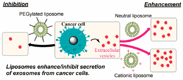
    Exosomes are tiny extracellular vesicles that are usually harvested in small quantities. Such small yield has been an obstacle for the expansion of the basic research regarding exosome analysis and applications in drug delivery. To increase exosome yield, we attempted to stimulate tumor cells via the addition of liposomes in vitro. Neutral, cationic-bare or PEGylated liposomes were incubated with four different tumor cell lines. The stimulatory effect of liposomal formulations on exosome secretion and cellular uptake propensity of the collected exosome by mother cells or different cells was evaluated. Both neutral and cationic-bare liposomes enhanced exosome secretion in a dose-dependent manner. Fluid cationic liposomes provided the strongest stimulation. Surprisingly, the PEGylation of bare liposomes diminished exosome secretion. Exosomes harvested in the presence of fluid cationic liposomes showed increased cellular uptake, but solid cationic liposomes did not. Our findings indicate that the physicochemical properties of liposomes determine whether they will act as a stimulant or as a depressant on exosome secretion from tumor cells. Liposomal stimulation may be a useful strategy to increase exosome yield, although further preparation to increase the purity of exosomes may be needed. In addition, fine-tuning of the biological properties of induced exosomes could be achieved via controlling the physicochemical properties of the stimulant liposomes.

    Graphical Abstract Fullsize Image
  • Yukitoshi Nagahara, Kei Kawakami, Abudubari Sikandan, Daiki Yagi, Ryo ...
    2018 Volume 41 Issue 5 Pages 743-748
    Published: May 01, 2018
    Released on J-STAGE: May 01, 2018
    JOURNAL FREE ACCESS FULL-TEXT HTML

    Sphingolipids are putative intracellular signal mediators in cell differentiation, growth inhibition, and apoptosis. Especially, sphingoid base-backbones of sphingolipids (sphingosine, sphinganine, and phytosphingosine) and their metabolites N-acyl-sphingoid bases (ceramides) are highly bioactive. In skin, one of the caspases, caspase-14, is expressed predominantly in cornifying epithelia, and caspase-14 plays an important role in keratinocyte differentiation. As ceramides were surrounding lipids in the keratinocytes and ceramides stimulate keratinocyte differentiation, we therefore examined the upregulation of caspase-14 by various sphingoid bases and ceramide. Sphingosine, sphinganine, phytosphingosine, and C2-ceramide treatment at the doses not damaging cells significantly increased caspase-14 mRNA and protein expression in dose-dependent manner on human keratinocyte HaCaT cells. These results indicated that sphingoid bases and ceramide upregulated caspase-14 mRNA to increase intracellular caspase-14 protein level. We next examined the caspase-14 upregulation mechanism by sphingoid bases. We used the most effective sphingoid base, phytosphingosine, and revealed that specific inhibitors of the mitogen-activated protein kinase, p38 and c-jun N-terminal protein kinase (JNK), blocked caspase-14 expression. This indicates that phytosphingosine upregulation of caspase-14 is involved of p38 and JNK activation. Moreover, phytosphingosine induced caspase-14 upregulation in vivo, suggesting that sphingoid bases were involved in keratinocyte differentiation by affecting caspase-14.

    Graphical Abstract Fullsize Image
  • Takeyoshi Wada, Takashi Hanyu, Kota Nozaki, Kosuke Kataoka, Tomoro Kaw ...
    2018 Volume 41 Issue 5 Pages 749-753
    Published: May 01, 2018
    Released on J-STAGE: May 01, 2018
    Advance online publication: March 02, 2018
    JOURNAL FREE ACCESS FULL-TEXT HTML
    Supplementary material

    Ge-132 is a synthetic organic germanium that is used as a dietary supplement. The antioxidant activity of Ge-132 on cultured mammalian cells was investigated in this study. First, Ge-132 cytotoxicity on mammalian cultured cells was determined by measuring lactate dehydrogenase (LDH) levels. Ge-132 had no cytotoxic effect on three different cell lines. Second, the cell proliferative effect of Ge-132 was determined by measuring ATP content of whole cells and counting them. Ge-132 treatment of Chinese hamster ovary (CHO-K1) and SH-SY5Y cells promoted cell proliferation in a dose-dependent manner. Finally, antioxidant activity of Ge-132 against hydrogen peroxide-induced oxidative stress was determined by measuring the levels of intracellular reactive oxygen species (ROS) and carbonylated proteins. Pre-incubation of CHO-K1 and SH-SY5Y cells with Ge-132 suppressed intracellular ROS production and carbonylated protein levels induced by hydrogen peroxide. Our results suggest that Ge-132 has antioxidant activity against hydrogen peroxide-induced oxidative stress.

    Graphical Abstract Fullsize Image
  • Yuri Ikeuchi-Takahashi, Ayaka Kobayashi, Chizuko Ishihara, Takumi Mats ...
    2018 Volume 41 Issue 5 Pages 754-760
    Published: May 01, 2018
    Released on J-STAGE: May 01, 2018
    JOURNAL FREE ACCESS FULL-TEXT HTML

    The aim of the present study was to investigate the influence of polysorbate 60 (Tween 60) on the development of morin-loaded nanoemulsions to improve the oral bioavailability of morin. Nanoemulsions were prepared using Tween 60 and polyvinyl alcohol (PVA) as emulsifiers, and medium chain triglycerides (MCT) as the lipid base. Low-saponification-degree PVA (LL-810) was also added to stabilize dispersed droplets. MCT-LL810 nanoemulsion containing LL-810 was prepared with a reduced amount of Tween 60. However, the area under the blood concentration–time curve (AUC) of MCT-LL810 (0.18) nanoemulsion containing a small amount of Tween 60 did not increase because the absorption of morin was limited by P-glycoprotein (P-gp)-mediated efflux. MCT-LL810 (0.24) nanoemulsion containing a large amount of Tween 60 showed the highest AUC, dispersed droplets containing Tween 60 may have been transported into epithelial cells in the small intestine, and P-gp transport activity appeared to be suppressed by permeated Tween 60. Based on the plasma concentration profile, dispersed droplets in MCT-LL810 (0.24) nanoemulsion permeated more rapidly through the mucus layer and the intestinal membrane than MCT (0.24) nanoemulsion without LL-810. In conclusion, a novel feature of Tween 60 incorporated into the dispersed droplets of a nanoemulsion interacting with P-gp was demonstrated herein. Dispersed droplets in MCT-LL810 (0.24) nanoemulsion containing LL-810 permeated rapidly through the mucus layer and intestinal membrane, and Tween 60 incorporated in dispersed droplets interacted with P-gp-mediated efflux, increasing the bioavailability of morin.

    Graphical Abstract Fullsize Image
  • Toshiyuki Takasu, Shoji Takakura
    2018 Volume 41 Issue 5 Pages 761-769
    Published: May 01, 2018
    Released on J-STAGE: May 01, 2018
    JOURNAL FREE ACCESS FULL-TEXT HTML

    Ipragliflozin is a selective sodium glucose cotransporter 2 (SGLT2) inhibitor that increases urinary glucose excretion and subsequently improves hyperglycemia in patients with type 2 diabetes mellitus (T2DM). To assess the beneficial effect of ipragliflozin on the mass and function of pancreatic β-cells under diabetic conditions, obese T2DM db/db mice were treated with ipragliflozin for 5 weeks. Glucose and lipid metabolism parameters, pathological changes in pancreatic islet cells and insulin content were evaluated. Pathological examination of pancreatic islet cells comprised measuring the ratios of insulin- and glucagon-positive cells and levels of oxidative stress markers. Hemoglobin A1c, plasma glucose, non-esterified fatty acid and triglyceride levels in ipragliflozin-treated groups were reduced compared to the diabetic control (DM-control) group. Histopathological examination of pancreatic islet cells revealed strong insulin staining and reduced glucagon staining in the ipragliflozin 10 mg/kg-treated group compared with the DM-control group. The ratio of α- to β-cell mass was lower in the ipragliflozin 10 mg/kg-treated group than the DM-control group and was similar to that of the non-diabetic control group. The density of immunostaining for 4-hydroxy-2-nonenal, an oxidative stress marker, in pancreatic islets was significantly lower in the ipragliflozin 10 mg/kg-treated group than the DM-control group. Pancreatic insulin content tended to be higher in the ipragliflozin-treated groups than the DM-control group. Our findings demonstrate the benefit of ipragliflozin treatment in improving glucolipotoxicity and reducing oxidative stress in pancreatic islet cells. Treatment with ipragliflozin may protect against the progressive loss of islet β-cells in patients with T2DM.

    Graphical Abstract Fullsize Image
    Editor's pick

    Ipragliflozin is a selective sodium glucose cotransporter 2 inhibitor that increases urinary glucose excretion, and subsequently improves glucolipotoxicity. The article by Takasu et al. demonstrated that the treatment with ipragliflozin decreased pancreatic cells positive for 4-hydroxy-2-nonenal (4HNE), an oxidative stress marker, in obese type 2 diabetes mellitus (DM) db/db mice. Histopathological examination of pancreatic islet cells revealed strong insulin staining, whereas reduced glucagon staining, accordingly pancreatic insulin content tended to be higher in the ipragliflozin 10 mg/kg-treated group compared with the DM-control group. It was demonstrated that ipragliflozin has a protective effect on the pancreas by reducing oxidative stress.

  • Atsuyoshi Nishina, Airi Miura, Masaharu Goto, Kahori Terakado, Daisuke ...
    2018 Volume 41 Issue 5 Pages 770-776
    Published: May 01, 2018
    Released on J-STAGE: May 01, 2018
    JOURNAL FREE ACCESS FULL-TEXT HTML
    Supplementary material

    Many natural products that inhibit melanogenesis, freckles, and hyperpigmentation have been selectively used in cosmetics because melanogenesis is linked to the multiple biogenesis cascades of melanin synthesis. However, some of these compounds have side effects that may result in their restriction in the future. We report here the isolation and structural elucidation of compounds extracted from Mansonia gagei and evaluate their activity on melanogenesis inhibition. We isolated five known compounds from M. gagei and identified them as mansonone E (1), mansorin I (2), populene F (3), mansonone G (4), and mansorin B (5). After evaluating the five compounds for cytotoxicity against B16 cells and inhibitory activity on α-melanocyte-stimulating hormone (α-MSH) induced melanogenesis, we determined that the cytotoxicity and melanogenesis-inhibitory effect of 1 were relatively low and high, respectively. Next, the effect of 1 on the expression of melanogenesis-related proteins was assessed; it was confirmed that 1 dose-dependently inhibited the expression levels of tyrosinase, tyrosinase-related protein 1 (TRP-1), TRP-2, cAMP response element binding protein (CREB), and microphthalmia-associated transcription factor (MITF) which were increased after stimulation by α-MSH. Furthermore, the effects of 1 on the phosphorylation levels of intracellular signaling pathway-related proteins were evaluated, and it was found that 1 dose-dependently rescued the phosphorylation of Akt and p38 mitogen-activated protein kinases (MAPK), which were up- or down-regulated after stimulation by α-MSH. In contrast, treatment with the phosphoinositide 3-kinase (PI3K)/Akt inhibitor wortmannin enhanced melanogenesis inhibition by mansonone E. Cumulatively, the data suggest that 1 suppresses α-MSH-induced melanogenesis in B16 cells by inhibiting both phosphorylation in the PI3K/Akt pathway and the expression of melanogenesis-related proteins.

    Graphical Abstract Fullsize Image
  • Asami Muraishi, Emi Haneta, Yoshiro Saito, Yutaka Hitomi, Mamoru Sano, ...
    2018 Volume 41 Issue 5 Pages 777-785
    Published: May 01, 2018
    Released on J-STAGE: May 01, 2018
    JOURNAL FREE ACCESS FULL-TEXT HTML
    Supplementary material

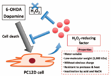
    PC12D cells, a subline of rat adrenal pheochromocytoma PC12 cells, extend neurites rapidly in response to differentiation stimuli and are used to investigate the molecular mechanisms of neurite extension. In the present study, we found significant tolerance of PC12D cells against Parkinson’s disease-related stimuli such as dopamine and 6-hydroxydopamine; this tolerance was significantly decreased by a change in the medium. Conditioned medium from PC12D cells induced tolerance against oxidative stress, which suggests that cytoprotective factor may be released by PC12D cells into the culture medium. Conditioned medium-induced tolerance was not found for PC12 cells or human neuroblastoma SH-SY5Y cells. A cytoprotective factor generated by PC12D cells exhibited hydrogen peroxide-reducing activity. Chemical characterization showed that this cytoprotective factor is water soluble and has a molecular weight about 1000 Da, and that its activity is inhibited by sodium cyanide. Release of this cytoprotective factor was increased by differentiation stimuli and oxidative stress. Taken together, these results suggest that release of a hydrogen peroxide-reducing factor by PC12D cells increases cell tolerance against oxidative stress. This study provides new insights into the antioxidative properties of factors in extracellular fluid.

    Graphical Abstract Fullsize Image
  • Maya Fujita, Takaki Yagi, Umi Okura, Jun’ichi Tanaka, Naohide Hirashim ...
    2018 Volume 41 Issue 5 Pages 786-796
    Published: May 01, 2018
    Released on J-STAGE: May 01, 2018
    JOURNAL FREE ACCESS FULL-TEXT HTML
    Supplementary material

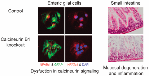
    Although calcineurin is abundantly expressed in the nervous system and involved in neurite extension and synaptic plasticity in neurons, little is known about its roles in glial cells. To investigate the roles of calcineurin in glial cells, we generated glial calcineurin B1-conditional knockout (CKO) mice and analyzed the abnormalities in the small intestine. The CKO mice were generated by crossing floxed calcineurin B1 mice with glial fibrillary acidic protein (GFAP)-Cre mice. The CKO mice exhibited growth retardation approximately from the third postnatal week and died mostly within the fourth postnatal week. The small intestine of the CKO mice was thin and hemorrhagic. The mucosal layer was degenerated and GFAP expression was reduced in the CKO small intestine. These pathological changes were associated with inflammation and increased intestinal permeability. In contrast, no apparent abnormalities were observed in the large intestine of the CKO mice. Nuclear factor of activated T cells failed to translocate into the nucleus after stimulation in enteric glial cells of the CKO small intestine. In conclusion, the calcineurin B1 deficiency in glial cells impairs the small intestine and leads to malnutrition and eventual death in mice, suggesting that calcineurin plays a novel and important role in enteric glial cells.

    Graphical Abstract Fullsize Image
  • Satomi Suzuki, Ai Tanaka, Hiroyuki Nakamura, Toshihiko Murayama
    2018 Volume 41 Issue 5 Pages 797-805
    Published: May 01, 2018
    Released on J-STAGE: May 01, 2018
    JOURNAL FREE ACCESS FULL-TEXT HTML

    Sphingolipids and their metabolic enzymes are implicated in ulcerative colitis. Ceramide kinase (CerK) catalyzes the phosphorylation of ceramide to ceramide-1-phosphate (C1P). Previous studies showed the activation of CerK by the pro-inflammatory cytokine interleukin-1β, the C1P-induced up-regulation of prostanoids exerting protective effects against colitis, and the C1P-induced down-regulation of the pro-inflammatory cytokine tumor necrosis factor-α. In order to elucidate CerK/C1P functions in colitis, we examined the severity of dextran sodium sulfate (DSS)-induced colitis in wild-type (WT) and CerK deletion (CerK(−/−)) mice. Lethal responses were observed in C57BL/6 mice treated with DSS in dose- and time-dependent manners. The depletion of CerK enhanced DSS-induced lethal responses without affecting the onset of these responses. In colons from mice treated with 2.5% DSS for 10 d, epithelial damage was significantly enhanced by the depletion of CerK by day 5, whereas decreases in occluding and E-cadherin levels were similar in both mice. On day 5, the DSS treatment increased spleen weights and colonic levels of cyclooxygenase-2, but not cytosolic phospholipase A2α, and induced a contractile dysfunction in the colons of both mice. The DSS-induced increase in the damage activity index score between days 5 and 10 was slightly enhanced and the decrease in this score from day 10 was slower in CerK(−/−) mice than in WT mice. On day 7 after the DSS treatment, spleen weights slightly decreased and increased in WT and CerK(−/−) mice, respectively. These results indicate that the depletion of CerK enhances the pathology of colitis and lethal responses in DSS-treated mice.

    Graphical Abstract Fullsize Image
Notes
  • Hiroko Maruyama, Fumitaka Kawakami, Thet-Thet Lwin, Motoki Imai, Fazel ...
    2018 Volume 41 Issue 5 Pages 806-810
    Published: May 01, 2018
    Released on J-STAGE: May 01, 2018
    JOURNAL FREE ACCESS FULL-TEXT HTML

    In this study, we examined the inhibitory effects of ferulic acid and caffeic acid on melanin production using a murine B16 melanoma cell line. The mechanisms by which the two acids inhibit melanin production were investigated by evaluating their effects on the activity of tyrosinase, which is involved is the first step of melanin biosynthesis. Ferulic acid showed no toxicity against the melanoma cells at any dose, whereas caffeic acid exerted cellular toxicity at concentrations higher than 0.35 mM. Both ferulic and caffeic acids effectively inhibited melanin production in the B16 melanoma cells. Ferulic acid reduced tyrosinase activity by directly binding to the enzyme, whereas no binding was observed between caffeic acid and tyrosinase. Both ferulic acid and caffeic acid inhibited casein kinase 2 (CK2)-induced phosphorylation of tyrosinase in a dose-dependent manner in vitro. Ferulic acid was found to be a more effective inhibitor of melanin production than caffeic acid; this difference in the inhibitory efficacy between the two substances could be attributable to the difference in their tyrosine-binding activity. Our analysis revealed that both substances also inhibited the CK2-mediated phosphorylation of tyrosinase.

    Graphical Abstract Fullsize Image
  • Yu Fukuoka, El-Sayed Khafagy, Takahiro Goto, Noriyasu Kamei, Kozo Taka ...
    2018 Volume 41 Issue 5 Pages 811-814
    Published: May 01, 2018
    Released on J-STAGE: May 01, 2018
    JOURNAL FREE ACCESS FULL-TEXT HTML

    In previous studies we showed that the complexation hydrogels based in poly(methacrylic acid-g-ethylene glycol) [P(MAA-g-EG)] rapidly release insulin in the intestine owing to their pH-dependent complexation properties; they also exhibit a high insulin-loading efficiency, enzyme-inhibiting properties, and mucoadhesive characteristics. Cell-penetrating peptides (CPPs), such as oligoarginines [hexa-arginine (R6), comprising six arginine residues], have been employed as useful tools for the oral delivery of therapeutic macromolecules. The aim of our study was to investigate the combination strategy of using P(MAA-g-EG) hydrogels with R6-based CPPs to improve the intestinal absorption of insulin. A high efficiency of loading into crosslinked P(MAA-g-EG) hydrogels was observed for insulin (96.1±1.4%) and R6 (46.6±3.8%). In addition, immediate release of the loaded insulin and R6 from these hydrogels was observed at pH 7.4 (80% was released in approximately 30 min). Consequently, a strong hypoglycemic response was observed (approximately 18% reduction in blood glucose levels) accompanied by an improvement in insulin absorption after the co-administration of insulin-loaded particles (ILP) and R6-loaded particles (ALP) into closed rat ileal segments compared with that after ILP administration alone. These results indicate that the combination of P(MAA-g-EG) hydrogels with CPPs may be a promising strategy for the oral delivery of various insulin preparations as an alternative to conventional parenteral routes.

    Graphical Abstract Fullsize Image
  • Makoto Ando, Takayuki Matsumoto, Shota Kobayashi, Maika Iguchi, Kumiko ...
    2018 Volume 41 Issue 5 Pages 815-819
    Published: May 01, 2018
    Released on J-STAGE: May 01, 2018
    JOURNAL FREE ACCESS FULL-TEXT HTML

    Hypertension is one of the most prevalent diseases worldwide and can cause harmful complications within the vascular system. Further research on vascular responsiveness to different ligands and diverse receptors in various arteries is required to understand the mechanisms underlying the development of these vascular complications. Here, we investigated the vasorelaxant effect of the protease-activated receptor 2 (PAR2) agonist 2-furoyl-LIGRLO-amide (2-Fly) and two commonest agents, namely endothelium-dependent dilator acetylcholine (ACh) and endothelium-independent dilator sodium nitroprusside (SNP), on the thoracic aorta isolated from aged spontaneously hypertensive rats (SHR) (age, 52±1 weeks). The effects of these agents were compared between aortas isolated from SHR and age-matched normotensive Wistar Kyoto (WKY) rats. Compared with the WKY group, in the SHR group, 2-Fly-induced relaxation was impaired, ACh-induced relaxation was slightly decreased at low concentrations, and SNP-induced relaxation was similar. In addition, 2-Fly-induced aortic relaxation was largely decreased by a PAR2 antagonist (FSLLRY), endothelial denudation, and a nitric oxide (NO) synthase inhibitor NG-nitro-L-arginine (L-NNA) but not by an Akt inhibitor. These results suggested that PAR2-induced relaxations of aortas of aged SHR was impaired, and this impaired aortic relaxation may be attributed to decreased NO bioavailability rather than altered NO sensitivity unrelated to the Akt activity.

    Graphical Abstract Fullsize Image
  • Tatsuo Shiba, Atsuko Yokota, Shuji Gamoh, Naoko Tanaka-Totoribe, Masac ...
    2018 Volume 41 Issue 5 Pages 820-823
    Published: May 01, 2018
    Released on J-STAGE: May 01, 2018
    Advance online publication: February 10, 2018
    JOURNAL FREE ACCESS FULL-TEXT HTML

    Studies indicate that 5-hydroxytryptamine (5-HT) released from activated platelets in coronary artery bypass grafting (CABG) induces 5-HT2A receptor-mediated graft spasm. We previously reported that 5-HT-induced constriction of human endothelium-denuded saphenous vein (SV) was significantly augmented in patients with diabetes mellitus (DM) than in patients without DM (non-DM), without changes in the levels of the membrane-bound 5-HT2A receptor of their smooth muscle cells. Although the internal thoracic artery (ITA) is the key graft conduit for CABG, the effect of DM on the ITA graft spasm is still unclear. Therefore, in this study, we investigated the effect of DM on 5-HT-induced vasoconstriction and the level of membrane-bound 5-HT2A receptor in ITA grafts. 5-HT-induced constriction of the isolated human endothelial-denuded ITA was significantly higher in patients with DM than in patients without DM. In addition, the level of the 5-HT2A receptor in the membrane fraction of human ITA smooth muscle cells was significantly higher in patients with DM than in those without DM. These results demonstrate that DM is a risk factor for CABG in both venous and arterial conduits, and that it differentially affects the level of the membrane-bound 5-HT2A receptor in the venous and arterial smooth muscle cells.

    Graphical Abstract Fullsize Image
  • Yushi Katsuyama, Norihisa Taira, Masato Yoshioka, Yuri Okano, Hitoshi ...
    2018 Volume 41 Issue 5 Pages 824-827
    Published: May 01, 2018
    Released on J-STAGE: May 01, 2018
    JOURNAL FREE ACCESS FULL-TEXT HTML

    The formation of skin pigmentation requires multiple steps, namely the activation of melanocytes, the synthesis of melanin, the transport of melanosomes to the tips of melanocyte dendrites and the transfer of melanosomes from melanocytes to surrounding keratinocytes. Recently, we reported that melanosomes accumulate in melanocytes when melanosome transport is disrupted and that they are then degraded by the autophagy system. In this study, we examined whether 3-O-glyceryl-2-O-hexyl ascorbate (VC-HG) suppresses melanogenesis through the activation of autophagy since VC-HG interferes with melanosome transport through the down-regulated expression of MyosinVa and Kinesin. The results demonstrate that VC-HG-treated B16 cells show an activation of autophagy through an increased expression level of Microtubule-associated protein 1 light chain 3 (LC3)-II and a decreased expression level of p62. Furthermore, the decrease of melanin content elicited by VC-HG was partially abolished by hydroxychloroquine or pepstatin A which are inhibitors of autophagy. Taken together, we conclude that VC-HG suppresses melanogenesis by activating the autophagy system.

    Graphical Abstract Fullsize Image
  • Yoshiro Saito, Hirofumi Misu, Hiroaki Takayama, Shin-ichiro Takashima, ...
    2018 Volume 41 Issue 5 Pages 828-832
    Published: May 01, 2018
    Released on J-STAGE: May 01, 2018
    JOURNAL FREE ACCESS FULL-TEXT HTML
    Supplementary material

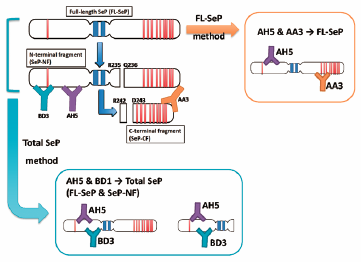
    Selenoprotein P (SeP) is a selenium (Se)-rich extracellular protein. SeP is identified as a hepatokine, causing insulin resistance in type 2 diabetes. Thus, the measurement of SeP in serum has received much attention, and several enzyme-linked immunosorbent assay (ELISA) kits for SeP determination are now commercially available. In the present study, we determined the serum SeP levels by our original ELISA and sol particle homogeneous immunoassay (SPIA) methods and also by commercially available kits, and these determinants were compared. We found a kit-dependent correlation of the determinants with our methods. These results suggest that the selection of kit is critical for comparison with our previous reports and for discussing the relationship between the serum SeP levels and disease condition.

    Graphical Abstract Fullsize Image
Errata
feedback
Top