Biological and Pharmaceutical Bulletin
Online ISSN : 1347-5215
Print ISSN : 0918-6158
ISSN-L : 0918-6158
Volume 43, Issue 9
Displaying 1-20 of 20 articles from this issue
Review
  • Hiroaki Yuasa, Tomoya Yasujima, Katsuhisa Inoue
    2020 Volume 43 Issue 9 Pages 1293-1300
    Published: September 01, 2020
    Released on J-STAGE: September 01, 2020
    JOURNAL FREE ACCESS FULL-TEXT HTML

    It has long been suggested that a Na+-dependent carrier-mediated transport system is involved in the absorption of nucleobases and analogs, including some drugs currently in therapeutic use, for their uptake at the brush border membrane of epithelial cells in the small intestine, mainly based on studies in non-primate experimental animals. The presence of this transport system was indeed proved by the recent identification of sodium-dependent nucleobase transporter 1 (SNBT1/Slc23a4) as its molecular entity in rats. However, this transporter has been found to be genetically deficient in humans and higher primates. Aware of this deficiency, we need to revisit the issue of the absorption of these compounds in the human small intestine so that we can understand the mechanisms and gain information to assure the more rational use and development of drugs analogous to nucleobases. Here, we review the current understanding of the intestinal absorption of nucleobases and analogs. This includes recent knowledge about the efflux transport of those compounds across the basolateral membrane when exiting epithelial cells, following brush border uptake, in order to complete the overall absorption process; the facilitative transporters of equilibrative nucleoside transporter 1 (ENT1/SLC29A1) and equilibrative nucleobase transporter 1 (ENBT1/SLC43A3) may be involved in that in many animal species, including human and rat, without any major species differences.

    Graphical Abstract Fullsize Image
    Editor's pick

    Knowledge of the intestinal absorption of nucleobases and analogs has accumulated, as is generally the case, mainly from studies using non-primate experimental animals. However, the recent identification of sodium-dependent nucleobase transporter 1, which plays a major role in their absorption in such animals, has led to exposing the fact that this important transporter is genetically missing in humans. To help to embark on efforts now needed to elucidate the mechanism of intestinal absorption of nucleobases and analogs in humans, which could be totally different from that in non-primate experimental animals, this article presents a comprehensive review on relevant knowledge and issues.

Communication to the Editor
  • Haitao Feng, Le Thanh Nam, Takuma Yoshikawa, Akihiro Kishimura, Takesh ...
    2020 Volume 43 Issue 9 Pages 1301-1305
    Published: September 01, 2020
    Released on J-STAGE: September 01, 2020
    JOURNAL FREE ACCESS FULL-TEXT HTML
    Supplementary material

    Enhancing blood flow to tumors is a prominent strategy for improving the tumor accumulation of macromolecular drugs through the enhanced permeability and retention (EPR) effect. IRL-1620 is an agonist of the endothelin B receptor, and is a promising molecule to enhance tumor blood flow by activating endothelial nitric oxide synthase. However, contradictory effects on tumor blood flow modulation have been reported because the effects of IRL-1620 may differ in different animal models. Here, we examined for the first time the effect of IRL-1620 on the EPR effect for PEGylated liposomes in a CT-26 murine colon cancer model. Co-injection of IRL-1620 at an optimum dose (3 nmol/kg) nearly doubled the tumor accumulation of liposomes compared with controls, indicating that IRL-1620 enhanced the EPR effect in the present colon cancer model. Co-injection of IRL-1620 is a promising strategy to improve the therapeutic effects of macromolecular drugs while reducing their side effects.

    Graphical Abstract Fullsize Image
Regular Articles
  • Chaoxin Fan, Fang Tian, Xin Zhao, Yi Sun, Xiaogai Yang, Hongbin Han, X ...
    2020 Volume 43 Issue 9 Pages 1306-1314
    Published: September 01, 2020
    Released on J-STAGE: September 01, 2020
    JOURNAL FREE ACCESS FULL-TEXT HTML
    Supplementary material

    The extracellular space (ECS) is the space between the neurons and the capillaries in the brain. The volume fraction (α) and the tortuosity (λ) are the main parameters used to describe its characteristics. Thymoquinone has been proved to possess anti-oxidant and anti-inflammatory activity. In this study, we used a gadolinium-diethylenetriaminepentacetate (Gd-DTPA)-enhanced magnetic resonance imaging (MRI) system to determine the effects of thymoquinone on ECS parameters in transient middle cerebral artery occlusion rats (tMCAO) to prove the neuroprotective effect of thymoquinone on brain tissue damage caused by ischemic stroke. Neurological examinations, 2,3,5-triphenyltetrazolium chloride (TTC) staining, hematoxylin–eosin (H&E) staining and assaying of ECS parameters using MRI were performed 24 h after surgery. We found that thymoquinone could improve the behavioural performance by neurological examinations. TTC staining indicated that thymoquinone significantly decreased the percentage of hemi-cerebral infarction. Also, H&E staining showed that thymoquinone could inhibit the neuron necrosis in the hippocampal CA1 region. We found that thymoquinone treatment could inhibit the changes in ECS diffusion parameters, which might prove that thymoquinone might protect brain tissue damage caused by ischemic stroke. Thymoquinone can protect the brain against cerebral ischemia–reperfusion injury, effectively ameliorate abnormalities in characteristics of ECS and decrease cerebral infarction in tMCAO rats.

    Graphical Abstract Fullsize Image
  • Yiwei Liu, Xia Chen, Qihai Gong, Jingshan Shi, Fei Li
    2020 Volume 43 Issue 9 Pages 1315-1323
    Published: September 01, 2020
    Released on J-STAGE: September 01, 2020
    JOURNAL FREE ACCESS FULL-TEXT HTML

    Vascular dementia (VD) is a common neurodegenerative disease, and the cognitive dysfunction is a major manifestation of VD. Lots of evidences showed that beta-amyloid (Aβ) deposition and neuroinflammation act as vital elements in the progress of VD. The previous studies showed that osthole (OST) can improve the cognitive function of VD and Alzheimer’s disease (AD). However, the effect of OST on Aβ in VD brain is still unclear. Chronic cerebral hypoperfusion (CCH) of rats were used to investigate the effect of OST on Aβ through nod-like receptor protein 3 (NLRP3) inflammasome in this study. Morris Water Maze and Y-maze were used to test the spatial learning, memory and working abilities. Hematoxylin–eosin (H&E) and Nissl staining were used to observe the morphology and number of hippocampal neurons. Immunofluorescence staining was used to observe the number of microglia activated. Western blot was used to detect the expression of proteins. The study results showed that OST obviously enhanced the spatial learning, memory and working abilities induced by modified bilateral common carotid artery occlusion (BCCAO) in rats, improved the pathological damage of hippocampal neurons induced by BCCAO in rats, inhibited the activation of microglia induced by BCCAO in rats. Furthermore, this study also discovered that OST reduced Aβ deposition in VD hippocampus via inhibition the NLRP3 inflammasome. Together, these results suggest that OST reduces Aβ deposition via inhibition NLRP3 inflammasome in microglial in VD.

    Graphical Abstract Fullsize Image
    Editor's pick

    Vascular dementia (VD) is a common neurodegenerative disease, in the progress of which neuroinflammation and beta-amyloid (Aβ) deposition act as vital elements. In this study, lots of Aβ and nod-like receptor protein 3 (NLRP3) inflammasome were found in VD rats’ brains induced by bilateral common carotid artery occlusion. A traditional Chinese medicine-Shechuangzi (osthole) extracted from the fruit of Cnidium monnieri (L.) possesses multiple pharmacological characteristics. This study has been proved the effect of osthole on VD rat evidenced by improving the behavior function, inhibiting the activation of microglia and reducing NLRP3 content, as well as decreasing the Aβ formation.

  • Ayako Tokunaga, Hirotaka Miyamoto, Shintaro Fumoto, Koyo Nishida
    2020 Volume 43 Issue 9 Pages 1324-1330
    Published: September 01, 2020
    Released on J-STAGE: September 01, 2020
    JOURNAL FREE ACCESS FULL-TEXT HTML
    Supplementary material

    The pharmacokinetics of some hepatically cleared drugs have been reported to fluctuate in patients with renal impairment, but the definitive factors have not been clarified. We compared the pharmacokinetics of some drugs with different hepatic elimination processes in a chronic kidney disease (CKD) rat model, to optimize their administration during kidney injury. We chose indocyanine green (ICG), midazolam (MDZ), and acetaminophen (APAP) as reference drugs to determine changes in hepatic clearance pathways in presence of CKD. Drugs were intravenously administered via the jugular vein to the CKD model rats, previously established by adenine administration, and then, blood, bile, and urine samples were collected. The plasma concentration of ICG, which is eliminated into the bile without biotransformation, increased; and its total body clearance (CLtot) significantly decreased in the CKD group compared to the control group. Moreover, the plasma concentrations of MDZ and APAP, metabolized in the liver by CYP3A and Ugt1a6 enzymes, respectively, were higher in the CKD group than in the control group. The biliary clearances of APAP and its derivative APAP-glucuronide increased in the CKD group, whereas their renal clearances were markedly decreased with respect to those in the control group. Altogether, plasma concentrations of some hepatically eliminated drugs increased in the CKD rat model, but depending on their pharmacokinetic characteristics. This study provides useful information for optimizing the administration of some hepatically cleared drugs in CKD patients.

    Graphical Abstract Fullsize Image
  • Atsuki Hosoda, Yoshio Matsumoto, Yuuki Toriyama, Takumi Tsuji, Yuya Yo ...
    2020 Volume 43 Issue 9 Pages 1331-1337
    Published: September 01, 2020
    Released on J-STAGE: September 01, 2020
    JOURNAL FREE ACCESS FULL-TEXT HTML

    Cisplatin (CDDP; cis-diamine dichloroplatinum)-induced nephrotoxicity is the main reason for dose limitations, which can reduce the efficacy of cancer treatment. Lower blood pressure and administration of renin angiotensin system (RAS) inhibitors have been reported as factors that exacerbate CDDP-induced nephrotoxicity; however, the detailed mechanisms remain unknown and the results of previous studies are conflicting. In this study, we examined the influence of various hypotensive drugs, including RAS inhibitors and calcium channel blockers, on CDDP-induced nephrotoxicity in BALB/c mice. The mice were divided into nine groups: (1) CDDP group (15 mg/kg CDDP), (2) AML group (5 mg/kg amlodipine), (3) ENA group (2.5 mg/kg enalapril), (4) telmisartan (TEL) group (10 mg/kg telmisartan), (5) LOS group (10 mg/kg losartan), (6) CDDP + AML group, (7) CDDP + ENA group, (8) CDDP + TEL group, and (9) CDDP + LOS group. Nephrotoxicity was evaluated by measuring serum creatinine (CRE) and blood urea nitrogen (BUN) levels. In addition, the kidney sections were stained with Masson’s trichrome and terminal deoxynucleotidyl transferase-mediated deoxyuridine triphosphate nick-end labeling (TUNEL) to assess the renal fibrosis area and apoptotic area. Serum CRE and BUN levels were increased in the CDDP + ENA, CDDP + LOS, and CDDP + TEL groups compared to those in the CDDP alone group, and the CDDP + AML group showed an increasing trend. However, there was no correlation between ∆CRE or ∆BUN levels and ∆ systolic blood pressure. The CDDP + TEL group showed a significant increase in the renal fibrosis area. These results suggest that exacerbation of CDDP-induced nephrotoxicity is not correlated with systolic blood pressure but is associated with administration of RAS inhibitors, particularly TEL.

    Graphical Abstract Fullsize Image
  • Takehiro Yamada, Shuhei Ishikawa, Nobuhisa Ishiguro, Masaki Kobayashi, ...
    2020 Volume 43 Issue 9 Pages 1338-1345
    Published: September 01, 2020
    Released on J-STAGE: September 01, 2020
    Advance online publication: June 23, 2020
    JOURNAL FREE ACCESS FULL-TEXT HTML
    Supplementary material

    Daptomycin, a cyclic lipopeptide antibiotic, has bactericidal activity against Gram-positive organisms and is especially effective against methicillin-resistant Staphylococcus aureus. Although daptomycin causes unique adverse drug reactions such as elevation of creatine phosphokinase or rhabdomyolysis, the detailed mechanisms underlying these adverse drug reactions in skeletal muscle are unclear. This study aimed to elucidate whether daptomycin causes direct skeletal muscle cell toxicity and investigate the relationship between daptomycin exposure and musculoskeletal toxicity. First, we evaluated the relationship between daptomycin exposure and skeletal muscle toxicity. Of the 38 patients who received daptomycin intravenously, an elevation in creatine phosphokinase levels was observed in five. The median plasma trough concentration of daptomycin in patients with elevated creatine phosphokinase levels was significantly higher than that in patients whose creatine phosphokinase levels were within the normal range, suggesting that increased exposure to daptomycin is related to elevation in creatine phosphokinase levels. In an in vitro study using human rhabdomyosarcoma cells, daptomycin reduced cell viability and increased membrane damage. These effects were more marked under hypoxic conditions. A necroptotic pathway seemed to be involved because phosphorylated mixed lineage kinase domain-like protein expression was enhanced following daptomycin exposure, which was significantly enhanced under hypoxic conditions. These findings indicate that daptomycin elicits cytotoxic effects against skeletal muscle cells via the necroptotic pathway, and the extent of toxicity is enhanced under hypoxic conditions.

    Graphical Abstract Fullsize Image
  • Min Cheol Pyo, Hye Soo Shin, Gyeong Yun Jeon, Kwang-Won Lee
    2020 Volume 43 Issue 9 Pages 1346-1355
    Published: September 01, 2020
    Released on J-STAGE: September 01, 2020
    JOURNAL FREE ACCESS FULL-TEXT HTML
    Supplementary material

    Ochratoxin A (OTA) is a mycotoxin produced by Aspergillus and Penicillium, and it is found in many foods. Acrylamide (AA) can be produced in foods treated at high temperatures. In this study, we investigated the combined toxicity of OTA and AA against human renal and hepatic cells in vitro. The concentration at which the synergistic effect of OTA and AA occurs was determined using the combination index obtained from the cell viability results for OTA and AA individually or in combination. The synergistic toxicity of both substances was evaluated by cell viability and the production of reactive oxygen species. In addition, apoptosis-related markers were significantly upregulated by OTA and AA individually or in combination. To determine the combined toxic effects of OTA and AA on the cells, the levels of enzymes involved in the phase I reaction and apoptosis-related markers were determined using quantitative (q)PCR and Western blot. The expression levels of CYP enzymes CYP1A1 and CYP1A2 involved in the phase I reaction significantly increased when the cells were treated with OTA and AA in combination. The expression of apoptosis-related markers, Bcl2-associated X protein (Bax) and caspase 3, also increased when the cells were treated with OTA and AA in combination. Therefore, the synergistic toxicity of OTA and AA suggests that such effects may contribute to nephrotoxicity and hepatotoxicity.

    Graphical Abstract Fullsize Image
    Editor's pick

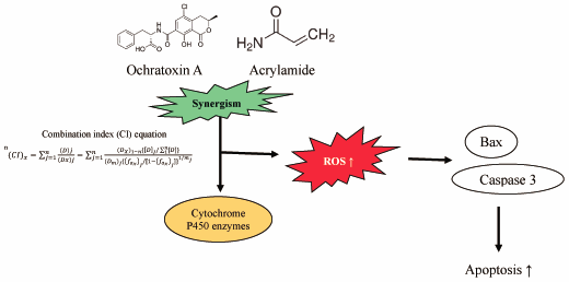
    The toxicity of each ochratoxin A and acrylamide is known but there is uncertainty about the cumulative toxicity of two compounds. In this research, Pyo et al. demonstrated that there is a synergistic relationship between ochratoxin A and acrylamide that raises oxidative stress, reduces antioxidant enzymes and causes apoptosis, exacerbating liver and kidney toxicity. These findings indicate that the risk could be increased further by the food-borne toxicant 's interaction with the toxicant produced during processing.

  • Kotaro Yamashiro, Megumi Aoki, Nobuyoshi Matsumoto, Yuji Ikegaya
    2020 Volume 43 Issue 9 Pages 1356-1360
    Published: September 01, 2020
    Released on J-STAGE: September 01, 2020
    JOURNAL FREE ACCESS FULL-TEXT HTML
    Supplementary material

    Polyherbal medicines are composed of multiple herbs and have traditionally been used in East Asian countries for the remedy of physiological symptoms. Although the effects of polyherbal formulations have been investigated at the molecular and behavioral levels, less is known about whether and how medicinal herbs affect the central nervous system in terms of neurophysiology. We introduced a novel blended herbal formulation that consisted of 35% linden, 21% mulberry, 20% lavandin, 20% butterfly pea, and 4% tulsi. After intraperitoneal administration of this formulation or saline, we simultaneously recorded epidural electrocorticograms (ECoGs) from the olfactory bulb (OB), primary somatosensory cortex (S1), and primary motor cortex (M1), along with electromyograms (EMGs) and electrocardiograms (ECGs), of rats exploring an open field arena. Using the EMGs and OB ECoGs, we segmented the behavioral states of rats into active awake, quiet awake, and sleeping states. Compared to saline, herbal medicine significantly shortened the total sleep time. Moreover, we converted the ECoG signal into a frequency domain using a fast Fourier transform (FFT) and calculated the powers at various ECoG oscillation frequencies. In the sleeping state, a slow component (0.5–3 Hz) of S1 ECoGs was significantly enhanced following the administration of the formulation, which suggests a region- and frequency-specific modulation of extracellular field oscillations by the polyherbal medicine.

    Graphical Abstract Fullsize Image
  • Tomoko Ishida, Mohammad Shahriar Khan, Honami Kodama, Yukiko Uejima, Y ...
    2020 Volume 43 Issue 9 Pages 1361-1366
    Published: September 01, 2020
    Released on J-STAGE: September 01, 2020
    JOURNAL FREE ACCESS FULL-TEXT HTML
    Supplementary material

    We examined the association of biological components in airborne particles, i.e., proteins and endotoxins, in outdoor air with asthma exacerbation in the Fukuoka metropolitan area, Fukuoka, Japan. Data on emergency department (ED) visits for asthma in children (age, 0–14 years) and adults (age, 15–64 years) were collected at a medical center from December 2014 to November 2015. One hundred eighty-one children and 143 adults visited the ED for asthma, and the weekly number of ED visits in children increased in autumn, i.e., September (second week) to November (first week). Fine (aerodynamic diameter ≤2.5 µm) and coarse (≥2.5 µm) particles were collected for 3 or 4 weeks per month, and protein and endotoxin concentrations were analyzed. Protein was largely prevalent in fine particles (0.34–7.33 µg/m3), and concentrations were high in April, May, June, and October. In contrast, endotoxin was mainly included in coarse particles (0.0010–0.0246 EU/m3), and concentrations were high in September (third week), October (first, second, and fourth weeks), February (fourth week), and July (first week). The results of a Poisson regression analysis indicated that endotoxin (in fine and coarse particles alike) was a significant factor for ED visits related to asthma in children, even after adjusting for meteorological factors, i.e., temperature, relative humidity, and wind speed. However, there was no association between environmental factors and ED visits for asthma in adults. These results suggest that endotoxin in outdoor air is significantly associated with an increased risk of asthma exacerbation in children.

    Graphical Abstract Fullsize Image
  • Zhifeng Zhao, Bin Zheng, Jinghan Li, Ziheng Wei, Sijie Chu, Xue Han, L ...
    2020 Volume 43 Issue 9 Pages 1367-1374
    Published: September 01, 2020
    Released on J-STAGE: September 01, 2020
    JOURNAL FREE ACCESS FULL-TEXT HTML

    Crocetin is a major bioactive ingredient in saffron (Crocus sativus L.) and has favorable cardiovascular effects. Here, the effects of crocetin on L-type Ca2+ current (ICa-L), contractility, and the Ca2+ transients of rat cardiomyocytes, were investigated via patch-clamp technique and the Ion Optix system. A 600 µg/mL dose of crocetin decreased ICa-L 31.50 ± 2.53% in normal myocytes and 35.56 ± 2.42% in ischemic myocytes, respectively. The current voltage nexus of the calcium current, the reversal of the calcium current, and the activation/deactivation of the calcium current was not changed. At 600 µg/mL, crocetin abated cell shortening by 28.6 ± 2.31%, with a decrease in the time to 50% of the peak and a decrease in the time to 50% of the baseline. At 600 µg/mL, crocetin abated the crest value of the ephemeral Ca2+ by 31.87 ± 2.57%. The time to half maximal of Ca2+ peak and the time constant of decay of Ca2+ transient were both reduced. Our results suggest that crocetin inhibits L-type Ca2+ channels, causing decreased intracellular Ca2+ concentration and contractility in adult rat ventricular myocytes. These findings reveal crocetin's potential use as a calcium channel antagonist for the treatment of cardiovascular disease.

    Graphical Abstract Fullsize Image
    Editor's pick

    Crocetin is a major bioactive component in saffron (Crocus sativus L.) and it has favorable cardiovascular protective effects. This study investigated the regulative effects of crocetin on L-type Ca2+ current (ICa-L), contractility, and the Ca2+ transients in rat ventricular cardiomyocytes using patch-clamp technique and Ion Optix system. The results indicated that crocetin inhibited ICa-L, intracellular Ca2+ concentration and contractility of cardiomyocytes. Crocetin (600 μg/ml) reduced cell shortening and the crest value of the ephemeral Ca2+ by 28.6 ± 2.31%, 31.87 ± 2.57%, respectively. These findings reveal that crocetin could be a potential calcium blocker for the treatment of cardiovascular disease.

  • Emiko Yoda, Keiko Hachisu, Hiroshi Kuwata, Yoshihito Nakatani, Shuntar ...
    2020 Volume 43 Issue 9 Pages 1375-1381
    Published: September 01, 2020
    Released on J-STAGE: September 01, 2020
    JOURNAL FREE ACCESS FULL-TEXT HTML

    Adipogenic differentiation is a complex process by which fibroblast-like undifferentiated cells are converted into cells that accumulate lipid droplets. We here investigated the effect of gene deletion of calcium-independent phospholipase A2γ (iPLA2γ), a membrane-bound PLA2 enzyme, on adipogenic differentiation in mice. Since iPLA2γ knockout (KO) mice showed reduced fat volume and weight, we prepared mouse embryonic fibroblasts (MEF) from wild-type (WT) and iPLA2γ KO mice and examined the effect of iPLA2γ deletion on in vitro adipogenic differentiation. iPLA2γ increased during adipogenic differentiation in WT mouse-derived MEFs, and the differentiation was partially abolished in iPLA2γ KO-derived MEFs. In KO-derived MEFs, the inductions of peroxisome proliferator activator receptor γ (PPARγ) and CAAT/enhancer-binding protein α (C/EBPα) were also reduced during adipogenic differentiation, and the reductions in PPARγ and C/EBPα expressions and the defect in adipogenesis were restored by treatment with troglitazone, a PPARγ ligand. These results indicate that iPLA2γ might play a critical role in adipogenic differentiation by regulating PPARγ expression.

    Graphical Abstract Fullsize Image
  • Takashi Ide, Izumi Origuchi
    2020 Volume 43 Issue 9 Pages 1382-1392
    Published: September 01, 2020
    Released on J-STAGE: September 01, 2020
    JOURNAL FREE ACCESS FULL-TEXT HTML

    The effects of different dietary fats on hepatic fatty acid oxidation were compared in male ICR mice and Sprague-Dawley rats. Animals were fed diets containing 100 g/kg of either palm oil (saturated fat), safflower oil (rich in linoleic acid), an oil of evening primrose origin (γ-linolenic acid, GLA oil), perilla oil (α-linolenic acid) or fish oil (eicosapentaenoic and doxosahexaenoic acids) for 21 d. GLA, perilla and fish oils, compared with palm and safflower oils, increased the activity of fatty acid oxidation enzymes in both mice and rats, with some exceptions. In mice, GLA and fish oils greatly increased the peroxisomal palmitoyl-CoA oxidation rate, and the activity of acyl-CoA oxidase and enoyl-CoA hydratase to the same degree. The effects were much smaller with perilla oil. In rats, enhancing effects were more notable with fish oil than with GLA and perilla oils, excluding the activity of enoyl-CoA hydratase, and were comparable between GLA and perilla oils. In mice, strong enhancing effects of GLA oil, which were greater than with perilla oil and comparable to those of fish oil, were confirmed on mRNA levels of peroxisomal but not mitochondrial fatty acid oxidation enzymes. In rats, the effects of GLA and perilla oils on mRNA levels of peroxisomal and mitochondrial enzymes were indistinguishable, and lower than those observed with fish oil. Therefore, considerable diversity in the response to dietary polyunsaturated fats, especially the oil rich in γ-linolenic acid and fish oil, of hepatic fatty acid oxidation pathway exists between mice and rats.

    Graphical Abstract Fullsize Image
  • Nehal E. Elsadek, Sherif E. Emam, Amr S. Abu Lila, Taro Shimizu, Hiden ...
    2020 Volume 43 Issue 9 Pages 1393-1397
    Published: September 01, 2020
    Released on J-STAGE: September 01, 2020
    JOURNAL FREE ACCESS FULL-TEXT HTML

    Protein-based therapeutics are beginning to be widely used in various clinical settings. Conjugation of polyethylene glycol (PEGylation) to protein therapeutics improves their circulation half-lives in the body. However, we and other groups observed that the initial dose of some PEGylated protein-based therapeutics may induce anti-PEG antibodies (primarily immunoglobulin M (IgM)), resulting in the accelerated clearance of a second dose. The mechanism behind the induction of anti-PEG IgM by PEGylated protein-based therapeutics is still unclear. In this study, we found that Pegfilgrastim (PEG-G-CSF, the PEGylated form of the recombinant human granulocyte colony-stimulating factor) induced anti-PEG IgM in mice when administered via either intravenous or subcutaneous administration. However, the anti-PEG IgM induction is diminished both in athymic nude mice lacking T cells and in splenectomized mice. In addition, anti-PEG IgM production was significantly diminished in the cyclophosphamide-treated mice depleted of B-cells. These results indicate that anti-PEG IgM production by Pegfilgrastim occurs in spleen in a T cell-dependent manner, which differs from anti-PEG IgM induced by PEGylated liposomes. However, B cells, both marginal zone and follicular, are essential for anti-PEG IgM production in both PEGylated preparations.

    Graphical Abstract Fullsize Image
  • Anna Iwahori, Masamitsu Maekawa, Aya Narita, Akie Kato, Toshihiro Sato ...
    2020 Volume 43 Issue 9 Pages 1398-1406
    Published: September 01, 2020
    Released on J-STAGE: September 01, 2020
    Advance online publication: June 25, 2020
    JOURNAL FREE ACCESS FULL-TEXT HTML
    Supplementary material

    Early diagnosis of Niemann–Pick diseases (NPDs) is important for better prognosis of such diseases. N-Palmitoyl-O-phosphocholine-serine (PPCS) is a new NPD biomarker possessing high sensitivity, and with its combination with sphingosylphosphocholine (SPC) it may be possible to distinguish NPD-C from NPD-A/B. In this study, a rapid liquid chromatography-tandem mass spectrometry (LC-MS/MS) method (method 1) and a validated LC-MS/MS analysis (method 2) of PPCS and SPC were developed, and we have proposed a diagnostic screening strategy for NPDs using a combination of serum PPCS and SPC concentrations. Nexera and API 5000 were used as LC-MS/MS systems. C18 columns with lengths of 10 and 50 mm were used for method 1 and 2, respectively. 2H3-Labeled PPCS and nor-SPC were used as internal standards. Selective reaction monitoring in positive-ion mode was used for MS/MS. Run times of 1.2 and 8 min were set for methods 1 and 2, respectively. In both methods 1 and 2, two analytes showed high linearity in the range of 1–4000 ng/mL. Method 2 provided high accuracy and precision in method validation. Serum concentrations of both analytes were significantly higher in NPD-C patients than those of healthy subjects in both methods. Serum PPCS correlated between methods 1 and 2; however, it was different in the case of SPC. The serum PPCS/SPC ratio was different in healthy subjects, NPD-C, and NPD-A/B. These results suggest that using a combination of the two LC-MS/MS analytical methods for PPCS and SPC is useful for diagnostic screening of NPDs.

    Graphical Abstract Fullsize Image
    Editor's pick

    Niemann-Pick diseases are classified into types A/B and C and early definitive diagnosis of them is important for better prognosis of the diseases. The authors developed a novel diagnostic screening strategy for Niemann-Pick diseases using a combination of serum concentrations of N-Palmitoyl-O-phosphocholine-serine and sphingosylphosphocholine based on analyses by liquid chromatography/tandem mass spectrometry. In this study, a rapid method and a validated analysis were developed. The former was useful for screening and the latter were useful for differentiation of Niemann–Pick diseases. This strategy may be useful for screening of Niemann-Pick diseases in clinical practice.

  • Tomoyasu Fujii, Mizuho Ogasawara, Jun Kamishikiryo, Tetsuo Morita
    2020 Volume 43 Issue 9 Pages 1407-1412
    Published: September 01, 2020
    Released on J-STAGE: September 01, 2020
    JOURNAL FREE ACCESS FULL-TEXT HTML

    The role of β-estradiol (E2) in lipoprotein metabolism in mammary tumors is unclear, therefore, we investigated the effect of E2 on the secretion of lipoprotein lipase (LPL) from mouse mammary tumor FM3A cells. E2-treated cells increased the secretion of active LPL from FM3A cells in a time- and dose-dependent manner. Activity of mitogen-activated protein kinase (MAPK) was increased in the tumor cells treated with E2, and enhanced secretion of LPL was suppressed by MAPK kinase 1/2 inhibitor, PD98059, extracellular signal-regulated kinase (ERK) 1/2 inhibitor, FR180204, p38 MAPK inhibitor, SB202190, and phosphatidyl inositol 3-kinase (PI3K) inhibitor, LY294002. In addition, the effect of E2 on LPL secretion was markedly suppressed by an inhibitor of mammalian target of rapamycin complex (mTORC) 1 and 2, KU0063794, but were not by a mTORC1 inhibitor, rapamycin. Furthermore, a small interfering RNA (siRNA)-mediated decrease in the expression of rapamycin-insensitive companion of mTOR (Rictor), a pivotal component of mTORC2, suppressed secretion of LPL by E2. These results suggest that the stimulatory secretion of LPL by E2 from the tumor cells is closely associated with an activation of mTORC2 rather than mTORC1 possibly via the MAPK cascade.

    Graphical Abstract Fullsize Image
  • Yunmei Mu, Jinting Li, Jeong-Hun Kang, Hinako Eto, Khadijah Zai, Akihi ...
    2020 Volume 43 Issue 9 Pages 1413-1420
    Published: September 01, 2020
    Released on J-STAGE: September 01, 2020
    JOURNAL FREE ACCESS FULL-TEXT HTML
    Supplementary material

    The gut–liver axis may be involved in non-alcoholic steatohepatitis (NASH) progression. Pathogen-associated molecular patterns leak through the intestinal barrier to the liver via the portal vein to contribute to NASH development. Active vitamin D3 (1,25(OH)2D3) is a potential therapeutic agent to enhance the intestinal barrier. Active vitamin D3 also suppresses inflammation and fibrosis in the liver. However, the adverse effects of active vitamin D3 such as hypercalcemia limit its clinical use. We created a nano-structured lipid carrier (NLC) containing active vitamin D3 to deliver active vitamin D3 to the intestine and liver to elicit NASH treatment. We found a suppressive effect of the NLC on the lipopolysaccharide-induced increase in permeability of an epithelial layer in vitro. Using mice in which NASH was induced by a methionine and choline-deficient diet, we discovered that oral application of the NLC ameliorated the permeability increase in the intestinal barrier and attenuated steatosis, inflammation and fibrosis in liver at a safe dose of active vitamin D3 at which the free form of active vitamin D3 did not show a therapeutic effect. These data suggest that the NLC is a novel therapeutic agent for NASH.

    Graphical Abstract Fullsize Image
Notes
  • Kojiro Kobayashi, Koji Higai, Takanori Mukozu, Daigo Matsui, Makoto Am ...
    2020 Volume 43 Issue 9 Pages 1421-1425
    Published: September 01, 2020
    Released on J-STAGE: September 01, 2020
    JOURNAL FREE ACCESS FULL-TEXT HTML

    Tivantinib, a mesenchymal–epithelial transition factor (cMET) inhibitor, is a molecular targeting drug that kills hepatocellular carcinoma (HCC) cells. Tivantinib alone does not affect the overall survival of patients with HCC, and combination treatment with tivantinib and other therapies has not been evaluated. This study was conducted to clarify the effect of the tivantinib in regulating breast cancer therapy-resistant protein (BCRP), a key transporter of 5-fluorouracil (5-FU), and dihydropyridine dehydrogenase (DPYD), a major metabolic enzyme of 5-FU. To this end, cMET gene expression was determined by RT-PCR in HepG2 (human hepatoma) cells. The transcriptional start sites of BCRP were determined by 5′-rapid amplification of cDNA ends (5′-RACE). BCRP and DPYD mRNA levels were determined by real-time RT-PCR, and promoter activities were measured by dual-luciferase assays. Results show that hepatocyte growth factor (HGF) upregulated the mRNA level of BCRP, but not DPYD, in HepG2 cells. The upregulation of BCRP expression by HGF was down-regulated by tivantinib. We also identified two transcriptional start sites (E1α, E1β) in BCRP by 5′-RACE. The transcriptional activity of the region −287 to E1α of BCRP was upregulated by HGF, which was decreased by tivantinib, whereas activity of the region −297 to E1βo f BCRP was not affected by tivantinib. Therefore, tivantinib regulates BCRP expression upstream of exon 1α. Combination treatment of tivantinib and 5-FU should be further evaluated for HCC therapy.

    Graphical Abstract Fullsize Image
  • Mai Hashimoto, Kazuaki Taguchi, Shuhei Imoto, Keishi Yamasaki, Hiroaki ...
    2020 Volume 43 Issue 9 Pages 1426-1429
    Published: September 01, 2020
    Released on J-STAGE: September 01, 2020
    JOURNAL FREE ACCESS FULL-TEXT HTML

    A nucleoside analog, 4′-cyano-2′-deoxyguanosine (CdG), which was developed as an inhibitor of the chronic hepatitis B virus (HBV), exhibited a superior antiviral activity against both wild-type and drugs-resistant HBV to marketed nucleoside analogs. In addition to previous pharmacokinetic studies of CdG in healthy rats, this study reports on an evaluation of the pharmacokinetic characteristics of CdG in a rat model of viral liver injury (VLI) induced by treatment with concanavalin A. Following an intravenous administration of CdG at a dose of 1 mg/kg, the plasma concentration profile of CdG in VLI model rats was found to be similar to that of healthy rats with no significant difference in kinetic parameters. However, when CdG was orally administered at a dose of 1 mg/kg, the maximum blood concentration was much lower in VLI model rats than in healthy rats. Interestingly, the amount of residual food in the stomachs in VLI model rats was significantly larger than that in healthy rats, indicating that the adsorption of CdG in the gastrointestinal tract was inhibited in the presence of food as well as other marketed nucleoside analogs. As observed in healthy rats, CdG was largely distributed to the liver compared to the kidney in the VLI model. These results suggest that liver pathology has only a minor effect on the pharmacokinetic properties of CdG, but the influence of food on CdG absorption needs to be considered.

    Graphical Abstract Fullsize Image
  • Masato Mashimo, Kana Murakami, Arisa Inoue, Takeshi Fujii
    2020 Volume 43 Issue 9 Pages 1430-1433
    Published: September 01, 2020
    Released on J-STAGE: September 01, 2020
    JOURNAL FREE ACCESS FULL-TEXT HTML

    Hippocampal cholinergic neurostimulating peptide (HCNP) is a secreted undecapeptide produced through proteolytic cleavage of its precursor protein, HCNPpp. Within hippocampal neurons, HCNP increases gene expression of choline acetyltransferase (ChAT), which catalyzes acetylcholine (ACh) synthesis, thereby modulating neural activity. HCNPpp also appears to be expressed in various immune cells. In the present study, we observed that HCNPpp is expressed in U937 human macrophage-like cells and that HCNP exposure suppresses lipopolysaccharide (LPS)-induced gene expression of ChAT. The opposite action is also seen in T lymphocytes, which suggest that HCNP appear to suppress cholinergic system in immune cells. In addition, HCNP suppresses LPS-induced gene expression of inflammatory enzymes including cyclooxygenase 2 (COX2) and inducible nitric oxide (NO) synthase (iNOS). The suppressive effect of HCNP may reflect suppression of mitogen-activated protein kinase (MAPK)/extracellular signal-regulated kinase (ERK) signaling activated by LPS. Thus, HCNP may have therapeutic potential as an anti-inflammatory drug.

    Graphical Abstract Fullsize Image
feedback
Top