Biological and Pharmaceutical Bulletin
Online ISSN : 1347-5215
Print ISSN : 0918-6158
ISSN-L : 0918-6158
Volume 45, Issue 11
Displaying 1-18 of 18 articles from this issue
Regular Articles
  • Tomoka Kawase, Anusuya Debnath, Tamaki Mizuno, Yui Miyake
    2022 Volume 45 Issue 11 Pages 1596-1601
    Published: November 01, 2022
    Released on J-STAGE: November 01, 2022
    JOURNAL FREE ACCESS FULL-TEXT HTML

    Vibrio vulnificus is a Gram-negative estuarine bacterium that causes infection in immuno-compromised patients, eels, and shrimp. V. vulnificus NCIMB2137, a metalloprotease-negative strain isolated from a diseased eel, produces a 45-kDa chymotrypsin-like alkaline serine protease known as VvsA. The gene encoding vvsA also includes another gene, vvsB with an unknown function; however, it is assumed to be an essential molecular chaperone for the maturation of VvsA. In the present study, we used an in vitro cell-free translation system to examine the maturation pathway of VvsA. We individually expressed the vvsA and vvsB genes and detected their mRNAs. However, the sample produced from vvsA did not exhibit protease activity. A sodium dodecyl sulfate (SDS) analysis detected the VvsB protein, but not the VvsA protein. A Western blotting analysis using a histidine (His)-tag at the amino terminus of proteins also showed no protein production by vvsA. These results suggested the translation, but not the transcription of vvsA. Factors derived from Escherichia coli were used in the in vitro cell-free translation system employed in the present study. The operon of the serine protease gene containing vvsA and vvsB was expressed in E. coli. Although serine proteases were produced, they were cleaved at different sites and no active mature forms were detected. These results indicate that the operon encoding vvsA and vvsB is a gene constructed to be specifically expressed in V. vulnificus.

  • Miyuki Yuda, Shin Aizawa, Isao Tsuboi, Yoko Hirabayashi, Tomonori Hara ...
    2022 Volume 45 Issue 11 Pages 1602-1608
    Published: November 01, 2022
    Released on J-STAGE: November 01, 2022
    JOURNAL FREE ACCESS FULL-TEXT HTML
    Supplementary material

    Lipopolysaccharide (LPS) treatment induced hemophagocytic lymphohistiocytosis in senescence-accelerated mice (SAMP1/TA-1), but not in senescence-resistant control mice (SAMR1). SAMP1/TA-1 treated with LPS exhibited functional impairment of the hematopoietic microenvironment, which disrupted the dynamics of hematopoiesis. Macrophages are a major component of the bone marrow (BM) hematopoietic microenvironment, which regulates hematopoiesis. Qualitative and quantitative changes in activated macrophages in LPS-treated SAMP1/TA-1 are thought to contribute to the functional deterioration of the hematopoietic microenvironment. Thus, we examined the polarization of pro-inflammatory (M1) and anti-inflammatory (M2) macrophages, and the dynamics of macrophage production in the BM of SAMP1/TA-1 and SAMR1 after LPS treatment. After LPS treatment, the proportions of M1 and M2 macrophages and the numbers of macrophage progenitor (CFU-M) cells increased in both SAMP1/TA-1 and SAMR1. However, compared to the SAMR1, the increase in the M1 macrophage proportion was prolonged, and the increase in the M2 macrophage proportion was delayed. The increase in the number of CFU-M cells was prolonged in SAMP1/TA-1 after LPS treatment. In addition, the levels of transcripts encoding an M1 macrophage-inducing cytokine (interferon-γ) and macrophage colony-stimulating factor were markedly increased, and the increases in the levels of transcripts encoding M2 macrophage-inducing cytokines (interleukin (IL)-4, IL-10, and IL-13) were delayed in SAMP1/TA-1 when compared to SAMR1. Our results suggest that LPS treatment led to the severely imbalanced polarization of activated M1/M2 macrophages accompanied by a prolonged increase in macrophage production in the BM of SAMP1/TA-1, which led to the impairment of the hematopoietic microenvironment, and disrupted the dynamics of hematopoiesis.

    Editor's pick

    Lipopolysaccharide (LPS) treatment induces hemophagocytic lymphohistiocytosis(HLH)-like features, including pancytopenia, in senescence-accelerated mice (SAMP1/TA-1). Prolonged hyper-inflammation in LPS-treated SAMP1/TA-1 severely impaired the hematopoietic microenvironment in the bone marrow (BM), disrupting the dynamics of hematopoiesis. Macrophages are major components of hematopoietic microenvironment, and the balance of pro-inflammatory macrophages (M1) and anti-inflammatory macrophages (M2) governs the inflammatory process. In this study, the authors showed that LPS treatment led to severely imbalanced M1 and M2 macrophage polarization and prolonged monocyte-macrophage hyper-production in the BM of SAMP1/TA-1, resulting in severe and persistent inflammation in the BM hematopoietic microenvironment, and disruption of the dynamics of hematopoiesis.

  • Yukiya Ohnishi, Daisuke Tsuji, Kohji Itoh
    2022 Volume 45 Issue 11 Pages 1609-1615
    Published: November 01, 2022
    Released on J-STAGE: November 01, 2022
    JOURNAL FREE ACCESS FULL-TEXT HTML
    Supplementary material

    Autophagy is a highly conserved intracellular degrading system and its dysfunction is considered related to the cause of neurodegenerative disorders. A previous study showed that the inhibition of endocytosis transport attenuates soluble N-ethylmaleimide-sensitive factor attachment protein receptor (SNARE) protein transport to lysosomes and block autophagy. The other studies demonstrated oxidative stress, one of the inducers of neurodegenerative diseases inhibits endocytosis transport. Thus, we hypothesized that oxidative stress-induced endocytosis inhibition causes alteration of SNARE protein transport to lysosomes and impairs autophagy. Here, we demonstrated that oxidative stress inhibits endocytosis and decreased the lysosomal localization of VAMP8, one of the autophagy-related SNARE proteins in a human neuroblastoma cell line. Moreover, this oxidative stress induction blocked the autophagosome-lysosome fusion step. Since we also observed decreased lysosomal localization of VAMP8 and inhibition of autophagosome-lysosome fusion in endocytosis inhibitor-treated cells, oxidative stress may inhibit VAMP8 trafficking by suppressing endocytosis and impair autophagy. Our findings suggest that oxidative stress-induced inhibition of VAMP8 trafficking to lysosomes is associated with the development of neurodegenerative diseases due to the blocked autophagosome-lysosome fusion, and may provide a new therapeutic target for restoring the autophagic activity.

  • Ting Zhang, Wan Lun Wang, Tong Jia Liu, Shuang Lu, Yan Chao Bian, Rui ...
    2022 Volume 45 Issue 11 Pages 1616-1626
    Published: November 01, 2022
    Released on J-STAGE: November 01, 2022
    JOURNAL FREE ACCESS FULL-TEXT HTML
    Supplementary material

    The dysregulation of certain long non-coding RNAs (lncRNAs) has been considered to be involved in neuropsychiatric disorders such as depression, implying the vital role of these transcripts. We have previously identified many differentially expressed lncRNAs in chronic unpredictable mild stress (CUMS) induced mice. Among them, lncRNA Gm16638-201 was highly expressed in the hippocampus (HIP) of CUMS, but the specific role and the underlying mechanisms remain unclear. Here, we reported that lncRNA Gm16638-201 was highly expressed in the prefrontal cortex (PFC) of CUMS induced depressive mice. Bioinformatic analysis shows that Gm16638-201 is mainly located in the cytoplasm. Nine neurological-related genes (Elmo2, Satb1, Hnrnpul1, Sipa1l3, Mapt, Tada3, Sgip1, IL-16, and StarD5) were predicted to be regulated in cis or trans by Gm16638-201 and involved into the 14-3-3Ɛ neurotrophic signaling pathway. We further confirmed the down-regulation of 14-3-3Ɛ and the nine predicted target genes in the PFC of CUMS mice except for Sgip1 and IL-16. In addition, they were also down-regulated in the primary cortical cell cultures with overexpression of Gm16638-201 constructed using an adenoviral-medicated gene expression system. In conclusion, we found that overexpression of Gm16638-201 negatively regulated several target genes and inhibited the 14-3-3Ɛ pathway in the PFC of CUMS induced depressive mice. This promising result suggests that Gm16638-201 may be a potential novel therapeutic target for depression.

  • Mana Azumi, Mikihiro Yoshie, Wataru Takano, Akari Ishida, Kazuya Kusam ...
    2022 Volume 45 Issue 11 Pages 1627-1635
    Published: November 01, 2022
    Released on J-STAGE: November 01, 2022
    JOURNAL FREE ACCESS FULL-TEXT HTML

    Eribulin, an inhibitor of microtubule dynamics, is used for treating breast cancers and sarcomas. The microtubule-destabilizing protein stathmin may modulate the antiproliferative activity of eribulin on breast cancer cells and leiomyosarcoma cells. The antitumor activity of eribulin in ovarian cancers has not been fully explored, so the present study aimed to determine the antitumor efficacy of eribulin and the involvement of stathmin in ovarian cancers. In a xenograft model of ovarian cancer, eribulin treatment reduced the tumor weight, which was accompanied by an increased level of phosphorylated stathmin. Eribulin stimulated the phosphorylation of stathmin in cultured cancer cell lines. The eribulin-induced phosphorylation of stathmin was inhibited by treatment with FTY720, an activator of protein phosphatase 2A (PP2A), and eribulin downregulated the expression of PP2A subunits. Furthermore, stathmin knockdown abrogated the inhibitory effects of eribulin on cell viability. Eribulin enhanced the antiproliferative effects of paclitaxel and concomitantly decreased stathmin expression. These results suggest that eribulin-induced phosphorylation of stathmin, mediated in part by PP2A downregulation, reduces stathmin activity and enhances the antiproliferative effects of paclitaxel in ovarian cancer. Collectively, the results of this study indicate that eribulin may suppress the proliferation of ovarian cancer cells partly by regulating the activity of stathmin.

    Editor's pick

    Stathmin, a microtubule destabilizing protein, may modulate the antiproliferative activity of eribulin, a microtubule dynamics inhibitor, in ovarian cancer. The authors investigated the function of stathmin in the antitumor effect of eribulin in ovarian cancer. In the cancer xenograft model and cultured cancer cells, eribulin treatment decreased tumor weight and increased phosphorylated stathmin mediated in part by downregulation of protein phosphatase 2A. Eribulin-induced phosphorylation of stathmin may also enhance the antiproliferative effect of paclitaxel. These results suggest that eribulin may inhibit proliferation of ovarian cancer cells in part by modulating stathmin activity.

  • Akimasa Sanagawa, Yuji Hotta, Rara Sezaki, Natsumi Tomita, Tomoya Kata ...
    2022 Volume 45 Issue 11 Pages 1636-1643
    Published: November 01, 2022
    Released on J-STAGE: November 01, 2022
    JOURNAL FREE ACCESS FULL-TEXT HTML
    Supplementary material

    In the field of cosmetic research, there is a growing interest in alternatives to animal experiments, such as in vitro models using cultured cells. The trend is spreading to the field of food and drugs. Although various types of cells are used as in vitro models, the effect of cellular senescence on the expression and function of transporters in these models is unclear. In the present study, we examined the effect of replicative senescence (by passage culture) on the expression and function of transporters in renal proximal tubular epithelial cells (RPTECs). The increase in senescence-associated-β-galactosidase (SA-β-gal)-positive cells, cell cycle arrest markers, and senescence-associated secretory phenotype (SASP) markers was associated with an increase in passage numbers of RPTECs. Gene expression of various transporters in RPTEC was also altered. The mRNA level of organic cation transporter 2 decreased most rapidly with passage numbers among the transporters. The uptake of fluorescent cationic substrates in SA-β-gal-positive RPTECs was less than that in SA-β-gal-negative RPTECs. However, these changes in the expression of transporters seem to be significantly different from those observed in rodents and human kidneys in many aspects. As cellular senescence is observed in various situations, especially in RPTECs, it may be necessary to exclude it from toxicological and pharmacokinetic evaluations using in vitro models as much as possible. Additionally, when discussing cellular senescence, it is important to note the differences between aging in cells and aging and senescence in individuals.

  • Iyuki Namekata, Haruhito Hiiro, Ryosuke Odaka, Taro Saito, Shogo Hamag ...
    2022 Volume 45 Issue 11 Pages 1644-1652
    Published: November 01, 2022
    Released on J-STAGE: November 01, 2022
    JOURNAL FREE ACCESS FULL-TEXT HTML

    The effect of blocking the persistent component of the sodium channel current (late INa) on the automatic activity of the isolated guinea pig pulmonary vein myocardium was examined. NCC-3902 blocked late INa, but did not affect other major ion channel currents stably expressed in cell lines. In isolated pulmonary vein cardiomyocytes, NCC-3902 blocked the late INa induced by a ramp depolarizing voltage clamp pulse similar to that of the pacemaker depolarization observed in the pulmonary vein myocardium. In isolated pulmonary vein tissue, NCC-3902 decreased the frequency of automatic firing of the myocardium through a reduction of the pacemaker depolarization slope. In isolated pulmonary vein cardiomyocytes, NCC-3902 significantly reduced the firing frequency of Ca2+ transients, but had no effect on Ca2+ sparks. NCC-3902 affected neither the spontaneous beating rate of the right atrium nor the contractile force of the ventricular myocardium. Selective blockers of late INa like NCC-3902, which inhibit the automatic activity of the pulmonary vein myocardium, appear to be promising as drugs for the pharmacological treatment of atrial fibrillation.

  • Xiaoyu Zhang, Zongtao Ren, Bin Liu, Shufei Wei
    2022 Volume 45 Issue 11 Pages 1653-1659
    Published: November 01, 2022
    Released on J-STAGE: November 01, 2022
    JOURNAL FREE ACCESS FULL-TEXT HTML
    Supplementary material

    Runt-related transcription factor 2 (RUNX2), a specific transcription factor of osteocytes, has been confirmed to be involved in the malignant biological behavior of various tumor cells, including renal cell carcinoma. However, the mechanism of action of RUNX2 in renal cell carcinoma cells is not yet fully understood. In this study, RUNX2-negative A498 cells and strongly positive ACHN cells were selected as the study subjects. An invasion chamber assay was used to detect the invasive ability of the cells. The expression of each protein was detected by Western blotting or immunofluorescence assays. The invasive ability of A498 cells was enhanced after the expression of RUNX2 protein was upregulated, whereas ACHN cells decreased after the expression of RUNX2 protein was silenced. The expression of calcium-activated neutral protease 2 (Calpain2) and fibronectin (FN) proteins was upregulated in A498 cells overexpressing RUNX2 protein, whereas it was downregulated after the downregulation of RUNX2 protein expression in ACHN cells. It was found that Calpain2 small interfering RNA (siRNA) or calpain inhibitor calpeptin could inhibit the expression of FN in ACHN and A498 cells overexpressing RUNX2. Calpain2 siRNA or calpeptin inhibited the invasion of A498 cells overexpressing RUNX2. Similarly, in ACHN cells, Calpain2 siRNA or calpeptin inhibited cell invasion. RUNX2 upregulates FN protein expression via Calpain2, thereby mediating renal cell carcinoma invasion.

  • Masamichi Inoue, Kyosuke Muta, Ahmed Fouad Abdelwahab Mohammed, Risako ...
    2022 Volume 45 Issue 11 Pages 1660-1668
    Published: November 01, 2022
    Released on J-STAGE: November 01, 2022
    JOURNAL FREE ACCESS FULL-TEXT HTML
    Supplementary material

    Hereditary amyloidgenic transthyretin (ATTR) amyloidosis is caused by a genetic point-mutated transthyretin such as TTR Val30Met (TTR V30M), since it forms protein aggregates called amyloid resulting in the tissue accumulation and functional disorders. In particular, ATTR produced by retinal pigment epithelial cells often causes ATTR ocular amyloidosis, which elicits deterioration of ocular function and ultimately blindness. Therefore, development of novel therapeutic agents is urgently needed. Genome-editing technology using Clustered Regularly Interspaced Short Palindromic Repeats-CRISPR associated proteins (CRISPR-Cas9) system is expected to be a therapeutic approach to treat genetic diseases, such as ATTR amyloidosis caused by a point mutation in TTR gene. Previously, we reported that glucuronylglucosyl-β-cyclodextrin conjugated with a polyamidoamine dendrimer (CDE) had excellent gene transfer ability and that underlying dendrimer inhibited TTR aggregation. Conversely, folate receptors are known to be highly expressed in retina; thus, folate has potential as a retinal target ligand. In this study, we prepared a novel folate-modified CDE (FP-CDE) and investigated its potential as a carrier for the retinal delivery of TTR-CRISPR plasmid DNA (pDNA). The results suggested that FP-CDE/TTR-CRISPR pDNA could be taken up by retinal pigment epithelial cells via folate receptors, exhibited TTR V30M amyloid inhibitory effect, and suppressed TTR production via the genome editing effect (knockout of TTR gene). Thus, FP-CDE may be useful as a novel therapeutic TTR-CRISPR pDNA carrier in the treatment of ATTR ocular amyloidosis.

    Editor's pick

    Hereditary amyloidogenic transthyretin (ATTR) ocular amyloidosis, an intractable disease, is caused by ATTR production from retinal cells. Therefore, development of novel therapeutic agents is urgently needed. In this study, folate-modified dendrimer/cyclodextrin conjugates (FP-CDE) were prepared and their ability to deliver plasmid DNA encoding the TTR-targeted genome editing CRISPR-Cas9 system (TTR-CRISPR pDNA) was investigated. As a result, FP-CDE/TTR-CRISPR pDNA complex was taken up by retinal pigment epithelial cells, exerted ATTR amyloid suppression, and inhibited TTR production through genome editing effect. Taken together, FP-CDE may be useful as a novel therapeutic TTR-CRISPR pDNA carrier in the treatment of ATTR ocular amyloidosis.

  • Bingyang Xue, Yasuyoshi Ishiwata, Yohei Kawano, Hiromitsu Takahashi, K ...
    2022 Volume 45 Issue 11 Pages 1669-1677
    Published: November 01, 2022
    Released on J-STAGE: November 01, 2022
    JOURNAL FREE ACCESS FULL-TEXT HTML

    Although paliperidone-related hyperglycemia has been extensively examined, the underlying mechanisms have not yet been elucidated. We investigated the effects of a single intravenous injection of paliperidone (0.2, 0.4, or 0.6 mg/kg) on serum concentrations of glucose and other endogenous metabolites in rats. We also examined the effects of a single intravenous injection of paliperidone (0.4 mg/kg) on AMP-activated protein kinase (AMPK) activity in the hypothalamus and liver. To clarify the relationship between AMPK activity and adrenaline secretion, the effects of berberine, which inhibits hypothalamic AMPK, on paliperidone-induced hyperglycemia were assessed. Significant increases were observed in serum glucose, adrenaline, and insulin concentrations following intravenous injections of paliperidone at doses of 0.4 and 0.6 mg/kg. A propranolol pretreatment attenuated paliperidone-induced increases in serum concentrations of glucose, but not adrenaline. Significant increases were also noted in phosphorylated AMPK concentrations in the hypothalamus following the administration of paliperidone at a dose of 0.4 mg/kg. A berberine pretreatment attenuated paliperidone-induced increases in blood concentrations of glucose, adrenaline, and insulin and phosphorylated AMPK concentrations in the hypothalamus. Collectively, the present results demonstrated that an acute treatment with paliperidone induced hyperglycemia, which was associated with the effects of hypothalamic AMPK activation on the secretion of adrenaline.

  • Daisuke Uta, Yoshihiro Inami, Miki Fukushima, Toshiaki Kume
    2022 Volume 45 Issue 11 Pages 1678-1683
    Published: November 01, 2022
    Released on J-STAGE: November 01, 2022
    JOURNAL FREE ACCESS FULL-TEXT HTML

    The skin is an important barrier that protects against invasion by foreign substances, including irritants and harmful microorganisms, and holds water in the body. Washing the skin with cleansers and shampoos containing anionic surfactants, for example sodium dodecyl sulfate (SDS), is important for maintaining skin homeostasis. However, surfactants can cause dermatitis, cutaneous hypersensitivity (e.g., alloknesis), and pruritus in humans. Our previous studies revealed an alloknesis response in the skin with SDS-induced dermatitis in C57BL/6 mice. In addition, we found that alloknesis responses and afterdischarge responses following stimulation with light touch are related because they are observed contemporaneously. In this study, we used Hos:HR-1 hairless mice to establish a mouse model to evaluate long-term drug application for alloknesis responses. Alloknesis was observed in HR-1 mice with SDS-induced dermatitis. The mean number of c-Fos (a marker of neural activity) immunopositive neurons was increased in the lamina 1-2 (L1-2) spinal dorsal horn, but not in L3-4, of SDS-treated HR-1 mice compared to vehicle-treated mice. We also discovered that afterdischarge responses were observed in neurons in L1-2. There was also a correlation between the intensity of the afterdischarge responses and depth of the recording site. Thus, the following were suggested: 1) neurons that mediate these afterdischarge responses are located on the superficial layer of the spinal cord; 2) afterdischarge responses can be an index of alloknesis responses, and 3) the mouse model of SDS-induced dermatitis is an appropriate alloknesis model.

    Editor's pick

    Cutaneous hypersensitivity (e.g., alloknesis) was observed in mice with sodium dodecyl sulfate (SDS)-induced dermatitis. Repeated application of SDS increased the expression of c-Fos-positive neurons in the superficial spinal dorsal horn (SDH). In vivo extracellular recording revealed afterdischarge responses following stimulation with light touch were also observed in the superficial SDH neurons. Authors found: 1) relation between the alloknesis responses and the afterdischarge responses, and 2) correlation between the intensity of the afterdischarge responses and depth of the recording site. These findings suggest afterdischarge responses can be an index of alloknesis responses.

  • Taiki Amano, Aya Yamamura, Moe Fujiwara, Seiji Hirai, Rubii Kondo, Yos ...
    2022 Volume 45 Issue 11 Pages 1684-1691
    Published: November 01, 2022
    Released on J-STAGE: November 01, 2022
    Advance online publication: August 20, 2022
    JOURNAL FREE ACCESS FULL-TEXT HTML

    Pulmonary arterial hypertension (PAH) is characterized by vascular remodeling of the pulmonary artery, which is mainly attributed to the excessive proliferation of pulmonary arterial smooth muscle cells (PASMCs) comprising the medial layer of pulmonary arteries. The activity of ion channels associated with cytosolic Ca2+ signaling regulates the pathogenesis of PAH. Limited information is currently available on the role of Cl channels in PASMCs. Therefore, the functional expression of ClC3 channels/transporters was herein investigated in the PASMCs of normal subjects and patients with idiopathic pulmonary arterial hypertension (IPAH). Expression analyses revealed the upregulated expression of ClC3 channels/transporters at the mRNA and protein levels in IPAH-PASMCs. Hypoosmotic perfusion (230 mOsm) evoked swelling-activated Cl currents (ICl-swell) in normal-PASMCs, whereas 100 µM 4,4′-diisothiocyanatostilbene-2,2′-disulfonic acid (DIDS) exerted the opposite effects. The small interfering RNA (siRNA) knockdown of ClC3 did not affect ICl-swell. On the other hand, ICl-swell was larger in IPAH-PASMCs and inhibited by DIDS and the siRNA knockdown of ClC3. IPAH-PASMCs grew more than normal-PASMCs. The growth of IPAH-PASMCs was suppressed by niflumic acid and DIDS, but not by 9-anthracenecarboxylic acid or T16Ainh-A01. The siRNA knockdown of ClC3 also inhibited the proliferation of IPAH-PASMCs. Collectively, the present results indicate that upregulated ClC3 channels/transporters are involved in ICl-swell and the excessive proliferation of IPAH-PASMCs, thereby contributing to the pathogenesis of PAH. Therefore, ClC3 channels/transporters have potential as a target of therapeutic drugs for the treatment of PAH.

  • Naoki Kawata, Rubii Kondo, Yoshiaki Suzuki, Hisao Yamamura
    2022 Volume 45 Issue 11 Pages 1692-1698
    Published: November 01, 2022
    Released on J-STAGE: November 01, 2022
    Advance online publication: August 20, 2022
    JOURNAL FREE ACCESS FULL-TEXT HTML

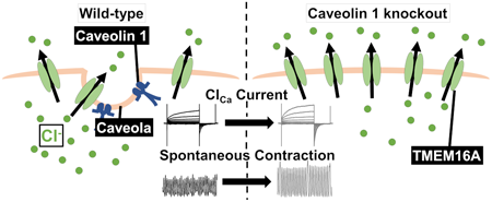
    Ca2+-activated Cl (ClCa) channels regulate membrane excitability and myogenic tone in vascular smooth muscles. TMEM16A-coding proteins are mainly responsible for functional ClCa channels in vascular smooth muscles, including portal vein smooth muscles (PVSMs). Caveolae are cholesterol-rich and Ω-shaped invaginations on the plasma membrane that structurally contributes to effective signal transduction. Caveolin 1 (Cav1) accumulates in caveolae to form functional complexes among receptors, ion channels, and kinases. The present study examined the functional roles of Cav1 in the expression and activity of ClCa channels in the portal vein smooth muscle cells (PVSMCs) of wild-type (WT) and Cav1-knockout (KO) mice. Contractile experiments revealed that the amplitude of spontaneous PVSM contractions was larger in Cav1-KO mice than WT mice. Under whole-cell patch-clamp configurations, ClCa currents were markedly inhibited by 1 µM Ani9 (a selective TMEM16A ClCa channel blocker) in WT and Cav1-KO PVSMCs. However, Ani9-sensitive ClCa currents were significantly larger in Cav1-KO PVSMCs than in WT PVSMCs. Expression analyses showed that TMEM16A expression levels were higher in Cav1-KO PVSMs than in WT PVSMs. Therefore, the caveolar structure formed by Cav1 negatively regulates the expression and activity of TMEM16A-mediated ClCa channels in vascular smooth muscle cells.

  • Yara Atef, Keita Kinoshita, Yusei Ichihara, Keisuke Ushida, Yuki Kurau ...
    2022 Volume 45 Issue 11 Pages 1699-1705
    Published: November 01, 2022
    Released on J-STAGE: November 01, 2022
    JOURNAL FREE ACCESS FULL-TEXT HTML

    Hydrogen sulfide and polysulfides are increasingly recognized as bioactive signaling molecules to produce various actions and regulate (patho)physiological processes. Here we examined the effects of sodium sulfide (Na2S) and sodium trisulfide (Na2S3) on an experimental model of intracerebral hemorrhage (ICH) in mice. Na2S or Na2S3 (25 µmol/kg, intraperitoneally (i.p.)) was administered 30 min before ICH induction by intrastriatal injection of collagenase. We found that Na2S significantly ameliorated sensorimotor functions of mice after ICH. Histopathological examinations revealed that Na2S inhibited neuron loss in the striatum, prevented axon degeneration in the internal capsule, and ameliorated axonal transport dysfunction in the striatum and the cerebral cortex where the edge of hematoma was located. Although Na2S did not suppress accumulation of activated microglia/macrophages in the peri-hematoma region, it suppressed ICH-induced upregulation of inflammatory mediators such as C-X-C motif ligand 2. On the other hand, Na2S3 did not ameliorate ICH-induced sensorimotor dysfunction. Although the effect of Na2S3 on several parameters such as axon degeneration and axonal transport dysfunction was comparable to that of Na2S, Na2S3 did not significantly inhibit neuron loss and upregulation of inflammatory mediators. These results suggest that the regulation of multiple pathological events is involved in the effect of Na2S leading to amelioration of neurological symptoms associated with ICH.

    Editor's pick

    Reactive sulfur species including monosulfides and persulfides/polysulfides are increasingly recognized to play important roles in (patho)physiological events in various organ systems. Authors compared the effects of monosulfide and trisulfide as therapeutic agents for intracerebral hemorrhage (ICH). Monosulfide alleviated neurological deficits after ICH and prevented ICH-induced neuronal death, axon degeneration and chemokine production. Trisulfide partially mimicked the effect of monosulfide and was more effective than monosulfide in suppressing recruitment of inflammatory cells, while having no effect on neurological functions. These findings underscore different pharmacological properties of individual sulfur species in regulation of brain pathology.

  • Marin Ikuse, Tatsuaki Tagami, Koki Ogawa, Tetsuya Ozeki
    2022 Volume 45 Issue 11 Pages 1706-1715
    Published: November 01, 2022
    Released on J-STAGE: November 01, 2022
    JOURNAL FREE ACCESS FULL-TEXT HTML
    Supplementary material

    Wear-resistant polymers and ceramics-based media have been used to pulverize the bulk powder of poorly water-soluble drugs to nanoscale size in conventional milling; however, contamination of such media is still an issue in the context of drug formulation manufacturing. In the present study, we developed a novel method for pulverizing the particles of a poorly water-soluble drug, ketoprofen, to nanoscale size by mixing mannitol and polypropylene glycol as a safe pulverizing medium. The ketoprofen nanoparticles were prepared using a Hoover automatic muller, equipment that traditionally has been used for the mixing of paint and ink. This process represents a novel application of this machine for the on-demand preparation of nanoparticulate formulations for use in the clinical setting. The optimal composition of the drug formulation was determined by designing an experiment consisting of the central composite design and responsive surface method. We obtained a design space that yielded ketoprofen nanoparticles with targeted particle size, poly-dispersity index, and drug release properties. We validated the manufacturing conditions by preparing ketoprofen nanoparticles in four compositions. Thus, the present study provided useful information regarding not only simple and effective contamination-free milling but also the experimental conditions need to produce nanoparticles of a poorly water-soluble drug.

Notes
  • Ayano Iwazaki, Kimie Imai
    2022 Volume 45 Issue 11 Pages 1716-1719
    Published: November 01, 2022
    Released on J-STAGE: November 01, 2022
    JOURNAL FREE ACCESS FULL-TEXT HTML

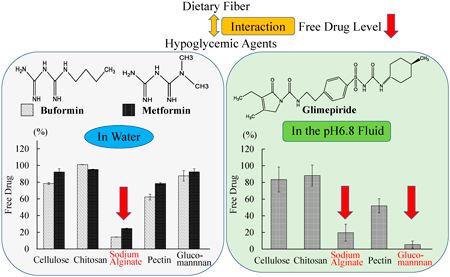
    We studied the effect of dietary fibers (DFs) on the levels of free hypoglycemic agents in vitro, i.e., glimepiride and the biguanides buformin and metformin. The levels of free buformin and free metformin were not affected by mixtures of DFs, i.e., cellulose, chitosan, pectin (PE), and glucomannan (GM), in fluids of pH 1.2 and 6.8 (similar to the pH of the stomach and intestines, respectively). However, the free biguanide level was significantly reduced by mixing with PE or sodium alginate (AL), in water. The free glimepiride level was reduced in the mixture of AL, PE, and GM (in a solution with a pH of 6.8). The changes in aqueous AL solution pH seemed to reflect the free metformin levels. Therefore, the effects of DFs on free drug levels were dependent on drug type, hypoglycemic agent, and mixing solution. In this study, the oral regimen concentrations of the drug and DFs were used. Based on these results, it is important to consider the interactions between hypoglycemic agents and DFs.

  • Shigeru Yamada, Hiroyuki Masuno, Hiroyuki Kagechika, Aya Tanatani, Yas ...
    2022 Volume 45 Issue 11 Pages 1720-1724
    Published: November 01, 2022
    Released on J-STAGE: November 01, 2022
    JOURNAL FREE ACCESS FULL-TEXT HTML

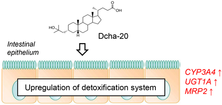
    Vitamin D is a fat-soluble micronutrient that plays essential roles in a range of biological processes, including cell proliferation, inflammation, and metabolism. In this study, we investigated the effects of a novel synthetic lithocholic acid derivative with vitamin D activity (Dcha-20) on pharmacokinetic gene expression in human induced pluripotent stem cell-derived intestinal organoids. Compared with vitamin D3 treatment, Dcha-20 was found to upregulate the expression and enzyme activity of the drug-metabolizing enzyme CYP3A4, an indicator of intestinal functional maturation. In addition, Dcha-20 specifically increased expression levels of the xenobiotic detoxification enzyme UGT1A and excretion transporter MRP2. These results suggest that Dcha-20 promotes activity of the intrinsic defense system of the intestinal epithelium.

  • Naoki Fujitani, Masayoshi Saito, Tomoya Akashi, Masashi Morita, Takano ...
    2022 Volume 45 Issue 11 Pages 1725-1727
    Published: November 01, 2022
    Released on J-STAGE: November 01, 2022
    JOURNAL FREE ACCESS FULL-TEXT HTML
    Supplementary material

    X-linked Adrenoleukodystrophy (X-ALD) is a rare genetic neurological disorder caused by a mutation of the ABCD1 gene that encodes a peroxisomal ABC protein ABCD1. ABCD1 has a role in transporting very long chain fatty acid (VLCFA)-CoA into the peroxisome for β-oxidation. ABCD1 dysfunction leads to reduced VLCFA β-oxidation and in turn increased VLCFA levels in the plasma and the cells of all tissues; these increased plasma levels have been used to diagnose X-ALD. It has been reported that plasma VLCFA is not correlated with the severity and disease phenotype of X-ALD. Therefore, we cannot predict the disease progression by the plasma VLCFA level. Cerebrospinal fluid (CSF) is constantly produced by brain, and thus levels of lipids containing VLCFA in CSF might be informative in terms of assessing X-ALD pathology. LC-MS/MS-based analysis showed that phosphatidylcholine (PC) containing VLCFA signals, such as PC 40 : 0(24 : 0/16 : 0), PC 42 : 0(26 : 0/16 : 0), PC 44 : 4(24 : 0/20 : 4) and PC 46 : 4(26 : 0/20 : 4) were characteristically detected only in the CSF from patients with X- ALD. In the present study, we analyzed limited number of patient’s CSF samples (2 patients with X-ALD) due to the limitations of the availability for CSF samples from this rare disease. However, our finding would offer helpful information for studying the disease progression biomarkers in X-ALD. To our knowledge, this is the first report of analyzing lipids containing VLCFA in CSF from patients with X-ALD.

feedback
Top