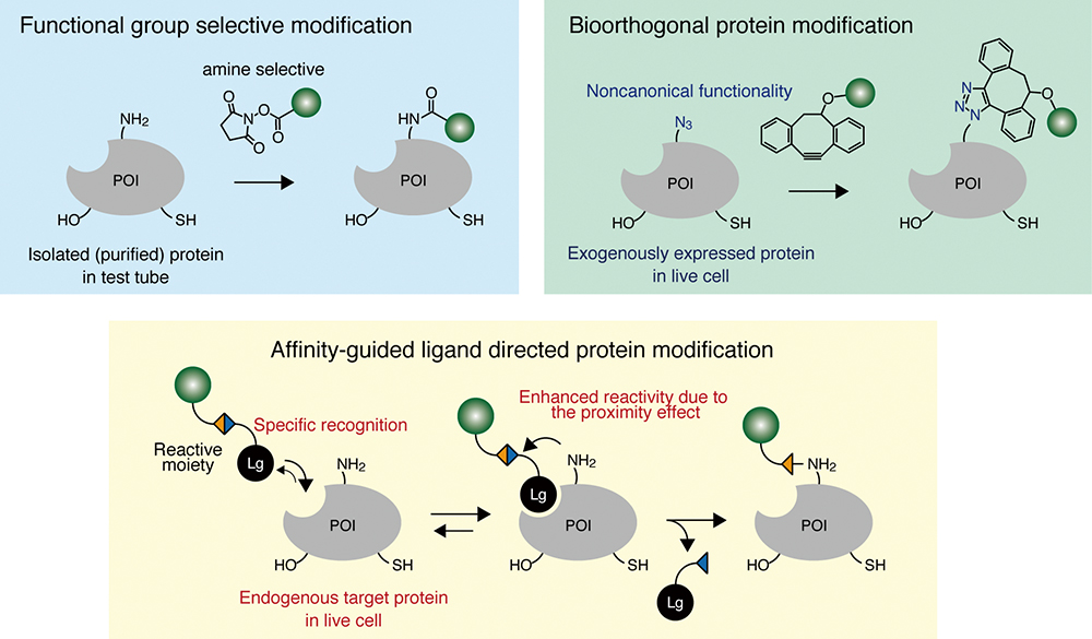
Chemical modification of proteins is important for creating a myriad of engineered proteins and for elucidating the function and dynamics of proteins in live cells. A wide variety of chemical protein modification methods have been developed and can be categorized into three classes: (i) modification of proteins using the reactivity of naturally occurring amino acids; (ii) modification by bioorthogonal reactions using unnatural amino acids, most of which can be site-selectively incorporated into proteins-of-interest using genetic codon expansion techniques; and (iii) recognition driven chemical modification, which is the only approach that allows modification of endogenous proteins without any genetic manipulation even under heavily crowded and multi-molecular conditions, as in live cells and organisms. All of these approaches have merits and limitations. In this review, we summarize these approaches and discuss their characteristics with respect to specificity, reaction rate and versatility.
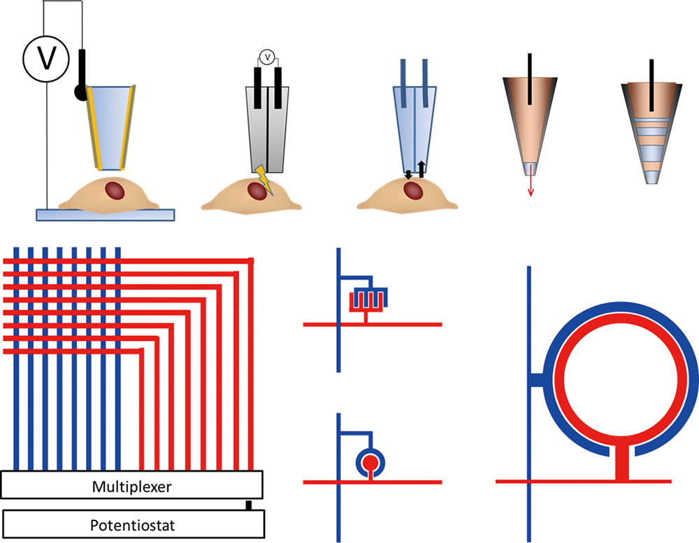
Applications of electrochemical biosensing for surveying intact cells and tissues have been focus of attention. Two experimental approaches have been used when performing amperometric measurements on biological cells, the stylus-type microelectrode probes and the electrode-integrated microdevices based on lithographic technologies. For the probe scanning approach, various types of microsensors were developed to monitor localized physical or chemical natures at a variety of surfaces in situ under wet conditions. Scanning electrochemical microscopy (SECM) has been applied for monitoring local oxygen, enzyme activity, and collection of transcripts. For the non-scanning type of approach, electrode array devices allow very rapid response, parallel monitoring, and multi-analyte assay. Sveral topics of on-chip-culture system were introduced especially concerning on gene expression monitoring by reporter system and reconstruction of in vivo-like nature by controlling microenvironments. Electrochemical reporter assay has been demonstrated to monitor the gene expression process of the gene-modified cultured cells. Long-term monitoring of cellular function of spheroids and three dimensionally-cultured cells were carried out by controlling microenvironments on the cellular chip.
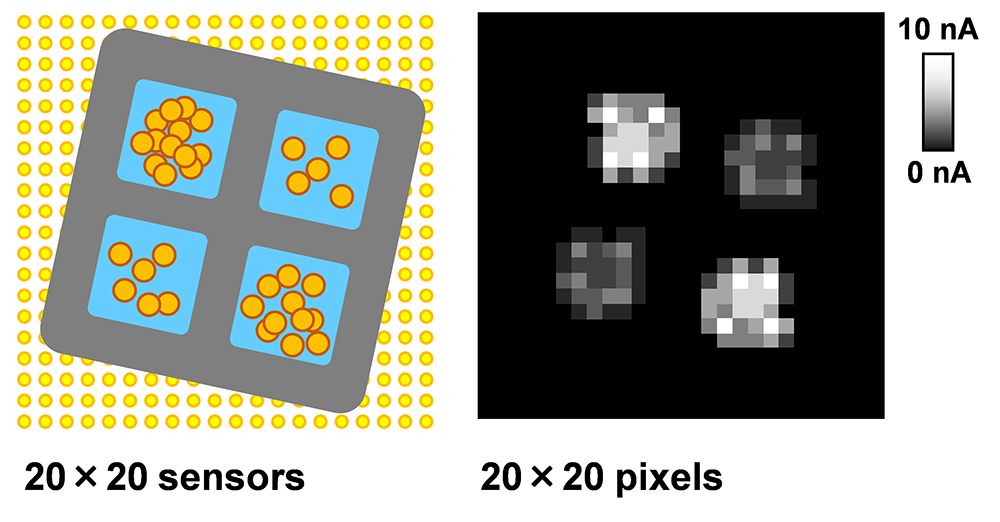
Tissue engineering requires analytical methods to monitor cell activity in hydrogels. Here, we present a method for the electrochemical imaging of cell activity in hydrogels embedded in printed polycaprolactone (PCL) scaffolds. Because a structure made of only hydrogel is fragile, PCL frameworks are used as a support material. A grid-shaped PCL was fabricated using an excluder printer. Photocured hydrogels containing cells were set at each grid hole, and cell activity was monitored using a large-scale integration-based amperometric device. The electrochemical device contains 400 microelectrodes for biomolecule detection, such as dissolved oxygen and enzymatic products. As proof of the concept, alkaline phosphatase and respiration activities of embryonic stem cells in the hydrogels were electrochemically monitored. The results indicate that the electrochemical imaging is useful for evaluating cells in printed scaffolds.
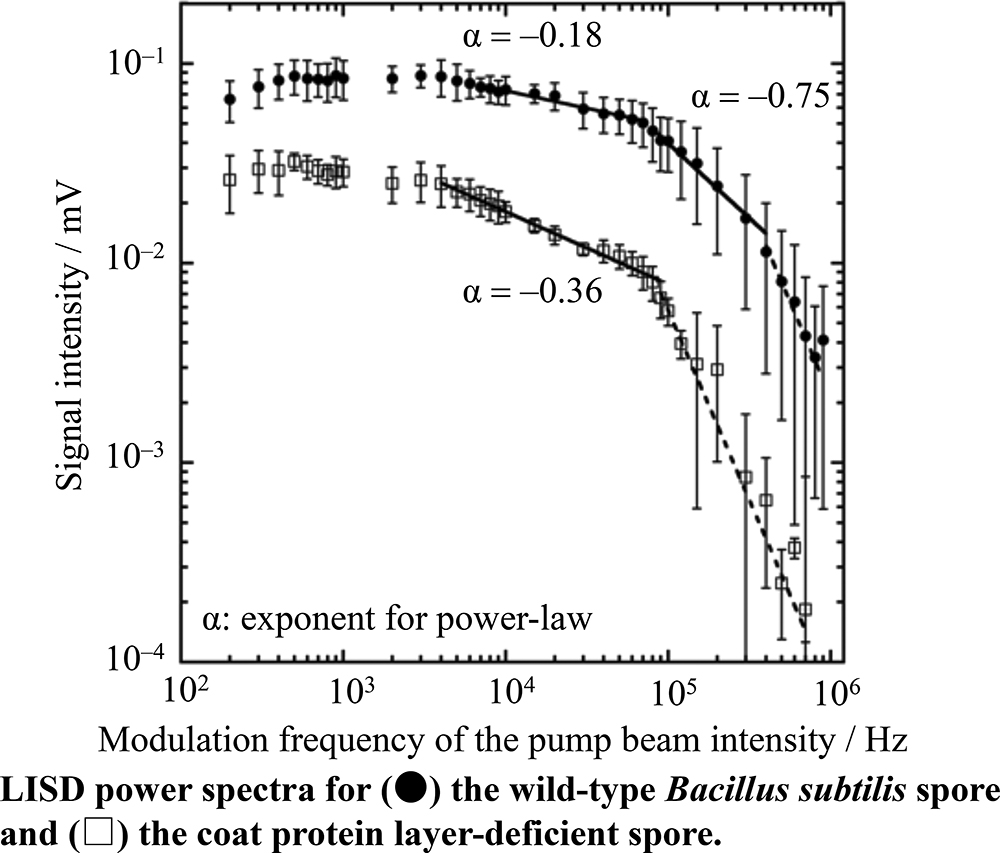
The differences in the mechanical properties between the cortex and coat protein layer in a Bacillus subtilis spore were clarified using an atomic force microscope (AFM) and an originally developed laser-induced surface deformation (LISD) microscope. AFM force curve measurements show that the Young’s modulus of the coat protein layer is ca. 66% lower compared with that of the cortex. It has been experimentally clarified that the cortex makes a greater contribution to the rigidity of spores than the coat protein layer. From comparisons of the LISD power spectra, it is revealed that the coat protein layer has two different viscoelastic regions, and that the cortex relatively has a higher viscous nature than the coat protein layer. Furthermore, the LISD power spectrum above 1 × 10⁵ Hz in the coat protein layer suggests that the local region in the coat protein layer behaves as a more elastic body compared with the cortex.
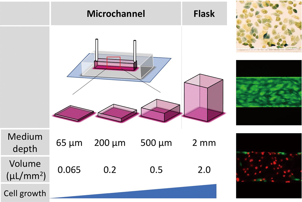
Microfluidic devices have emerged as a new cell culture tool, which can mimic the structure and physiology of living human organs. However, no standardized culture method for a microfluidic device has yet been established. Here, we describe the effects of various conditions on cell proliferation in a microchannel with a depth smaller than 100 μm. Primary endothelial cell proliferation was suppressed with a decrease in the culture medium volume per cell culture area. Moreover, cell growth was compared with or without medium flow, and the optimum culture condition was determined to be 1 μL/h flow in a 65-μm-deep microchannel. In addition, glucose consumption was greater under fluidic conditions than under static conditions, and the ability of tumor (HeLa) cells to convert glucose into lactate appeared to be higher in a static culture than that in a fluidic culture. Overall, our results will serve as a useful guide for designing a microfluidic cell culture platform in a channel smaller than 100 μm.
Membrane dynamic structures such as filopodia, lamellipodia, and ruffles have important cellular functions in phagocytosis and cell motility, and in pathological states, such as cancer metastasis. Phosphatidylinositol 3,4,5-trisphosphate (PIP3) is a crucial lipid that regulates PIP3 dynamics. Investigations of how PIP3 is involved in these functions have mainly relied on pharmacological interventions, and therefore have not generated detailed spatiotemporal information concerning membrane dynamics upon PIP3 production. In the present study, we applied an optogenetic approach using the CRY2–CIBN system. Using this system, we revealed that local PIP3 generation induced directional cell motility and membrane ruffles in COS7 cells. Furthermore, combined with structured illumination microscopy (SIM), membrane dynamics were investigated with high spatial resolution. We observed PIP3-induced apical ruffles and unique actin fiber behavior in that a single actin fiber protruded from the plasma membrane was taken up into the plasma membrane without depolymerization. This system has the potential to investigate other high-level cell motility and dynamic behaviors, such as cancer cell invasion and wound healing with high spatiotemporal resolution, and could provide new insights of biological sciences for membrane dynamics.
Epithelial-mesenchymal transition (EMT), phenotypic changes in cell adhesion and migration, is involved in cancer invasion and metastasis, hence becoming a target for anti-cancer drugs. In this study, we report a method for the evaluation of EMT inhibitors by using a photoactivatable gold substrate, which changes from non-cell-adhesive to cell-adhesive in response to light. The method is based on the geometrical confinement of cell clusters and the subsequent migration induction by controlled photoirradiation of the substrate. As a proof-of-concept experiment, a known EMT inhibitor was successfully evaluated in terms of the changes in cluster area or leader cell appearance, in response to biochemically and mechanically induced EMT. Furthermore, an application of the present method for microbial secondary metabolites identified nanaomycin H as an EMT inhibitor, potentially killing EMTed cells in disseminated conditions. These results demonstrate the potential of the present method for screening new EMT inhibitors.
As protein–protein interactions (PPI) have been mostly investigated in cellulo or in vivo, it is unclear whether the PPI-based imaging schemes are practically valid in a bioanalytical means in vitro. The present study exemplifies the PPI in vitro inside a unique single-chain probe, named TP2.4, which carries a full-length artificial luciferase (ALuc) sandwiched in between two model proteins of interest, e.g., FKBP and FRB, expressed in E. coli, and purified. We found that the TP2.4 efficiently recognizes its ligand in vitro and varies its molecular kinetics: i.e., rapamycin boosts the enzymatic affinities (Km) of TP2.4 to its substrates, but does not or only weakly influences the turnover rates (Kcat) and the maximal velocity (Vmax). The corresponding circular dichroism (CD) study shows that rapamycin weakly contributes to the enhancement of the α-helical contents in TP2.4. Kinetic constants according to the substrates revealed that a coelenterazine derivative, 6-N3-CTZ, exerted the best catalytic efficiency and the greatest variance in the total photon counts. The present study is the first in vitro example that demonstrates how intramolecular PPI works in a purified single-chain bioluminescent probe and what factors practically influence the biochemistry.
The conjugation of biomolecules, such as protein, sugar, and DNA, with metal nanoparticles is an important technique for bioassay and biomaterial preparation. In this study, we aim to enzymatically immobilize a functional peptide on gold nanoparticles (AuNPs) using a single-step reaction. We used tyrosinase, a catechol oxidase, to immobilize an enzymatic peptide. We performed immobilization experiments of a fluorescent compound-linked caspase-3 substrate peptide using tyrosinase on chitosan-coated AuNPs. Peptides were effectively immobilized onto the AuNPs depending on the presence of tyrosine within the sequence, which suggests the DOPA-quinone produced from tyrosine, via tyrosinase, is connected to the chitosan amino group. Although fluorescent emission from the immobilized capase-3 substrate was quenched by AuNPs, fluorescence intensity recovery occurred due to the addition of caspase-3. Thus, we were able to easily prepare functional AuNPs that can be used for a caspase-3 activity assay. Our results indicate that the tyrosinase-mediated peptide link to chitosan-coated particles is a useful technique for preparing functionalized nanoparticles.
A peptide-oligonucleotide conjugate (1) was synthesized by the attachment of FAM, TAMRA, and biotin moieties to a telomere DNA sequence of 5′-TAG GGT TAG GGT TAG GGT TAG GG-3′. This conjugate was induced to be an anti-parallel structure in the presence of sodium ion (Na+), whereas a hybrid one was formed under potassium ion (K+) as a monitoring by circular dichromic spectra. The conformation change of this conjugate gave an effective FRET signal change upon the addition of NaCl, compared with the case of KCl. Under 5 mM KCl as an extracellular condition, a FRET change was observed upon addition of NaCl and quantitative FRET change was observed in 0 – 250 mM NaCl. This conjugate was immobilized on the cell surface through a sugar chain on the cell, biotinyl concanavallin A and streptavidin. This conjugate was utilized for Na+ sensing based on anti-parallel tetraplex formation with Na+.
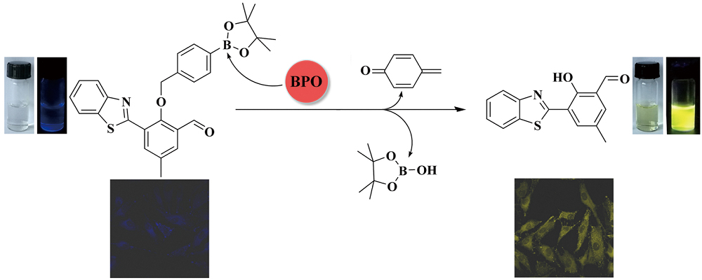
We present herein a novel ratiometric fluorescent probe (1) for benzoyl peroxide (BPO). Probe 1 was obtained by coupling the recognition unit of arylboronate to a benzothiazole-derived fluorophore. The probe solution is colorless and displays weak blue fluorescence at 460 nm. Upon the addition of BPO, the arylboronate substituent can be removed via oxidation and 1,4-elimination processes. The released fluorophore emits strong yellow-greenish fluorescence at 546 nm. The ratiometric response of the probe is highly selective and sensitive for BPO. The dynamic range was fitted over 1.0 – 75.0 μM with a detection limit of 0.26 μM. In addition, the probe was applied to quantitative detection of BPO in real samples of wheat flour and an antimicrobial agent. Cellular experiments further demonstrated that probe 1 can be effectively utilized for imaging BPO in living cells.
A differential array consisting of commercially available common fluorescent dyes was constructed for the identification of proteins and human cancer cells. Fluorescence of dyes was differently altered by mixing with proteins and human cancer cells, generating response patterns that are unique to the analytes. Linear discriminant analysis of the obtained patterns enabled the accurate identification of eight proteins and three human cancer cells. As this system can be easily prepared, it would offer a unique opportunity for array-based differential biosensing.
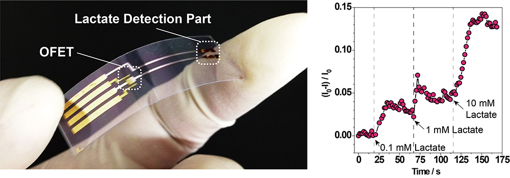
A novel flexible lactate sensor based on organic field-effect transistors (OFETs) is demonstrated. Because lactate is known as a biomarker for assessing our physical performance, wearable lactate sensors could contribute to the monitoring of human health conditions. The flexible and low-voltage operatable OFET possesses an extended-gate modified with enzymes and an osmium-redox polymer for the lactate detection, meaning that the continuous measurement of lactate levels (0 – 10 mM) has been successfully achieved. We believe that insight obtained will open up opportunities for applying OFETs in wearable biosensors.
We present an innovative concept of a screening tool for detecting free microcystin in cyanobacteria using a sandwich immunodetection format, based on Michael addition reaction between α,β-unsaturated carbonyl moiety of microcystin and thiol of coating substance. This proof-of-concept immunoassay was developed using bovine serum albumin as a microcystin-binding model, and was tested with toxic Microcystis samples. The preliminary results indicate that the proposed Michael addition-based immunodetection is promising and can be used as a platform for further development to become a useful tool for free microcystin analysis in various samples in the future.
Here, we demonstrated a strategy for developing signaling aptamers, based on screening of signaling aptamers from multiple aptamer candidates obtained by SELEX with next generation sequencing. Among aptamer candidates labelled by 6-carboxyfluorescein and quencher at both end termini, there is the possibility of discovering a potent signaling aptamer. In this study, we discovered DNA signaling aptamers against VEGFR-1. This strategy has the potential for signaling aptamer discovery without the extremely laborious task of optimization of oligodeoxynucleotide modifications.