All issues

Volume 32, Issue 1
Displaying 1-21 of 21 articles from this issue
- |<
- <
- 1
- >
- >|
-
Article type: Guest Editorial
2016 Volume 32 Issue 1 Pages 1
Published: January 10, 2016
Released on J-STAGE: January 10, 2016
JOURNAL FREE ACCESSDownload PDF (138K)
Reviews
-
Masatoshi MAEKI, Hiroshi YAMAGUCHI, Manabu TOKESHI, Masaya MIYAZAKIArticle type: Reviews
2016 Volume 32 Issue 1 Pages 3-9
Published: January 10, 2016
Released on J-STAGE: January 10, 2016
 JOURNAL FREE ACCESSThis review summarizes two microfluidic-based protein crystallization methods, protein crystallization behavior in the microfluidic devices, and their applications for X-ray crystal structure analysis. Microfluidic devices provide many advantages for protein crystallography; they require small sample volumes, provide high-throughput screening, and allow control of the protein crystallization. A droplet-based protein crystallization method is a useful technique for high-throughput screening and the formation of a single crystal without any complicated device fabrication process. Well-based microfluidic platforms also enable effective protein crystallization. This review also summarizes the protein crystal growth behavior in microfluidic devices as, is known from viewpoints of theoretical and experimental approaches. Finally, we introduce applications of microfluidic devices for on-chip crystal structure analysis.View full abstractDownload PDF (1014K)
 -
Akihide HIBARA, Mao FUKUYAMA, Myungwha CHUNG, Craig PRIEST, Mikhail A. ...Article type: Reviews
2016 Volume 32 Issue 1 Pages 11-21
Published: January 10, 2016
Released on J-STAGE: January 10, 2016
 JOURNAL FREE ACCESSFundamental aspects of rapidly advancing micro/nanofluidic devices are reviewed from the perspective of liquid interface chemistry and physics, including the influence of capillary pressure in microfluidic two-phase flows and phase transitions related to capillary condensation.View full abstractDownload PDF (5440K)
 -
Mladen FRANKO, Mingqiang LIU, Aleš BOŠKIN, Ambra DELNERI ...Article type: Reviews
2016 Volume 32 Issue 1 Pages 23-30
Published: January 10, 2016
Released on J-STAGE: January 10, 2016
 JOURNAL FREE ACCESSEfficient environment protection and human safety require high-throughput analysis techniques for pollutants or toxicants for large sample sets. State-of-the-art HPLC and GC coupled to various detecting strategies offer excellent sensitivity and selectivity, though they are quite time-extensive (2 – 3 samples/h or less when sample preparation is involved). Efforts are made towards screening techniques with high sample throughputs simultaneously providing detection limits below the maximum contaminant levels for the analyte. However, such approaches frequently sacrifice the selectivity or sensitivity (or just give a yes/no response). In this review, we demonstrate thermal-lens spectrometry and microscopy as highly sensitive spectrometric techniques in combination with flow-injection analysis (FIA) and microfluidic FIA along with lab-on-a-chip chemistry for fast screening (several samples/h and up to 20 samples/min) exemplified by organophosphates and carbamates as neurotoxigenic compounds. Various approaches to determining other topical toxicants, like microcystin and cyanopigments as its indicators, allergens, and carcinogenic chromate, are also discussed.View full abstractDownload PDF (2047K)
Original Papers
-
Kazuma OGAWA, Takashi KANETAArticle type: Original Papers
2016 Volume 32 Issue 1 Pages 31-34
Published: January 10, 2016
Released on J-STAGE: January 10, 2016
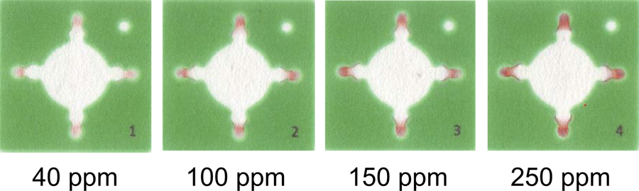 JOURNAL FREE ACCESSMicrofluidic paper-based analytical devices (μPADs) were used to detect the iron ion content in the water of a natural hot spring in order to assess the applicability of this process to the environmental analysis of natural water. The μPADs were fabricated using a wax printer after the addition of hydroxylamine into the detection reservoirs to reduce Fe3+ to Fe2+, 1,10-phenanthroline for the forming of a complex, and poly(acrylic acid) for ion-pair formation with an acetate buffer (pH 4.7). The calibration curve of Fe3+ showed a linearity that ranged from 100 to 1000 ppm in the semi-log plot whereas the color intensity was proportional to the concentration of Fe3+ and ranged from 40 to 350 ppm. The calibration curve represented the daily fluctuation in successive experiments during four days, which indicated that a calibration curve must be constructed for each day. When freshly prepared μPADs were compared with stored ones, no significant difference was found. The μPADs were applied to the determination of Fe3+ in a sample of water from a natural hot spring. Both the accuracy and the precision of the μPAD method were evaluated by comparisons with the results obtained via conventional spectrophotometry. The results of the μPADs were in good agreement with, but less precise than, those obtained via conventional spectrophotometry. Consequently, the μPADs offer advantages that include rapid and miniaturized operation, although the precision was poorer than that of conventional spectrophotometry.View full abstractDownload PDF (597K)
 -
Nathan J. OBORNY, Elton E. Melo COSTA, Leena SUNTORNSUK, Fabiane C. AB ...Article type: Original Papers
2016 Volume 32 Issue 1 Pages 35-40
Published: January 10, 2016
Released on J-STAGE: January 10, 2016
 JOURNAL FREE ACCESSA portable fluorescence detection system for use with microchip electrophoresis was developed and compared to a benchtop system. Using this system, six neuroactive amines commonly found in brain dialysate (arginine, citrulline, taurine, histamine, glutamate, and aspartate) were derivatized offline with naphthalene-2,3-dicarboxaldehyde/cyanide, separated electrophoretically, and detected by fluorescence. The limits of detection for the analytes of interest were 50 – 250 nM for the benchtop system and 250 nM – 1.3 μM for the portable system, both of which were adequate for most analyte detection in brain microdialysis samples. The portable system was then demonstrated for the detection of the same six amines in a rat brain microdialysis sample.View full abstractDownload PDF (895K)
 -
Yoshinori IIGUNI, Ayaka TANAKA, Shinya KITAGAWA, Hajime OHTANIArticle type: Original Papers
2016 Volume 32 Issue 1 Pages 41-48
Published: January 10, 2016
Released on J-STAGE: January 10, 2016
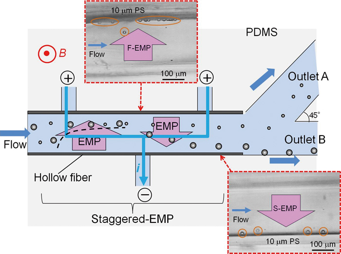 JOURNAL FREE ACCESSA novel microchip separation system for microparticles based on electromagnetophoresis (EMP) was developed. In this system, focusing and separation of flowing microparticles in a microchannel could be performed by staggered-EMP by controlling the electric current applied to the channel locally combined with the split-flow system for fractionation of eluates. To apply the electric current through the flushing medium in the microchannel, a hollow fiber-embedded microchip with multiple electrodes was fabricated. The hollow fiber was made by a semi-permeable membrane and could separate small molecules. This microchip allowed us to apply the electric current to a part of the microchannel without any pressure control device because a main channel contacted with the subchannels that had electrodes through the semi-permeable membrane. Moreover, the separation using this microchip was combined with the split-flow system at two outlets to improve separation efficiency. Using this system, with the split-flow ratio of 10:1, 87% of 3 μm polystyrene (PS) latex particles were isolated from a mixture of 3 and 10 μm particles. Even the separation of 6 and 10 μm PS particles was achieved with about 77% recovery and 100% purity. In addition, by controlling the applied current, size fractionation of polypropylene (PP) particles was demonstrated. Moreover, biological particles such as pollens could be separated with high separation efficiency by this technique.View full abstractDownload PDF (1066K)
 -
Masakazu MUTO, Makoto YAMAMOTO, Masahiro MOTOSUKEArticle type: Original Papers
2016 Volume 32 Issue 1 Pages 49-55
Published: January 10, 2016
Released on J-STAGE: January 10, 2016
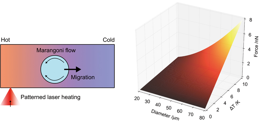 JOURNAL FREE ACCESSWe present a noncontact handling of droplets in a microfluidic platform by the Marangoni convection, interfacial tension driven flow, generated by a light-induced local temperature gradient in the surrounding liquid of the droplet. Droplets flowing in a microchannel experience a force due to the interfacial tension gradient when approaching the heated area. This method provides noncontact, selective and flexible manipulation for droplets flowing in microchannel network. In this study, an O/W emulsion system with oleic acid for the dispersed phase and a buffer solution for the continuous one was used. Trajectory control and trapping for droplets with 5 – 65 pL in volume was achieved by patterned laser irradiation. Also, we quantitatively evaluated the driving force exerted on droplets by measuring the fluidic temperature distribution around the droplet. From the balance of the drag force and the photo-induced Marangoni force, the driving force was determined using the measured temperature gradient of the droplet. From the results, the applicability of noncontact droplet manipulation using the photothermal Marangoni effect by continuous-phase heating has been demonstrated.View full abstractDownload PDF (3567K)
 -
Hirokazu WATANABE, Ryuji KAWANOArticle type: Original Papers
2016 Volume 32 Issue 1 Pages 57-60
Published: January 10, 2016
Released on J-STAGE: January 10, 2016
 JOURNAL FREE ACCESSThis study describes the pore-forming properties of magainin 1 in planar lipid bilayers. These bilayers were prepared by the droplet contact method, which was executed on a microfabricated device for a high-throughput study. We arrayed four droplet chambers parallelly in the single device, and the current measurements were carried out simultaneously. Using this system, we measured the channel current conductance of magainin 1. We determined the pore size and the number of assembling monomers in magainin pores in mammalian and bacterial model membranes. This system is a powerful tool for analyzing transmembrane peptides and their antimicrobial activities.View full abstractDownload PDF (729K)
 -
Manami ITO, Haruka SUGIURA, Shotaro AYUKAWA, Daisuke KIGA, Masahiro TA ...Article type: Original Papers
2016 Volume 32 Issue 1 Pages 61-66
Published: January 10, 2016
Released on J-STAGE: January 10, 2016
 JOURNAL FREE ACCESS
Supplementary materialRecently, micrometer-sized bacterial culture systems have attracted attention as useful tools for synthetic biology studies. Here, we present the development of a bacterial continuous culture system based on a microdroplet open reactor consisting of two types of water-in-oil microdroplets with diameters of several hundred micrometers. A continuous culture was realized the through supply of nutrient substrates and the removal of waste and excess bacterial cells based on repeated fusion and fission of droplets. The growth dynamics was controlled by the interval of fusion. We constructed a microfluidic system and quantitatively assessed the dynamics of the bacterial growth using a mathematical model. This system will facilitate the study of synthetic biology and metabolic engineering in the future.View full abstractDownload PDF (1159K) -
Soojeong SHIN, Ishtiaq AHMED, Jangsun HWANG, Youngmin SEO, Eunwon LEE, ...Article type: Original Papers
2016 Volume 32 Issue 1 Pages 67-73
Published: January 10, 2016
Released on J-STAGE: January 10, 2016
 JOURNAL FREE ACCESSIn recent years, a microfluidic technology has contributed a significant role in biological research, specifically for the study of biofilms. Bacterial biofilms are a source of infections and contamination in the environment due to an extra polymeric matrix. Inadequate uses of antibiotics make the bacterial biofilms antibiotic resistant. Therefore, it is important to determine the effective concentration of antibiotics in order to eliminate bacterial biofilms. The present microfluidic study was carried out to analyze the activities of tobramycin and sodium dodecyl sulfate (SDS) against Pseudomonas aeruginosa biofilms with a continuous flow in order to achieve a greater delivery of the agents. The results show that a co-treatment of tobramycin and SDS significantly reduced the biomass of biofilms (by more than 99%) after 24 h. Tobramycin and SDS killed and detached bacteria in the cores of biofilms. Evidently, our data suggest that a microchannel would be effective for both quantitative and qualitative evaluations in order to test combinatorial effect of drugs and chemicals on a complexed biological system including biofilm.View full abstractDownload PDF (1962K)
 -
Ling LIN, Kazuma MAWATARI, Kyojiro MORIKAWA, Takehiko KITAMORIArticle type: Original Papers
2016 Volume 32 Issue 1 Pages 75-78
Published: January 10, 2016
Released on J-STAGE: January 10, 2016
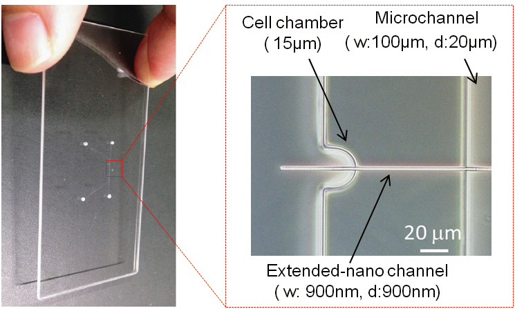 JOURNAL FREE ACCESSSingle cell analysis has been of great interest in recent years. In particular, to achieve living single cell analysis is the ultimate goal to study the dynamic process of the single cell. However, single cell volume is pL in scale, and it is difficult to realize living single cell analysis, even by microfluidic technology (nL-sub nL). Herein, a novel microfluidic platform was developed by integrating a single cell chamber and an extended-nano channel (aL-fL volume). A single cell was isolated and cultured for more than 12 h by pressure-driven flow control. In addition, an electric resistance measurement method was developed to monitor the cell viability without fluorescence labeling. This platform will provide a new method for living single cell analysis by utilizing the novel analytical functions of the extended-nano space.View full abstractDownload PDF (3407K)
 -
Kyojiro MORIKAWA, Takehiko TSUKAHARAArticle type: Original Papers
2016 Volume 32 Issue 1 Pages 79-83
Published: January 10, 2016
Released on J-STAGE: January 10, 2016
 JOURNAL FREE ACCESSIn the field of micro- and nanofluidics, various kinds of novel devices have been developed. For such devices, not only fluidic control but also surface control of micro/nano channels is essential. Recently, fluidic control by hydrophobic nanostructured surfaces have attracted much attention. However, conventional fabrication methods of nanostructures require complicated steps, and integration of the nanostructures into micro/nano channels makes fabrication procedures even more difficult and complicated. In the present study, a simple and easy fabrication method of nanostructures integrated into microchannels was developed. Various sizes of nanostructures were successfully fabricated by changing the plasma etching time and etching with a basic solution. Furthermore, it proved possible to construct highly hydrophobic nanostructured surfaces that could effectively control the fluid in microchannels at designed pressures. We believe that the fabrication method developed here and the results obtained are valuable contributions towards further applications in the field of micro- and nanofluidics.View full abstractDownload PDF (1044K)
 -
Jing WU, Nae Yoon LEEArticle type: Original Papers
2016 Volume 32 Issue 1 Pages 85-92
Published: January 10, 2016
Released on J-STAGE: January 10, 2016
 JOURNAL FREE ACCESSHere, we introduce a simple and facile technique for fabricating microfluidic optical cells by utilizing a micropatterned polymer mold, followed by imprinting on thermoplastic substrates. This process has reduced the surface roughness of the microchannel, making it suitable for microscale optical measurements. The micropatterned polymer mold was fabricated by first micromilling on a poly(methylmethacrylate) (PMMA) substrate, and then transferring the micropattern onto an ultraviolet (UV)-curable optical adhesive. After an anti-adhesion treatment of the polymer mold fabricated using the UV-curable optical adhesive, the polymer mold was used repeatedly for imprinting onto various thermoplastics, such as PMMA, polycarbonate (PC), and poly(ethyleneterephthalate) (PET). The roughness values for the PMMA, PC, and PET microchannels were approximately 11.3, 20.3, and 14.2 nm, respectively, as compared to those obtained by micromilling alone, which were 15.9, 76.8, and 207.5 nm, respectively. Using the imprint-molded thermoplastic optical cell, rhodamine B and copper ions were successfully quantified. The reduced roughness of the microchannel surface resulted in improved sensitivity and reduced noise, paving the way for integration of the detection module so as to realize totally integrated microdevices.View full abstractDownload PDF (24879K)
 -
Mina OKOCHI, Tomohiro KAMIYA, Takeshi OMASA, Masayoshi TANAKA, Hiroyuk ...Article type: Original Papers
2016 Volume 32 Issue 1 Pages 93-97
Published: January 10, 2016
Released on J-STAGE: January 10, 2016
 JOURNAL FREE ACCESSSimple and rapid tools for antibody detection are beneficial for therapeutic monoclonal antibody development. Using a synthetic cationic antibody-recognizing peptide, a label-free colorimetric assay for antibody detection was established, in which a solution containing silver nanoparticles (AgNPs) changes color depending on particle aggregation/dispersion. Among the peptide probes we previously screened as IgG binding, one (NKFRGKYK) has four cationic amino acids and a pI of 10.46. Hence, we hypothesized that the peptide would both bind IgG and induce anionic AgNP aggregation via electrostatic interactions. This dual functionality of the IgG binding peptide could be useful for colorimetric detection. The detection of IgG in a solution containing culture media was investigated, and IgG was successfully detected at 100 to 500 nM within 2 min. This method is promising for high-throughput selection of IgG-producing cells since it is fast, readily observable, and does not involve complicated nanoparticle functionalization.View full abstractDownload PDF (3528K)
 -
Yuto KAZAMA, Akihide HIBARAArticle type: Original Papers
2016 Volume 32 Issue 1 Pages 99-102
Published: January 10, 2016
Released on J-STAGE: January 10, 2016
 JOURNAL FREE ACCESS
Supplementary materialA method of embedding micro-optics into a microfluidic device was proposed and demonstrated. First, the usefulness of embedded right-angle prisms was demonstrated in microscope observation. Lateral-view microscopic observation of an aqueous dye flow in a 100-μm-sized microchannel was demonstrated. Then, the embedded right-angle prisms were utilized for multi-beam laser spectroscopy. Here, crossed-beam thermal lens detection of a liquid sample was applied to glucose detection.View full abstractDownload PDF (1233K) -
Frederik H. KRIEL, Craig PRIESTArticle type: Original Papers
2016 Volume 32 Issue 1 Pages 103-108
Published: January 10, 2016
Released on J-STAGE: January 10, 2016
 JOURNAL FREE ACCESSSpectroscopic analysis of solutions containing samples at high concentrations or molar absorptivity can present practical challenges. In absorbance spectroscopy, short optical path lengths or multiple dilution is required to bring the measured absorbance into the range of the Beer’s Law calibration. We have previously reported an open “pillar-cuvette” with a micropillar array that is spontaneously filled with a precise (nL or μL) volume to create the well-defined optical path of, for example, 10 to 20 μm. Evaporation should not be ignored for open cuvettes and, herein, the volume of loaded sample and the rate of evaporation from the cuvette are studied. It was found that the volume of loaded sample (between 1 and 10 μL) had no effect on the Beer’s Law calibration for methyl orange solutions (molar absorptivity of (2.42 ± 0.02)× 104 L mol−1 cm−1) for cuvettes with a 14.2 ± 0.2 μm path length. Evaporation rates of water from methyl orange solutions were between 2 and 5 nL s−1 (30 – 40% relative humidity; 23°C), depending on the sample concentration and ambient conditions. Evaporation could be reduced by placing a cover slip several millimeters above the cuvette. Importantly, the results show that a “drop-and-measure” method (measurement within ∼3 s of cuvette loading) eliminates the need for extrapolation of the absorbance–time data for accurate analysis of samples.View full abstractDownload PDF (979K)
Notes
-
Alternating Current Cloud Point Extraction on a Microfluidic Chip: the Use of Ferrocenyl SurfactantsYuya USUI, Naoki SASAKIArticle type: Notes
2016 Volume 32 Issue 1 Pages 109-111
Published: January 10, 2016
Released on J-STAGE: January 10, 2016
 JOURNAL FREE ACCESSAlternating current cloud point extraction (ACPE) is a preconcentration technique that can be employed in the analysis of membrane proteins on a microfluidic chip. However, the selectivity of ACPE relies on the hydrophobicity of the analytes. In this study, 11-ferrocenyltrimethylundecylammonium bromide (FTMA) was utilized to introduce electrostatic interaction as part of the ACPE technique. The use of ACPE with oxidized FTMA resulted in efficient concentration of fluorescently labeled anionic membrane proteins. We expect the approach outlined in this report to be useful in the preconcentration technique of microchip electrophoresis.View full abstractDownload PDF (3775K)
 -
Kiichi SATO, Sayaka KIKUCHI, Eri YOSHIDA, Reina ISHII, Naoki SASAKI, K ...Article type: Notes
2016 Volume 32 Issue 1 Pages 113-116
Published: January 10, 2016
Released on J-STAGE: January 10, 2016
 JOURNAL FREE ACCESS
Supplementary materialThe patterned coculture of different types of living cells in a microfluidic device is crucial for the analysis of cellular interactions and cell-cell communication. In the present study, cell patterning was achieved by photocrosslinking benzophenone derivatives in a microfluidic channel. Optimization of UV irradiation conditions enabled successful fixation of live cells. In addition, patterning and co-culture of non-adherent K562 cells and adherent RF-6A cells was achieved by successive rounds of patterning. The present approach is expected to be useful for the development of in vitro methods for studying cell signaling.View full abstractDownload PDF (1245K) -
Kenichi MAENO, Shoma AKI, Kenji SUEYOSHI, Hideaki HISAMOTO, Tatsuro EN ...Article type: Notes
2016 Volume 32 Issue 1 Pages 117-120
Published: January 10, 2016
Released on J-STAGE: January 10, 2016
 JOURNAL FREE ACCESS
Supplementary materialIn this study, a polymer-based two-dimensional photonic crystal (PhC) cavity for visible-light-based optical-sensing applications was designed and fabricated for the first time. The PhC cavity configuration was designed to operate at 650 nm, and fabricated with a polymer (resist) on a silicon substrate using electron-beam lithography. For investigating sensing applications based on shifting of condition exhibiting a photonic bandgap (PBG), the polymer monolayer deposition (layer-by-layer method) was monitored as the light-intensity change at the cavity position. Consequently, the monolayer-level detection of polyions was achieved.View full abstractDownload PDF (885K)
Announcements
-
Article type: Announcements
2016 Volume 32 Issue 1 Pages 121
Published: January 10, 2016
Released on J-STAGE: January 10, 2016
JOURNAL FREE ACCESSDownload PDF (3304K)
- |<
- <
- 1
- >
- >|