
- |<
- <
- 1
- >
- >|
-
吉村 雅満2017 年 38 巻 9 号 p. 447
発行日: 2017/09/10
公開日: 2017/09/20
ジャーナル フリーPDF形式でダウンロード (148K)
-
桜井 健次, 蒋 金星原稿種別: 論文
2017 年 38 巻 9 号 p. 448-454
発行日: 2017/09/10
公開日: 2017/09/20
 ジャーナル フリー
Exotic functions of thin films are quite often connected to the unique atomic and molecular features of buried layers and interfaces. In reality, the structures are far from uniform, but seeing such inhomogeneity is extremely difficult. The present research concerns how to solve the difficulty. The novel technique developed is the X-ray reflectivity imaging. While ordinary X-ray reflectivity gives very precise information on the electron density profile along the depth in thin films, the method can have some imaging capability, by combining with the image reconstruction scheme. Some practical applications will be reported.
抄録全体を表示PDF形式でダウンロード (1896K) -
今田 裕, 三輪 邦之, 今井 みやび, 河原 祥太, 木村 謙介, 金 有洙原稿種別: 研究紹介
2017 年 38 巻 9 号 p. 455-459
発行日: 2017/09/10
公開日: 2017/09/20
 ジャーナル フリー
Energy transfer is an essential process in photosynthesis and various energy-harvesting devices. So far, nanoscale features of energy transfers are unknown because of the limited spatial resolution of conventional methods. We conducted a molecular-level investigation of energy transfers in molecular dimers consisting of a free-base phthalocyanine and magnesium phthalocyanine (H2Pc and MgPc) by scanning tunneling luminescence spectroscopy. Local excitation of an MgPc with the tunneling current of a scanning tunneling microscope give rise to luminescence from a nearby H2Pc as a result of energy transfer. We demonstrate the possibility of a single-molecule valve device for energy transfer, which is revealed by a blinking behavior of energy transfer. Moreover, a back-and-forth energy transfer was observed, where the energy was transferred from the second singlet state (so-called Qy state) of H2Pc to the first singlet state (Q state) of MgPc and finally to the first singlet state (Qx state) of H2Pc.
抄録全体を表示PDF形式でダウンロード (836K) -
 高橋 隆, 菅原 克明, 一ノ倉 聖, 高山 あかり, 長谷川 修司原稿種別: 研究紹介
2017 年 38 巻 9 号 p. 460-465
発行日: 2017/09/10
公開日: 2017/09/20
 ジャーナル フリー
We have fabricated alkali metal (Li, Rb, Cs) and alkaline-earth metal (Ca) intercalated bilayer graphene on SiC substrate, and characterized them by low-energy electron diffraction, angle-resolved photoemission spectroscopy, and 4-point-probe measurements. We observed a free-electron-like state in the center of the Brillouin zone, called “interlayer state”, as well as the folded π/π* bands in Rb-, Cs-, and Ca-intercalated graphene, while it was absent in Li counterpart. Ca-intercalated bilayer graphene shows the zero-resistance below 4K, indicative of the two-dimensional superconductivity. These results suggest that the interlayer state plays an important role for the superconductivity in intercalated bilayer graphene.
抄録全体を表示Editor's pickPDF形式でダウンロード (1371K) -
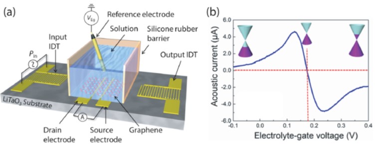
—Lab on a Graphene—小野 尭生, 金井 康, 奥田 聡志, 大野 恭秀, 前橋 兼三, 井上 恒一, 松本 和彦原稿種別: 研究紹介
2017 年 38 巻 9 号 p. 466-472
発行日: 2017/09/10
公開日: 2017/09/20
 ジャーナル フリー
Graphene has attracted much attention since its discovery and has been studied in variety of research fields from quantum physics to biomedical engineering due to its unique properties. The authors have focused on graphene’s keen electrical response to charged biomolecules and investigated biosensing application of graphene field-effect transistor,naming it “lab on a graphene”. In this article, their research progress is reported. The charged biomolecules induce hole or electron carriers to graphene channel and can be detected as a drain current change. Graphene’s high carrier mobility, two-dimensional structure and electrochemical stability achieve highly sensitive and speedy detection. While the bare graphene successfully detected pH changes, aptamer-modified graphene transistor realized specific detection of immunoglobulin E with the detection limit of 290 pM. The authors recently applied this high sensitivity to the detection of human-infectious avian influenza virus. In the preliminary results using pseudo-virus (lectin), glycan-modified graphene transistor shows high specificity of infectivity and sensitivity at subnanomolar range. Novel fabrication process for flexible graphene device and wireless operation of graphene transistor are also reported.
抄録全体を表示PDF形式でダウンロード (1293K)
-
角南 寛, 清水 雄介, 横田 育子, 五十嵐 靖之, 岸本 英博, 松下 正之原稿種別: 研究紹介
2017 年 38 巻 9 号 p. 473-478
発行日: 2017/09/10
公開日: 2017/09/20
 ジャーナル フリー
NIH-3T3 cells were adhered to three kinds of 3D micro-patterned scaffolds, placed face-down into culture medium in glass-bottomed dishes, and cell migration and the scaffolds were observed over 72 h. The three scaffolds differed only in terms of the unit shape of the repetitive pattern, namely a scale structure with equilateral triangular pores, a check structure with regular tetragonal pores, or a stripe structure with rectangular grooves. The angle that cells turn is influenced by the unit shape of the 3D patterned scaffold on which they are cultured. These differences in the angles that migrating cells turned correlated with differences in the angles they extended protrusions. In summary, the unit shape of the micro-patterned scaffold affects the angle at which cells extend, which in turn affects the angle at which migrating cells turn.
抄録全体を表示PDF形式でダウンロード (967K)
-
岩清水 晃2017 年 38 巻 9 号 p. 479-480
発行日: 2017/09/10
公開日: 2017/09/20
 ジャーナル フリー南部鉄器の「南部」の名称は約四〇〇年前,南部信直公が盛岡に城を構え,藩主としてこの地を持っていたことにはじまる。南部藩主が京都から盛岡に釜師を招き茶の湯釜を作らせたといわれる。盛岡には古くから砂鉄,岩鉄などの良質な鉄資源や,川砂,粘土,漆,木炭などの原料がすべて地元で産出され,鋳物産業にはもってこいの立地条件にありそのころから鉄器が製造されてきた。守るべき伝統は守りつつ新しいことへも挑戦していく南部鉄器を紹介いたします。抄録全体を表示PDF形式でダウンロード (486K)
-
岡 博文2017 年 38 巻 9 号 p. 481-482
発行日: 2017/09/10
公開日: 2017/09/20
ジャーナル フリーPDF形式でダウンロード (393K)
-
荻野 俊郎2017 年 38 巻 9 号 p. 483
発行日: 2017/09/10
公開日: 2017/09/20
 ジャーナル フリーPDF形式でダウンロード (233K)
 -
—表面・界面分析の基礎と応用—横田 泰之2017 年 38 巻 9 号 p. 484
発行日: 2017/09/10
公開日: 2017/09/20
 ジャーナル フリーPDF形式でダウンロード (246K)
-
2017 年 38 巻 9 号 p. 485
発行日: 2017/09/10
公開日: 2017/09/20
ジャーナル フリーPDF形式でダウンロード (165K)
- |<
- <
- 1
- >
- >|